SPARC, an acronym for “secreted protein, acidic and rich in cysteine”, is also known as osteonectin or BM-40 (1-5). It is the founding member of a family of secreted matricellular proteins with similar domain structure. The 286 amino acid (aa), 43 kDa protein contains an N-terminal acidic region that binds calcium, a follistatin domain that contains Kazal-like sequences, and a C-terminal extracellular calcium (EC) binding domain with two EF-hand motifs (1-5). Crystal structure modeling shows that residues implicated in cell binding, inhibition of cell spreading, and disassembly of focal adhesions cluster on one face of SPARC, while a collagen binding epitope and an N-glycosylation site are opposite this face (6). SPARC is produced by fibroblasts, capillary endothelial cells, platelets and macrophages, especially in areas of tissue morphogenesis and remodeling (3, 7). SPARC shows context-specific effects, but generally inhibits adhesion, spreading and proliferation, and promotes collagen matrix formation (3-5). For endothelial cells, SPARC disrupts focal adhesions and binds and sequesters PDGF and VEGF (3-5). SPARC is abundantly expressed in bone, where it promotes osteoblast differentiation and inhibits adipogenesis (5, 8). SPARC is potentially cleaved by metalloproteinases, producing an angiogenic peptide that includes the copper-binding sequence KGHK (7). Paradoxically, SPARC is highly expressed in many tumor types undergoing an endothelial to mesenchymal transition; its expression, however, mainly decreases the likelihood of metastasis and confers sensitivity to chemotherapy and radiation (4, 9-11). Stabilin-1, which is expressed on alternately activated macrophages, is the first SPARC receptor to be identified. It binds the SPARC EC domain and mediates endocytosis for degradation (12). Mature human SPARC shows 92%, 92%, 97%, 99%, 96% and 85% aa identity with mouse, rat, canine, bovine, porcine and chick SPARC, respectively.

Key Product Details
Species Reactivity
Validated:
Cited:
Applications
Validated:
Cited:
Label
Antibody Source
Product Specifications
Immunogen
Ala18-Ile303
Accession # P09486
Specificity
Clonality
Host
Isotype
Scientific Data Images for Human SPARC Antibody (122511)
Detection of SPARC in MG-63 cells by Flow Cytometry
MG-63 cells were stained with Mouse Anti-Human SPARC Monoclonal Antibody (Catalog # MAB941, filled histogram) or isotype control antibody (Catalog # MAB002, open histogram) followed by Allophycocyanin-conjugated Anti-Mouse IgG Secondary Antibody (Catalog # F0101B). To facilitate intracellular staining, cells were fixed with Flow Cytometry Fixation Buffer (Catalog # FC004) and permeabilized with Flow Cytometry Permeabilization/Wash Buffer I (Catalog # FC005). View our protocol for Staining Intracellular Molecules.
Detection of SPARC in HT1080 Human Cell Line by Flow Cytometry.
HT1080 human fibrosarcoma cell line was stained with Mouse Anti-Human SPARC Monoclonal Antibody (Catalog # MAB941, filled histogram) or isotype control antibody (Catalog # MAB002, open histogram), followed by Allophycocyanin-conjugated Anti-Mouse IgG Secondary Antibody (Catalog # F0101B). To facilitate intracellular staining, cells were fixed with Flow Cytometry Fixation Buffer (Catalog # FC004) and permeabilized with Flow Cytometry Permeabilization/Wash Buffer I (Catalog # FC005). View our protocol for Staining Intracellular Molecules.
SPARC/Osteonectin in Human Ovary Cancer Tissue.
SPARC/Osteonectin was detected in immersion fixed paraffin-embedded sections of human ovarian clear cell carcinoma tissue using Human SPARC/Osteonectin Monoclonal Antibody (Catalog # MAB941) at 25 µg/mL overnight at 4 °C. Tissue was stained using the Anti-Mouse HRP-DAB Cell & Tissue Staining Kit (brown; Catalog # CTS002) and counterstained with hematoxylin (blue). View our protocol for Chromogenic IHC Staining of Paraffin-embedded Tissue Sections.
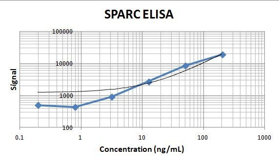
Human SPARC ELISA Standard Curve.
Recombinant Human VSIG8 protein was serially diluted 2-fold and captured by Mouse Anti-Human VSIG8 Monoclonal Antibody (Catalog # MAB9418) coated on a Clear Polystyrene Microplate (Catalog # DY990). Goat Anti-Human SPARC Antigen Affinity-purified Polyclonal Antibody (Catalog # AF941) was biotinylated and incubated with the protein captured on the plate. Detection of the standard curve was achieved by incubating Streptavidin-HRP (Catalog # DY998) followed by Substrate Solution (Catalog # DY999) and stopping the enzymatic reaction with Stop Solution (Catalog # DY994).Applications for Human SPARC Antibody (122511)
CyTOF-ready
ELISA
This antibody functions as an ELISA capture antibody when paired with Goat Anti-Human SPARC Antigen Affinity-purified Polyclonal Antibody (Catalog # AF941).
This product is intended for assay development on various assay platforms requiring antibody pairs. We recommend the Human SPARC DuoSet ELISA Kit (Catalog # DY941-05) for convenient development of a sandwich ELISA or the Human SPARC Quantikine ELISA Kit (Catalog # DSP00) for a complete optimized ELISA.
Immunohistochemistry
Sample: Immersion fixed paraffin-embedded sections of human ovarian clear cell carcinoma tissue
Intracellular Staining by Flow Cytometry
Sample: MG-63 human osteosarcoma cell ine or HT1080 human fibrosarcoma cell line fixed with Flow Cytometry Fixation Buffer (Catalog # FC004) and permeabilized with Flow Cytometry Permeabilization/Wash Buffer I (Catalog # FC005)
Reviewed Applications
Read 2 reviews rated 5 using MAB941 in the following applications:
Flow Cytometry Panel Builder
Bio-Techne Knows Flow Cytometry
Save time and reduce costly mistakes by quickly finding compatible reagents using the Panel Builder Tool.
Advanced Features
- Spectra Viewer - Custom analysis of spectra from multiple fluorochromes
- Spillover Popups - Visualize the spectra of individual fluorochromes
- Antigen Density Selector - Match fluorochrome brightness with antigen density

Formulation, Preparation, and Storage
Purification
Reconstitution
Reconstitute at 0.5 mg/mL in sterile PBS. For liquid material, refer to CoA for concentration.
Formulation
*Small pack size (-SP) is supplied either lyophilized or as a 0.2 µm filtered solution in PBS.
Shipping
Stability & Storage
- 12 months from date of receipt, -20 to -70 °C as supplied.
- 1 month, 2 to 8 °C under sterile conditions after reconstitution.
- 6 months, -20 to -70 °C under sterile conditions after reconstitution.
Calculators
Background: SPARC
References
- Lankat-Buttgereit, B. et al. (1988) FEBS Lett. 236:352.
- Sweetwyne, M. T. et al. (2004) J. Histochem. Cytochem. 52:723.
- Sage, H. et al. (1989) J. Cell Biol. 109:341.
- Framson, P. E. and E. H. Sage (2004) J. Cell. Biochem. 92:679.
- Alford, A. I. and K. D. Hankenson (2006) Bone 38:749.
- Hohenester, E et al. (1997) EMBO J. 16:3778.
- Sage, E. H. et al. (2003) J. Biol. Chem. 278:37849.
- Delany, A. M. et al. (2003) Endocrinology 144:2588.
- Robert, G. et al. (2006) Cancer Res. 66:7516.
- Koblinski, J. E. et al. (2005) Cancer Res. 65:7370.
- Tai, I. T. et al. (2005) J. Clin. Invest. 115:1492.
- Kzhyshkowska, J. et al. (2006) J. Immunol. 176:5825.
Long Name
Alternate Names
Gene Symbol
UniProt
Additional SPARC Products
Product Documents for Human SPARC Antibody (122511)
Certificate of Analysis
To download a Certificate of Analysis, please enter a lot or batch number in the search box below.
Note: Certificate of Analysis not available for kit components.
Product Specific Notices for Human SPARC Antibody (122511)
For research use only
Citations for Human SPARC Antibody (122511)
 Powered by Bioz
Customer Reviews for Human SPARC Antibody (122511) (2)
Have you used Human SPARC Antibody (122511)?
Submit a review and receive an Amazon gift card!
$25/€18/£15/$25CAN/¥2500 Yen for a review with an image
$10/€7/£6/$10CAN/¥1110 Yen for a review without an image
Submit a review
Customer Images
-
Application: ELISASample Tested: Serum and PlasmaSpecies: HumanVerified Customer | Posted 11/08/2019This antibody was used as capture with AF941 as the detection. It worked great in an ELISA to measure human serum and plasma samples.

-
Application: ELISASample Tested: Recombinant proteinSpecies: HumanVerified Customer | Posted 01/09/2018
There are no reviews that match your criteria.
Protocols
Find general support by application which include: protocols, troubleshooting, illustrated assays, videos and webinars.
- 7-Amino Actinomycin D (7-AAD) Cell Viability Flow Cytometry Protocol
- Antigen Retrieval Protocol (PIER)
- Antigen Retrieval for Frozen Sections Protocol
- Appropriate Fixation of IHC/ICC Samples
- Cellular Response to Hypoxia Protocols
- Chromogenic IHC Staining of Formalin-Fixed Paraffin-Embedded (FFPE) Tissue Protocol
- Chromogenic Immunohistochemistry Staining of Frozen Tissue
- ClariTSA™ Fluorophore Kits
- Detection & Visualization of Antibody Binding
- ELISA Sample Preparation & Collection Guide
- ELISA Troubleshooting Guide
- Extracellular Membrane Flow Cytometry Protocol
- Flow Cytometry Protocol for Cell Surface Markers
- Flow Cytometry Protocol for Staining Membrane Associated Proteins
- Flow Cytometry Staining Protocols
- Flow Cytometry Troubleshooting Guide
- Fluorescent IHC Staining of Frozen Tissue Protocol
- Graphic Protocol for Heat-induced Epitope Retrieval
- Graphic Protocol for the Preparation and Fluorescent IHC Staining of Frozen Tissue Sections
- Graphic Protocol for the Preparation and Fluorescent IHC Staining of Paraffin-embedded Tissue Sections
- Graphic Protocol for the Preparation of Gelatin-coated Slides for Histological Tissue Sections
- How to Run an R&D Systems DuoSet ELISA
- How to Run an R&D Systems Quantikine ELISA
- How to Run an R&D Systems Quantikine™ QuicKit™ ELISA
- IHC Sample Preparation (Frozen sections vs Paraffin)
- Immunofluorescent IHC Staining of Formalin-Fixed Paraffin-Embedded (FFPE) Tissue Protocol
- Immunohistochemistry (IHC) and Immunocytochemistry (ICC) Protocols
- Immunohistochemistry Frozen Troubleshooting
- Immunohistochemistry Paraffin Troubleshooting
- Intracellular Flow Cytometry Protocol Using Alcohol (Methanol)
- Intracellular Flow Cytometry Protocol Using Detergents
- Intracellular Nuclear Staining Flow Cytometry Protocol Using Detergents
- Intracellular Staining Flow Cytometry Protocol Using Alcohol Permeabilization
- Intracellular Staining Flow Cytometry Protocol Using Detergents to Permeabilize Cells
- Preparing Samples for IHC/ICC Experiments
- Preventing Non-Specific Staining (Non-Specific Binding)
- Primary Antibody Selection & Optimization
- Propidium Iodide Cell Viability Flow Cytometry Protocol
- Protocol for Heat-Induced Epitope Retrieval (HIER)
- Protocol for Liperfluo
- Protocol for Making a 4% Formaldehyde Solution in PBS
- Protocol for VisUCyte™ HRP Polymer Detection Reagent
- Protocol for the Characterization of Human Th22 Cells
- Protocol for the Characterization of Human Th9 Cells
- Protocol for the Preparation & Fixation of Cells on Coverslips
- Protocol for the Preparation and Chromogenic IHC Staining of Frozen Tissue Sections
- Protocol for the Preparation and Chromogenic IHC Staining of Frozen Tissue Sections - Graphic
- Protocol for the Preparation and Chromogenic IHC Staining of Paraffin-embedded Tissue Sections
- Protocol for the Preparation and Chromogenic IHC Staining of Paraffin-embedded Tissue Sections - Graphic
- Protocol for the Preparation and Fluorescent IHC Staining of Frozen Tissue Sections
- Protocol for the Preparation and Fluorescent IHC Staining of Paraffin-embedded Tissue Sections
- Protocol for the Preparation of Gelatin-coated Slides for Histological Tissue Sections
- Protocol: Annexin V and PI Staining by Flow Cytometry
- Protocol: Annexin V and PI Staining for Apoptosis by Flow Cytometry
- Quantikine HS ELISA Kit Assay Principle, Alkaline Phosphatase
- Quantikine HS ELISA Kit Principle, Streptavidin-HRP Polymer
- Sandwich ELISA (Colorimetric) – Biotin/Streptavidin Detection Protocol
- Sandwich ELISA (Colorimetric) – Direct Detection Protocol
- TUNEL and Active Caspase-3 Detection by IHC/ICC Protocol
- The Importance of IHC/ICC Controls
- Troubleshooting Guide: ELISA
- Troubleshooting Guide: Fluorokine Flow Cytometry Kits
- Troubleshooting Guide: Immunohistochemistry
- View all Protocols, Troubleshooting, Illustrated assays and Webinars
Associated Pathways
