Human toll-like receptor (TLR) family includes ten members that activate the innate immune response via an ability to recognize molecular structures found in a variety of microbial pathogens (1‑3). All TLR family members are type I transmembrane proteins with a large number of extracellular leucine-rich repeats (LRRs) and a cytoplasmic Toll/IL-1 receptor (TIR) domain. Human TLR2 is synthesized as a 784 amino acid (aa) precursor (2) that contains a signal sequence (aa 1-18), an extracellular domain (aa 19-588) with approximately 20 LRRs, a transmembrane segment (aa 589-609), and a cytoplasmic TIR domain (aa 610-784). The receptor is expressed on a number of cell types including monocytes, dendritic cells, neutrophils, B cells endothelial cells, and hepatocytes (1, 2, 4). TLR2 functions as part of a heterodimeric complex with either TLR1 or TLR6, and possibly other co-receptors (1). These complexes recognize lipoproteins and glycolipids from gram-positive and gram-negative bacteria as well as mycoplasma and yeast. TLR2/TLR1 heterodimers bind triacylated lipopeptides, while the TLR2/TLR6 heterodimer preferentially recognizes diacylated lipopeptides (5). Upon ligand recognition, TLR2 delivers an activating signal via the associated adapter molecules, MyD88 and TIRAP (1, 6). TLR2 signaling results in dendritic cell maturation characterized by increased surface expression of class II MHC and the T cell costimulators, CD80 and CD86 (1, 2). Activation via TLR2 also results in production of a number of pro-inflammatory cytokines including TNF-alpha, IL-2, IL-6, IL-12, and MIP-2 (1-3).

Key Product Details
Species Reactivity
Validated:
Human
Cited:
Human, Mouse, Porcine
Applications
Validated:
Neutralization, Flow Cytometry, CyTOF-ready
Cited:
Immunohistochemistry-Paraffin, Western Blot, Neutralization, Flow Cytometry, Immunocytochemistry, Blocking, ELISA Development, Functional Assay
Label
Unconjugated
Antibody Source
Monoclonal Mouse IgG2B Clone # 383936
Loading...
Product Specifications
Immunogen
NS0 mouse myeloma cell line transfected with human TLR2
Accession # O60603
Accession # O60603
Specificity
Stains human TLR2 transfectants but not irrelevant transfectants.
Clonality
Monoclonal
Host
Mouse
Isotype
IgG2B
Endotoxin Level
<0.10 EU per 1 μg of the antibody by the LAL method.
Scientific Data Images for Human TLR2 Antibody (383936)
IL‑8 Secretion Induced by Pam3CSK4and Neutralization by Human TLR2 Antibody.
The synthetic tripalmitoylated lipopeptide Pam3CSK4stimulates IL-8 secretion in the HEK293 human embryonic kidney cell line transfected with human TLR2, in a dose-dependent manner (orange line), as measured by the Human CXCL8/IL-8 Quantikine ELISA Kit (Catalog # D8000C). IL-8 secretion elicited by Pam3CSK4(0.5 µg/mL) is neutralized (green line) by increasing concentrations of Mouse Anti-Human TLR2 Monoclonal Antibody (Catalog # MAB2616). The ND50 is typically 0.03-0.15 µg/mL.
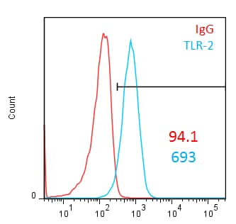
Detection of TLR2 in Human Monocytes by Flow Cytometry.
Human whole blood monocytes were stained with Mouse Anti-Human TLR2 Monoclonal Antibody (Catalog # MAB2616, filled histogram) or isotype control antibody (Catalog # MAB0041, open histogram), followed by Phycoerythrin-conjugated Anti-Mouse IgG F(ab')2Secondary Antibody (Catalog # F0102B).Applications for Human TLR2 Antibody (383936)
Application
Recommended Usage
CyTOF-ready
Ready to be labeled using established conjugation methods. No BSA or other carrier proteins that could interfere with conjugation.
Flow Cytometry
2.5 µg/106 cells
Sample: Human whole blood monocytes
Sample: Human whole blood monocytes
Neutralization
Measured by its ability to neutralize Pam3CSK4-induced IL‑8 secretion in the HEK293 human embryonic kidney cell line transfected with human TLR2. The Neutralization Dose (ND50) is typically 0.03-0.15 µg/mL in the presence of 0.5 µg/mL The synthetic tripalmitoylated lipopeptide Pam3CSK4.
Reviewed Applications
Read 3 reviews rated 4.7 using MAB2616 in the following applications:
Flow Cytometry Panel Builder
Bio-Techne Knows Flow Cytometry
Save time and reduce costly mistakes by quickly finding compatible reagents using the Panel Builder Tool.
Advanced Features
- Spectra Viewer - Custom analysis of spectra from multiple fluorochromes
- Spillover Popups - Visualize the spectra of individual fluorochromes
- Antigen Density Selector - Match fluorochrome brightness with antigen density

Formulation, Preparation, and Storage
Purification
Protein A or G purified from hybridoma culture supernatant
Reconstitution
Reconstitute at 0.5 mg/mL in sterile PBS. For liquid material, refer to CoA for concentration.
Loading...
Formulation
Lyophilized from a 0.2 μm filtered solution in PBS with Trehalose. *Small pack size (SP) is supplied either lyophilized or as a 0.2 µm filtered solution in PBS.
Shipping
Lyophilized product is shipped at ambient temperature. Liquid small pack size (-SP) is shipped with polar packs. Upon receipt, store immediately at the temperature recommended below.
Stability & Storage
Use a manual defrost freezer and avoid repeated freeze-thaw cycles.
- 12 months from date of receipt, -20 to -70 °C as supplied.
- 1 month, 2 to 8 °C under sterile conditions after reconstitution.
- 6 months, -20 to -70 °C under sterile conditions after reconstitution.
Calculators
Background: TLR2
References
- Wetzler, L. (2003) Vaccine 21:S2/55.
- Kirschning, C. and R. Schumann (2002) Curr. Top. microbiol. Immunol. 270:121.
- Netea, M. et al. (2004) J. Leukoc. Biol. 75:749.
- Flo, T. et al. (2001) J. Leukoc. Biol. 69:474.
- Akira, S. (2003) Curr. Opin. Immunol. 15:5.
- Yamamoto, M. et al. (2002) Nature 420:324.
Long Name
Toll-like Receptor 2
Alternate Names
CD282
Gene Symbol
TLR2
UniProt
Additional TLR2 Products
Product Documents for Human TLR2 Antibody (383936)
Certificate of Analysis
To download a Certificate of Analysis, please enter a lot or batch number in the search box below.
Note: Certificate of Analysis not available for kit components.
Product Specific Notices for Human TLR2 Antibody (383936)
For research use only
Citations for Human TLR2 Antibody (383936)
 Powered by Bioz
Customer Reviews for Human TLR2 Antibody (383936) (3)
4.7 out of 5
3 Customer Ratings
Have you used Human TLR2 Antibody (383936)?
Submit a review and receive an Amazon gift card!
$25/€18/£15/$25CAN/¥2500 Yen for a review with an image
$10/€7/£6/$10CAN/¥1110 Yen for a review without an image
Submit a review
Customer Images

Showing
1
-
3 of
3 reviews
Showing All
Filter By:
-
Application: MicroarraySample Tested: EDTA PlasmaSpecies: HumanVerified Customer | Posted 12/13/2019Antibody was printed on custom arrays and incubated with fluorescently labeled human EDTA plasma

-
Application: MicroarraysSample Tested: EDTA PlasmaSpecies: HumanVerified Customer | Posted 03/11/2019
-
Application: Flow CytometrySample Tested: KF6 human keratinocytes and THP-1 human acute monocytic leukemia cell lineSpecies: HumanVerified Customer | Posted 04/26/2016
There are no reviews that match your criteria.
Protocols
Find general support by application which include: protocols, troubleshooting, illustrated assays, videos and webinars.
- 7-Amino Actinomycin D (7-AAD) Cell Viability Flow Cytometry Protocol
- Extracellular Membrane Flow Cytometry Protocol
- Flow Cytometry Protocol for Cell Surface Markers
- Flow Cytometry Protocol for Staining Membrane Associated Proteins
- Flow Cytometry Staining Protocols
- Flow Cytometry Troubleshooting Guide
- Intracellular Flow Cytometry Protocol Using Alcohol (Methanol)
- Intracellular Flow Cytometry Protocol Using Detergents
- Intracellular Nuclear Staining Flow Cytometry Protocol Using Detergents
- Intracellular Staining Flow Cytometry Protocol Using Alcohol Permeabilization
- Intracellular Staining Flow Cytometry Protocol Using Detergents to Permeabilize Cells
- Propidium Iodide Cell Viability Flow Cytometry Protocol
- Protocol for Liperfluo
- Protocol for the Characterization of Human Th22 Cells
- Protocol for the Characterization of Human Th9 Cells
- Protocol: Annexin V and PI Staining by Flow Cytometry
- Protocol: Annexin V and PI Staining for Apoptosis by Flow Cytometry
- Troubleshooting Guide: Fluorokine Flow Cytometry Kits
- View all Protocols, Troubleshooting, Illustrated assays and Webinars


