Human UCH-L1/PGP9.5 Antibody (671108)
R&D Systems | Catalog # MAB6007

Key Product Details
Validated by
Knockout/Knockdown
Species Reactivity
Validated:
Human
Cited:
Human
Applications
Validated:
Knockout Validated, Immunohistochemistry, Western Blot, Simple Western, Immunoprecipitation
Cited:
Immunohistochemistry, Western Blot, Immunoprecipitation, Surface Plasmon Resonance (SPR
Label
Unconjugated
Antibody Source
Monoclonal Mouse IgG2A Clone # 671108
Loading...
Product Specifications
Immunogen
E. coli-derived recombinant human UCH-L1/PGP9.5
Gln2-Ala223
Accession # P09936
Gln2-Ala223
Accession # P09936
Specificity
Detects human UCH-L1/PGP9.5 in direct ELISAs and Western blots. In direct ELISAs and Western blots, no
cross-reactivity with recombinant human UCH-L3 is observed.
Clonality
Monoclonal
Host
Mouse
Isotype
IgG2A
Scientific Data Images for Human UCH-L1/PGP9.5 Antibody (671108)
Detection of Human UCH-L1/PGP9.5 by Western Blot.
Western blot shows lysates of human brain (cortex) tissue. PVDF membrane was probed with 2 µg/mL of Mouse Anti-Human UCH-L1/PGP9.5 Monoclonal Antibody (Catalog # MAB6007) followed by HRP-conjugated Anti-Mouse IgG Secondary Antibody (Catalog # HAF007). A specific band was detected for UCH-L1/PGP9.5 at approximately 29 kDa (as indicated). This experiment was conducted under reducing conditions and using Immunoblot Buffer Group 1.
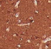
UCH-L1/PGP9.5 in Human Prostate.
UCH-L1/PGP9.5 was detected in immersion fixed paraffin-embedded sections of human prostate using Mouse Anti-Human UCH-L1/PGP9.5 Monoclonal Antibody (Catalog # MAB6007) at 15 µg/mL overnight at 4 °C. Before incubation with the primary antibody, tissue was subjected to heat-induced epitope retrieval using Antigen Retrieval Reagent-Basic (Catalog # CTS013). Tissue was stained using the Anti-Mouse HRP-DAB Cell & Tissue Staining Kit (brown; Catalog # CTS002) and counterstained with hematoxylin (blue). Specific staining was localized to cytoplasm of glandular epithelial cells. View our protocol for Chromogenic IHC Staining of Paraffin-embedded Tissue Sections.
Detection of Human and Mouse UCH-L1/PGP9.5 by Simple WesternTM.
Simple Western lane view shows lysates of human brain (cortex) tissue and mouse brain (cortex) tissue, loaded at 0.2 mg/mL. A specific band was detected for UCH-L1/PGP9.5 at approximately 32 kDa (as indicated) using 10 µg/mL of Mouse Anti-Human UCH-L1/PGP9.5 Monoclonal Antibody (Catalog # MAB6007). This experiment was conducted under reducing conditions and using the 12-230 kDa separation system.
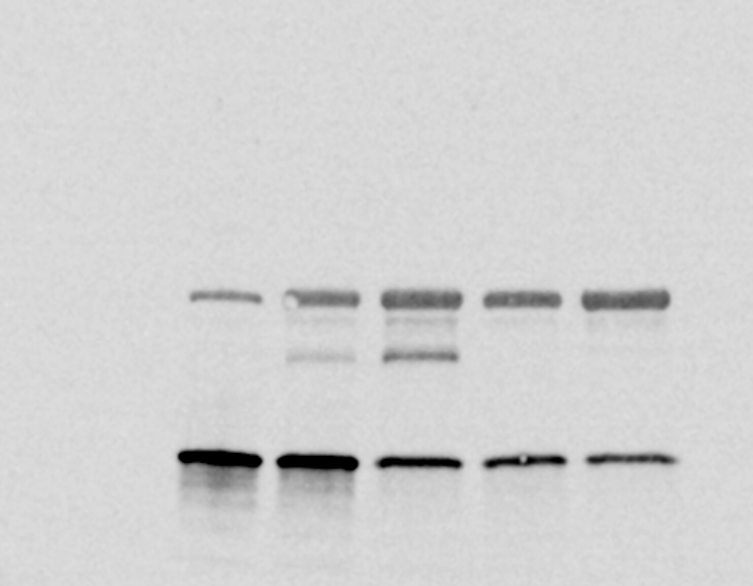
Western Blot Shows Human UCH-L1/PGP9.5 Specificity by Using Knockout Cell Line.
Western blot shows lysates of HEK293T human embryonic kidney parental cell line and UCH-L1/PGP9.5 knockout HEK293T cell line (KO). PVDF membrane was probed with 2 µg/mL of Mouse Anti-Human UCH-L1/PGP9.5 Monoclonal Antibody (Catalog # MAB6007) followed by HRP-conjugated Anti-Mouse IgG Secondary Antibody (Catalog # HAF018). A specific band was detected for UCH-L1/PGP9.5 at approximately 28 kDa (as indicated) in the parental HEK293T cell line, but is not detectable in knockout HEK293T cell line. GAPDH (Catalog # MAB5718) is shown as a loading control. This experiment was conducted under reducing conditions and using Immunoblot Buffer Group 1.
Detection of UCH-L1/PGP9.5 by Western Blot
UCHL1 was highly expressed in human leiomyoma. (A–C) Strong expression of UCHL1 was observed by (A) immunohistochemistry, (B) real-time PCR, and (C) Western blotting in leiomyoma. The scale bar shows 50 µm. The bar shows the standard error (* p < 0.05, ** p < 0.01). Image collected and cropped by CiteAb from the following open publication (https://pubmed.ncbi.nlm.nih.gov/36830563), licensed under a CC-BY license. Not internally tested by R&D Systems.
Detection of UCH-L1/PGP9.5 by Western Blot
UCHL1 was highly expressed in human leiomyoma. (A–C) Strong expression of UCHL1 was observed by (A) immunohistochemistry, (B) real-time PCR, and (C) Western blotting in leiomyoma. The scale bar shows 50 µm. The bar shows the standard error (* p < 0.05, ** p < 0.01). Image collected and cropped by CiteAb from the following open publication (https://pubmed.ncbi.nlm.nih.gov/36830563), licensed under a CC-BY license. Not internally tested by R&D Systems.Applications for Human UCH-L1/PGP9.5 Antibody (671108)
Application
Recommended Usage
Immunohistochemistry
8-25 µg/mL
Sample: Immersion fixed paraffin-embedded sections of human prostate
Sample: Immersion fixed paraffin-embedded sections of human prostate
Immunoprecipitation
25 µg/mL
Sample: Cell lysates spiked with Recombinant Human UCH-L1/PGP9.5 (Catalog # 6007-CY), see our available Western blot detection antibodies
Sample: Cell lysates spiked with Recombinant Human UCH-L1/PGP9.5 (Catalog # 6007-CY), see our available Western blot detection antibodies
Knockout Validated
UCH-L1/PGP9.5
is specifically detected in HEK293T human embryonic kidney parental cell line but is not detectable in
UCH-L1/PGP9.5 knockout HEK293T cell line.
Simple Western
10 µg/mL
Sample: Human brain (cortex) tissue and mouse brain (cortex) tissue
Sample: Human brain (cortex) tissue and mouse brain (cortex) tissue
Western Blot
2 µg/mL
Sample: Human brain (cortex) tissue
Sample: Human brain (cortex) tissue
Reviewed Applications
Read 3 reviews rated 4 using MAB6007 in the following applications:
Formulation, Preparation, and Storage
Purification
Protein A or G purified from hybridoma culture supernatant
Reconstitution
Sterile PBS to a final concentration of 0.5 mg/mL. For liquid material, refer to CoA for concentration.
Loading...
Formulation
Lyophilized from a 0.2 μm filtered solution in PBS with Trehalose. *Small pack size (SP) is supplied either lyophilized or as a 0.2 µm filtered solution in PBS.
Shipping
Lyophilized product is shipped at ambient temperature. Liquid small pack size (-SP) is shipped with polar packs. Upon receipt, store immediately at the temperature recommended below.
Stability & Storage
Use a manual defrost freezer and avoid repeated freeze-thaw cycles.
- 12 months from date of receipt, -20 to -70 °C as supplied.
- 1 month, 2 to 8 °C under sterile conditions after reconstitution.
- 6 months, -20 to -70 °C under sterile conditions after reconstitution.
Calculators
Background: UCH-L1/PGP9.5
Long Name
Ubiquitin C-terminal Hydrolase L1
Alternate Names
PARK5, PGP9.5, UCHL1
Gene Symbol
UCHL1
UniProt
Additional UCH-L1/PGP9.5 Products
Product Documents for Human UCH-L1/PGP9.5 Antibody (671108)
Certificate of Analysis
To download a Certificate of Analysis, please enter a lot or batch number in the search box below.
Note: Certificate of Analysis not available for kit components.
Product Specific Notices for Human UCH-L1/PGP9.5 Antibody (671108)
For research use only
Related Research Areas
Citations for Human UCH-L1/PGP9.5 Antibody (671108)
 Powered by Bioz
Customer Reviews for Human UCH-L1/PGP9.5 Antibody (671108) (3)
4 out of 5
3 Customer Ratings
Have you used Human UCH-L1/PGP9.5 Antibody (671108)?
Submit a review and receive an Amazon gift card!
$25/€18/£15/$25CAN/¥2500 Yen for a review with an image
$10/€7/£6/$10CAN/¥1110 Yen for a review without an image
Submit a review
Customer Images
Showing
1
-
3 of
3 reviews
Showing All
Filter By:
-
Application: Western BlotSample Tested: Cancer cell lysatesSpecies: HumanVerified Customer | Posted 06/25/2025This antibody has a few additional bands in some samples, likely due to interactions between antibodies or ubiquitination of other proteins in sample

-
Application: ImmunohistochemistrySample Tested: Brain tissueSpecies: HumanVerified Customer | Posted 11/10/2021
-
Application: Immunocytochemistry/ImmunofluorescenceSample Tested: Skin tissueSpecies: HumanVerified Customer | Posted 05/21/20211st PGP9.5 in 1:500, 2nd antibody Cy3 Red staining = laminin + Cy5 +Dapi mounting medium Collagen autofluorescence was a problem in some of the sections. Zamboni Fixed, 20% sucr.
There are no reviews that match your criteria.
Protocols
Find general support by application which include: protocols, troubleshooting, illustrated assays, videos and webinars.
- Antigen Retrieval Protocol (PIER)
- Antigen Retrieval for Frozen Sections Protocol
- Appropriate Fixation of IHC/ICC Samples
- Cellular Response to Hypoxia Protocols
- Chromogenic IHC Staining of Formalin-Fixed Paraffin-Embedded (FFPE) Tissue Protocol
- Chromogenic Immunohistochemistry Staining of Frozen Tissue
- ClariTSA™ Fluorophore Kits
- Detection & Visualization of Antibody Binding
- Fluorescent IHC Staining of Frozen Tissue Protocol
- Graphic Protocol for Heat-induced Epitope Retrieval
- Graphic Protocol for the Preparation and Fluorescent IHC Staining of Frozen Tissue Sections
- Graphic Protocol for the Preparation and Fluorescent IHC Staining of Paraffin-embedded Tissue Sections
- Graphic Protocol for the Preparation of Gelatin-coated Slides for Histological Tissue Sections
- IHC Sample Preparation (Frozen sections vs Paraffin)
- Immunofluorescent IHC Staining of Formalin-Fixed Paraffin-Embedded (FFPE) Tissue Protocol
- Immunohistochemistry (IHC) and Immunocytochemistry (ICC) Protocols
- Immunohistochemistry Frozen Troubleshooting
- Immunohistochemistry Paraffin Troubleshooting
- Immunoprecipitation Protocol
- Preparing Samples for IHC/ICC Experiments
- Preventing Non-Specific Staining (Non-Specific Binding)
- Primary Antibody Selection & Optimization
- Protocol for Heat-Induced Epitope Retrieval (HIER)
- Protocol for Making a 4% Formaldehyde Solution in PBS
- Protocol for VisUCyte™ HRP Polymer Detection Reagent
- Protocol for the Preparation & Fixation of Cells on Coverslips
- Protocol for the Preparation and Chromogenic IHC Staining of Frozen Tissue Sections
- Protocol for the Preparation and Chromogenic IHC Staining of Frozen Tissue Sections - Graphic
- Protocol for the Preparation and Chromogenic IHC Staining of Paraffin-embedded Tissue Sections
- Protocol for the Preparation and Chromogenic IHC Staining of Paraffin-embedded Tissue Sections - Graphic
- Protocol for the Preparation and Fluorescent IHC Staining of Frozen Tissue Sections
- Protocol for the Preparation and Fluorescent IHC Staining of Paraffin-embedded Tissue Sections
- Protocol for the Preparation of Gelatin-coated Slides for Histological Tissue Sections
- R&D Systems Quality Control Western Blot Protocol
- TUNEL and Active Caspase-3 Detection by IHC/ICC Protocol
- The Importance of IHC/ICC Controls
- Troubleshooting Guide: Immunohistochemistry
- Troubleshooting Guide: Western Blot Figures
- Western Blot Conditions
- Western Blot Protocol
- Western Blot Protocol for Cell Lysates
- Western Blot Troubleshooting
- Western Blot Troubleshooting Guide
- View all Protocols, Troubleshooting, Illustrated assays and Webinars
Loading...