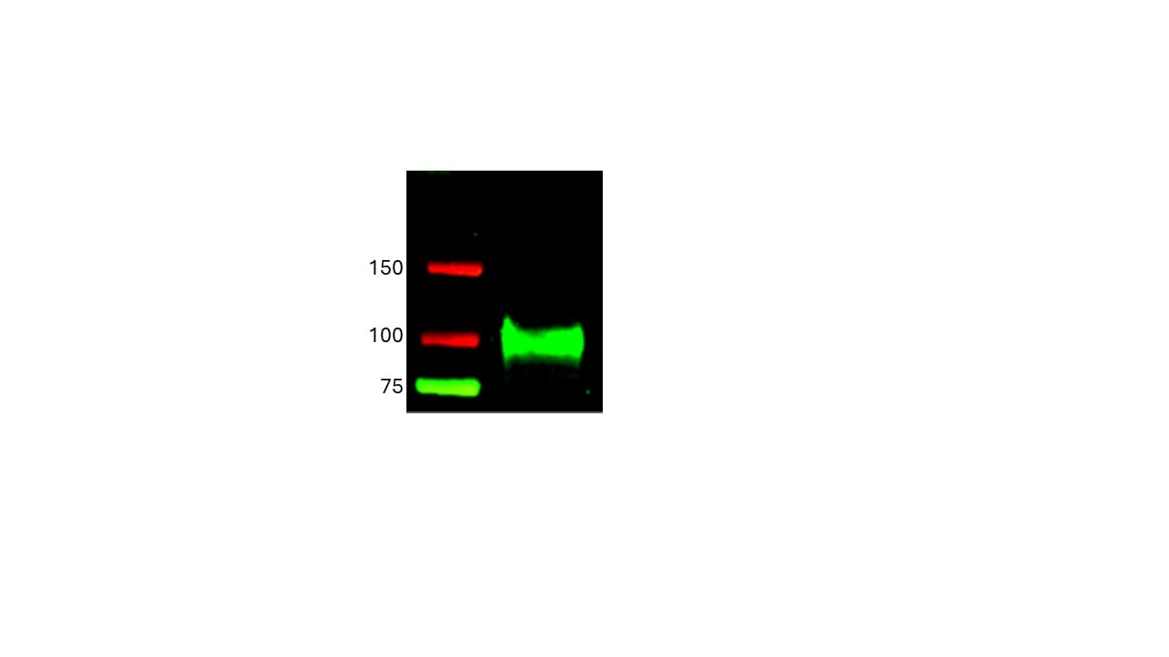
Detection of Mouse ICAM-1/CD54 by Western Blot
D-JNKI1 treatment rescues inflammation in delta / delta ep2 mice.Mice were treated with D-JNKI1 or TAT peptide (22 mg/kg i.p. at 10 days of age) and analyzed after 12 days (A) D-JNKI1 treatment prevents disease onset in delta / delta ep2 mice. Immunoblot of epidermal lysates showing the effect of D-JNKI1 on the phosphorylation and expression of the indicated proteins, quantified as in Figure 1F. ACTB is shown as a loading control. (B–D) Decreased eyelid inflammation, mast cells infiltration (B; TB+; quantified in the plot on the right), epidermal chemokine/cytokine levels (C) and activated T cells, B cells and dendritic cells in lymph nodes (D) in D-JNKI1-treated delta / delta ep2 mice. Scale bars, 25 µm. Data represent mean ± SEM (n = 3–5; p1 = 0.026, p2 = 0.042, p3 = 0.022, p4 = 0.048, p5 = 0.044, p6 = 0.020, p7 = 0.025, p8 = 0.018, p9 = 0.016, p10 = 0.014, p11 = 0.023, p12 = 0.011, p13 = 0.039, p14 = 0.049, p15 = 0.015, p16 = 0.003, p17 = 1.70E-4, p18 = 0.008, p19 = 0.008, p20 = 0.004, p21 = 0.003, p22 = 0.017, p23 = 0.026, p24 = 0.027, p25 = 0.005, p26 = 2.13E-6, p27 = 4.50E-8, p28 = 1.39E-5, p29 = 0.001, p30 = 0.001, p31 = 0.001, p32 = 0.023, p33 = 2.35E-4, p34 = 0.050, p35 = 0.002, p36 = 0.050 and p37 = 0.012).DOI:
https://dx.doi.org/10.7554/eLife.14012.015K6 expression and epidermal chemokine and cytokine levels in D-JNKI1-treated mice.(A) K6 expression is indistinguishable in TAT or D-JNKI1 treated F/F2 and △/△ep2 littermates. Scale bars, 25 µm. (B) Inflammatory chemokines and cytokines in epidermal lysates of TAT or D-JNKI1-treated mice. Data represent mean ± SEM (n = 3–5; p1 = 0.005, p2 = 0.001, p3 = 0.043, p4 = 0.026, p5 = 0.032, p6 = 0.016 and p7 = 0.051).DOI:
https://dx.doi.org/10.7554/eLife.14012.016The inflammatory phenotype of delta / delta ep2 mice is not rescued by MyD88, caspase 1/11, or TNF knockout.Macroscopic appearance, spleen and lymph node size and circulating blood cell analysis are shown for the indicated genotypes (A–C). (A) Representative pictures of 4 month old delta / delta ep2, delta / delta ep2 MyD88


 Powered by Bioz