Interleukin 10, also known as cytokine synthesis inhibitory factor (CSIF), is the charter member of the IL-10 family of alpha -helical cytokines that also includes IL-19, IL‑20, IL-22, and IL-24. IL-10 is secreted by many activated hematopoietic cell types as well as hepatic stellate cells, keratinocytes, and placental cytotrophoblasts. Mature mouse IL-10 shares 85% amino acid sequence identity with rat and 70%‑77% with bovine, canine, equine, feline, human, ovine, and porcine IL-10. Whereas human IL-10 is active on mouse cells, mouse IL-10 does not act on human cells. IL-10 is a 178 amino acid molecule that contains two intrachain disulfide bridges and is expressed as a 36 kDa noncovalently associated homodimer. The IL-10 dimer binds to two IL-10 R alpha /IL-10 R1 chains, resulting in recruitment of two IL‑10 R beta /IL‑10 R2 chains and activation of a signaling cascade involving JAK1, TYK2, and STAT3. IL-10 R beta does not bind IL-10 by itself but is required for signal transduction. IL-10 R beta also associates with IL-20 R alpha, IL-22 R alpha, or IL-28 R alpha to form the receptor complexes for IL‑22, IL-26, IL-28, and IL-29. IL-10 is a critical molecule in the control of viral infections and allergic and autoimmune inflammation. It promotes phagocytic uptake and Th2 responses but suppresses antigen presentation and Th1 proinflammatory responses.

Key Product Details
Species Reactivity
Validated:
Mouse
Cited:
Mouse
Applications
Validated:
Western Blot, Neutralization
Cited:
Western Blot, Neutralization
Label
Unconjugated
Antibody Source
Polyclonal Goat IgG
Loading...
Product Specifications
Immunogen
E. coli-derived recombinant mouse IL‑10
Ser19-Ser178
Accession # NP_034678
Ser19-Ser178
Accession # NP_034678
Specificity
Detects mouse IL-10 in direct ELISAs and Western blots. In direct ELISAs, approximately 30% cross-reactivity with recombinant rat IL‑10 and recombinant cotton rat IL-10 is observed, and less than 5% cross-reactivity with recombinant porcine IL-10, recombinant canine IL-10, recombinant feline IL-10, recombinant equine IL-10, recombinant viral IL-10, recombinant guinea pig IL-10, and recombinant human IL‑10 is observed.
Clonality
Polyclonal
Host
Goat
Isotype
IgG
Endotoxin Level
<0.10 EU per 1 μg of the antibody by the LAL method.
Scientific Data Images for Mouse IL-10 Antibody
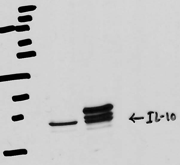
Detection of Recombinant Mouse IL‑10 by Western Blot.
Western blot shows 25 ng of Recombinant Mouse IL-10 (Catalog # 417-ML), Recombinant Human IL-10 (Catalog # 217-IL), Recombinant Rat IL-10 (Catalog # 522-RLB), and Recombinant Human IL-26/AK155 Monomer (Catalog # 1375-IL). PVDF Membrane was probed with 0.1 µg/mL of Goat Anti-Mouse IL-10 Antigen Affinity-purified Polyclonal Antibody (Catalog # AF-417-NA) followed by HRP-conjugated Anti-Goat IgG Secondary Antibody (Catalog # HAF109). A specific band was detected for IL-10 at approximately 16 kDa (as indicated). This experiment was conducted under reducing conditions and using Immunoblot Buffer Group 3.
Cell Proliferation Induced by IL‑10 and Neutralization by Mouse IL‑10 Antibody.
Recombinant Mouse IL-10 (Catalog # 417-ML) stimulates proliferation in the MC/9-2 mouse mast cell line in a dose-dependent manner (orange line). Proliferation elicited by Recombinant Mouse IL-10 (2.5 ng/mL) is neutralized (green line) by increasing concentrations of Goat Anti-Mouse IL-10 Antigen Affinity-purified Polyclonal Antibody (Catalog # AF-417-NA). The ND50 is typically 0.01-0.05 µg/mL.
Mouse IL-10 ELISA Standard Curve
Recombinant Mouse IL‑10 (Catalog # 417-ML) was serially diluted and captured by Rat Anti-Mouse IL‑10 Monoclonal Antibody (Catalog # MAB417) coated on a Clear Polystyrene Microplate (Catalog # DY990). Goat Anti-Mouse IL‑10 Antigen Affinity-purified Polyclonal Antibody (Catalog # AF-417-NA) was biotinylated and incubated with the protein captured on the plate. Detection of the standard curve was achieved by incubating Streptavidin-HRP (Catalog # DY998)Applications for Mouse IL-10 Antibody
Application
Recommended Usage
Western Blot
0.1 µg/mL
Sample: Recombinant Mouse IL‑10 (Catalog # 417-ML)
Sample: Recombinant Mouse IL‑10 (Catalog # 417-ML)
Neutralization
Measured by its ability to neutralize IL‑10-induced proliferation in the MC/9‑2 mouse mast cell line. Thompson-Snipes, L. et al. (1991) J. Exp. Med. 173:507. The Neutralization Dose (ND50) is typically 0.01-0.05 µg/mL in the presence of 2.5 ng/mL Recombinant Mouse IL‑10.
Reviewed Applications
Read 1 review rated 5 using AF-417-NA in the following applications:
Formulation, Preparation, and Storage
Purification
Antigen Affinity-purified
Reconstitution
Reconstitute at 0.2 mg/mL in sterile PBS. For liquid material, refer to CoA for concentration.
Loading...
Formulation
Lyophilized from a 0.2 μm filtered solution in PBS with Trehalose. *Small pack size (SP) is supplied either lyophilized or as a 0.2 µm filtered solution in PBS.
Shipping
Lyophilized product is shipped at ambient temperature. Liquid small pack size (-SP) is shipped with polar packs. Upon receipt, store immediately at the temperature recommended below.
Stability & Storage
Use a manual defrost freezer and avoid repeated freeze-thaw cycles.
- 12 months from date of receipt, -20 to -70 °C as supplied.
- 1 month, 2 to 8 °C under sterile conditions after reconstitution.
- 6 months, -20 to -70 °C under sterile conditions after reconstitution.
Calculators
Background: IL-10
Long Name
Interleukin 10
Alternate Names
CSIF, GVHDS, IL10, IL10A, TGIF
Entrez Gene IDs
Gene Symbol
IL10
UniProt
Additional IL-10 Products
Product Documents for Mouse IL-10 Antibody
Certificate of Analysis
To download a Certificate of Analysis, please enter a lot or batch number in the search box below.
Note: Certificate of Analysis not available for kit components.
Product Specific Notices for Mouse IL-10 Antibody
For research use only
Related Research Areas
Citations for Mouse IL-10 Antibody
 Powered by Bioz
Customer Reviews for Mouse IL-10 Antibody (1)
5 out of 5
1 Customer Rating
Have you used Mouse IL-10 Antibody?
Submit a review and receive an Amazon gift card!
$25/€18/£15/$25CAN/¥2500 Yen for a review with an image
$10/€7/£6/$10CAN/¥1110 Yen for a review without an image
Submit a review
Customer Images
Showing
1
-
1 of
1 review
Showing All
Filter By:
-
Application: Western BlotSample Tested: Aorta tissueSpecies: MouseVerified Customer | Posted 04/06/2021

There are no reviews that match your criteria.
Protocols
Find general support by application which include: protocols, troubleshooting, illustrated assays, videos and webinars.
- Cellular Response to Hypoxia Protocols
- R&D Systems Quality Control Western Blot Protocol
- Troubleshooting Guide: Western Blot Figures
- Western Blot Conditions
- Western Blot Protocol
- Western Blot Protocol for Cell Lysates
- Western Blot Troubleshooting
- Western Blot Troubleshooting Guide
- View all Protocols, Troubleshooting, Illustrated assays and Webinars
Loading...
Associated Pathways


