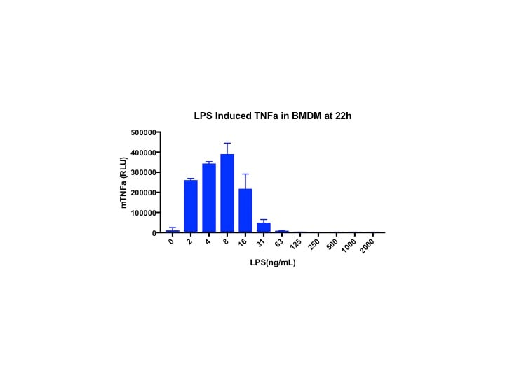
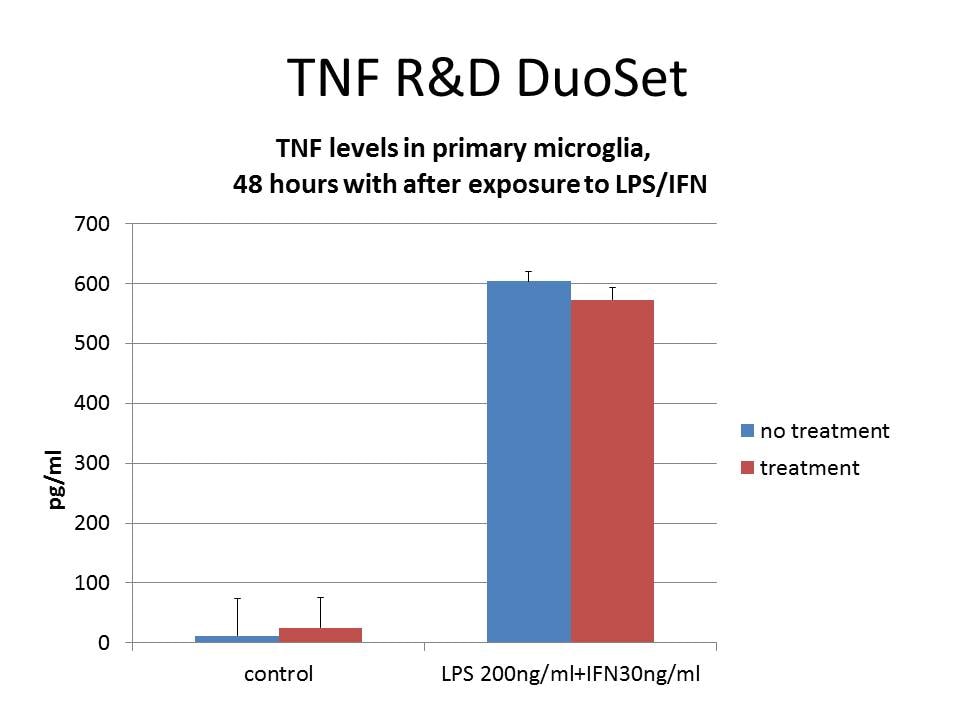
Mouse TNF-alpha DuoSet ELISA
R&D Systems | Catalog # DY410
Key Product Details
Assay Type
Assay Range
Sample Type
Note: Diluents for complex matrices, such as serum and plasma, should be evaluated prior to use in this DuoSet
Reactivity
Mouse TNF-alpha DuoSet ELISA Features
- Optimized capture and detection antibody pairings with recommended concentrations save lengthy development time
- Development protocols are provided to guide further assay optimization
- Assay can be customized to your specific needs
- Economical alternative to complete kits
Product Summary for Mouse TNF-alpha DuoSet ELISA
This DuoSet ELISA Development kit contains the basic components required for the development of sandwich ELISAs to measure natural and recombinant mouse TNF-alpha. The suggested diluent is suitable for the analysis of most cell culture supernate samples. Diluents for complex matrices, such as serum and plasma, should be evaluated prior to use in this DuoSet.
Product Specifications
Assay Format
Sample Volume Required
Detection Method
Conjugate
Specificity
Label
Scientific Data Images for Mouse TNF-alpha DuoSet ELISA
Mouse TNF-alpha ELISA Standard Curve
Kit Contents for Mouse TNF-alpha DuoSet ELISA
- Capture Antibody
- Detection Antibody
- Recombinant Standard
- Streptavidin conjugated to horseradish-peroxidase (Streptavidin-HRP)
Other Reagents Required
DuoSet Ancillary Reagent Kit 2 (5 plates): (Catalog # DY008C) containing 96 well microplates, plate sealers, substrate solution, stop solution, plate coating buffer (PBS), wash buffer, and Reagent Diluent Concentrate 2.
PBS: (Catalog # DY006), or 137 mM NaCl, 2.7 mM KCl, 8.1 mM Na2HPO4, 1.5 mM KH2PO4, pH 7.2 - 7.4, 0.2 µm filtered
Wash Buffer: (Catalog # WA126), or equivalent
Reagent Diluent*
Blocking Buffer*
Substrate Solution: ELISA TMB Substrate (Catalog # DY999B or DY999B-250)
Stop Solution: Methanesulfonic acid (Catalog # DY994B or DY994B-250)
Microplates: (Catalog # DY990), or equivalent
Plate Sealers: (Catalog # DY992), or equivalent
*For the recommended Reagent Diluent and Blocking Buffer for a specific DuoSet ELISA Development Kit, refer to the product datasheet.
Preparation and Storage
Shipping
Stability & Storage
Background: TNF-alpha
Long Name
Alternate Names
Entrez Gene IDs
Gene Symbol
Additional TNF-alpha Products
Product Documents for Mouse TNF-alpha DuoSet ELISA
Certificate of Analysis
To download a Certificate of Analysis, please enter a lot or batch number in the search box below.
Note: Certificate of Analysis not available for kit components.
Product Specific Notices for Mouse TNF-alpha DuoSet ELISA
For research use only
Related Research Areas
Citations for Mouse TNF-alpha DuoSet ELISA
Customer Reviews for Mouse TNF-alpha DuoSet ELISA (34)
Have you used Mouse TNF-alpha DuoSet ELISA?
Submit a review and receive an Amazon gift card!
$25/€18/£15/$25CAN/¥2500 Yen for a review with an image
$10/€7/£6/$10CAN/¥1110 Yen for a review without an image
Submit a review
Customer Images
-
Sample Tested: Mouse brain tissueSpecies: MouseVerified Customer | Posted 11/21/2025TNFa standard curveI performed an ELISA with mouse brain tissue following the supplier's protocol. We obtain a nice reference curve and very nice results.
-
Sample Tested: Hepa 1-6 mouse hepatoma cell lineVerified Customer | Posted 11/30/2024
-
Sample Tested: 3T3-L1 mouse embryonic fibroblast adipose-like cell lineVerified Customer | Posted 11/30/2024
-
Sample Tested: colon and PlasmaVerified Customer | Posted 07/05/2023
-
Sample Tested: Lung tissueVerified Customer | Posted 05/18/2023
-
Sample Tested: Mouse spleenocytesVerified Customer | Posted 02/07/2023
-
Sample Tested: murine lung tissueVerified Customer | Posted 11/11/2022
-
Sample Tested: Cell culture supernatantVerified Customer | Posted 11/07/2022
-
Sample Tested: Cell culture supernatant, primary macrophages and Bone marrow-derived primary macrophagesVerified Customer | Posted 08/08/2022Kit perfectly detected TNF-a in polarised M1 like macrophages
-
Sample Tested: Cell Culture SupernatesVerified Customer | Posted 05/23/2022
-
Sample Tested: Cell culture supernatantVerified Customer | Posted 12/09/2021RAW cells stimulated with 10ng/mL LPS for 24 hours
-
Sample Tested: Mice serumVerified Customer | Posted 09/23/2021The evaluation was conducted in seric samples from adult C57BL/6J mice after 12-weeks treatment with a high-fat diet (60 % calories from fat). Results were accurate and showed a potent TNF-a levels in the serum samples.
-
Sample Tested: Cell culture supernatantVerified Customer | Posted 04/09/2021
-
Sample Tested: Mouse spleenocytesVerified Customer | Posted 11/09/2020
-
Sample Tested: Peritoneal macrophagesVerified Customer | Posted 06/18/2020We tested TNFa production in culture supernatant of mouse macrophages induced by Staphylococcus aureus (SA) lipoprotein after 4 h treatment. As expected, SA lipoprotein induces significant production of TNFa.
-
Sample Tested: RAW 264.7 mouse monocyte/macrophage cell line and Cell culture supernatantVerified Customer | Posted 11/10/2019
-
Sample Tested: PlasmaVerified Customer | Posted 09/07/2019
-
Sample Tested: Lung tissueVerified Customer | Posted 05/22/2019Mouse BAL fluid diluted 1:50 and incubated overnight at 4 degrees C.
-
Sample Tested: mouse liver lysatesVerified Customer | Posted 04/05/2019
-
Sample Tested: Cell culture supernatantVerified Customer | Posted 01/03/2019
-
Sample Tested: astrocytesVerified Customer | Posted 11/08/2018Briefly, 96‐well microplates were incubated with the capture antibody overnight, at room temperature. Following coating, plates were blocked with 1% BSA in PBS and then washed with 0.05% Tween 20 in PBS. Samples and standards were then added and allowed to incubate for 2 hr while shaking at room temperature. Plates were then washed and allowed to incubate with the biotinylated goat anti‐mouse TNF alpha detection antibody for 2 hr while shaking at room temperature, then washed again. Finally, we incubated the plates with Streptavidin‐HRP and a substrate solution for 30‐min each. A stop solution of 1N H2SO4 was added just prior to plate reading. The optical‐density of each well was determined using a microplate reader, set to 450 nm.
-
Sample Tested: Adult lungVerified Customer | Posted 10/08/2018
-
Sample Tested: BMDM culture supernatant and Serum-free Cell Culture SupernatesVerified Customer | Posted 08/07/2018Mouse BMDMs treated with synthetic 150nM of ssRNA mimics.
-
Sample Tested: Serum and PlasmaVerified Customer | Posted 07/29/2018
-
Sample Tested: Cell culture supernatantVerified Customer | Posted 07/25/2018
-
Sample Tested: Microglial cellsVerified Customer | Posted 07/17/2018
-
Sample Tested: SerumVerified Customer | Posted 03/30/2018
-
Sample Tested: BMDMVerified Customer | Posted 03/28/2018
-
Sample Tested: primary microglia cellsVerified Customer | Posted 11/22/2017Measuring TNF -alpha in primary microglia cells
-
Sample Tested: Peritoneal resident macrophagesVerified Customer | Posted 09/27/2017
-
Sample Tested: Brain tissueVerified Customer | Posted 07/21/2017
-
Sample Tested: primary mouse macrophage supernatantVerified Customer | Posted 04/21/2017
-
Sample Tested: Mouse spleenocytesVerified Customer | Posted 06/03/2016
-
Sample Tested: Blood Serum and Mouse spleenocytesVerified Customer | Posted 02/10/2016The kit was fairly easy to use and the standard curve turned out well. However, the OD values were extremely low.
There are no reviews that match your criteria.
Protocols
View specific protocols for Mouse TNF-alpha DuoSet ELISA (DY410):
GENERAL ELISA PROTOCOL
Plate Preparation
- Dilute the Capture Antibody to the working concentration in PBS without carrier protein. Immediately coat a 96-well microplate with 100 μL per well of the diluted Capture Antibody. Seal the plate and incubate overnight at room temperature.
- Aspirate each well and wash with Wash Buffer, repeating the process two times for a total of three washes. Wash by filling each well with Wash Buffer (400 μL) using a squirt bottle, manifold dispenser, or autowasher. Complete removal of liquid at each step is essential for good performance. After the last wash, remove any remaining Wash Buffer by aspirating or by inverting the plate and blotting it against clean paper towels.
- Block plates by adding 300 μL Reagent Diluent to each well. Incubate at room temperature for a minimum of 1 hour.
- Repeat the aspiration/wash as in step 2. The plates are now ready for sample addition.
Assay Procedure
- Add 100 μL of sample or standards in Reagent Diluent, or an appropriate diluent, per well. Cover with an adhesive strip and incubate 2 hours at room temperature.
- Repeat the aspiration/wash as in step 2 of Plate Preparation.
- Add 100 μL of the Detection Antibody, diluted in Reagent Diluent, to each well. Cover with a new adhesive strip and incubate 2 hours at room temperature.
- Repeat the aspiration/wash as in step 2 of Plate Preparation.
- Add 100 μL of the working dilution of Streptavidin-HRP to each well. Cover the plate and incubate for 20 minutes at room temperature. Avoid placing the plate in direct light.
- Repeat the aspiration/wash as in step 2.
- Add 100 μL of Substrate Solution to each well. Incubate for 20 minutes at room temperature. Avoid placing the plate in direct light.
- Add 50 μL of Stop Solution to each well. Gently tap the plate to ensure thorough mixing.
- Determine the optical density of each well immediately, using a microplate reader set to 450 nm. If wavelength correction is available, set to 540 nm or 570 nm. If wavelength correction is not available, subtract readings at 540 nm or 570 nm from the readings at 450 nm. This subtraction will correct for optical imperfections in the plate. Readings made directly at 450 nm without correction may be higher and less accurate.
Find general support by application which include: protocols, troubleshooting, illustrated assays, videos and webinars.
- ELISA Sample Preparation & Collection Guide
- ELISA Troubleshooting Guide
- How to Run an R&D Systems DuoSet ELISA
- How to Run an R&D Systems Quantikine ELISA
- How to Run an R&D Systems Quantikine™ QuicKit™ ELISA
- Quantikine HS ELISA Kit Assay Principle, Alkaline Phosphatase
- Quantikine HS ELISA Kit Principle, Streptavidin-HRP Polymer
- Sandwich ELISA (Colorimetric) – Biotin/Streptavidin Detection Protocol
- Sandwich ELISA (Colorimetric) – Direct Detection Protocol
- Troubleshooting Guide: ELISA
- View all Protocols, Troubleshooting, Illustrated assays and Webinars
FAQs for Mouse TNF-alpha DuoSet ELISA
-
Q: Does the Mouse TNF-alpha DuoSet ELISA recognize the monomer and/or trimer form of TNF-alpha?
A: TNF-alpha self-dissociates to monomers in the picomolar range. The assay standard curve concentration is from approximately 2 - 118 femtomolar, the range in which self-dissociation is expected to occur. This supports that the assay detects monomeric TNF-alpha.