CD14 is a 55 kDa GPI-linked cell surface glycoprotein that is preferentially expressed on monocytes/macrophages. It is a pattern recognition protein that acts as a receptor for lipopolysaccharide (LPS) and other microbial cell wall components. CD14 also occurs as a secreted protein. Mature porcine CD14 shares 70‑75% aa identity with human, cow, sheep, water buffalo and horse CD14 and 61‑62% aa identity with mouse and rat CD14.

Key Product Details
Validated by
Biological Validation
Species Reactivity
Validated:
Porcine, Equine
Cited:
Equine
Applications
Validated:
Western Blot, Flow Cytometry, CyTOF-ready
Cited:
Flow Cytometry
Label
Unconjugated
Antibody Source
Monoclonal Mouse IgG2B Clone # 433423
Loading...
Product Specifications
Immunogen
E. coli-derived recombinant porcine CD14
Specificity
Detects porcine CD14 in direct ELISAs and Western blots. In direct ELISAs and Western blots, no cross-reactivity with recombinant human CD14 and recombinant mouse CD14 is observed.
Clonality
Monoclonal
Host
Mouse
Isotype
IgG2B
Scientific Data Images for CD14 Antibody (433423)
Detection of CD14 in Porcine PBMCs by Flow Cytometry.
Porcine peripheral blood mononuclear cells were stained with Mouse Anti-Porcine/Equine CD14 Monoclonal Antibody (Catalog # MAB4597, filled histogram) or isotype control antibody (Catalog # MAB0041, open histogram), followed by Phycoerythrin-conjugated Anti-Mouse IgG F(ab')2Secondary Antibody (Catalog # F0102B).
Detection of CD14 in Equine PBMCs by Flow Cytometry.
Equine peripheral blood mononuclear cells (PBMCs) treated with PMA and Calcium Ionomycin for 24 hours were stained with Mouse Anti-Porcine/Equine CD14 Monoclonal Antibody (Catalog # MAB4597, filled histogram) or isotype control antibody (Catalog # MAB004, open histogram), followed by Phycoerythrin-conjugated Anti-Mouse IgG Secondary Antibody (Catalog # F0102B).Applications for CD14 Antibody (433423)
Application
Recommended Usage
CyTOF-ready
Ready to be labeled using established conjugation methods. No BSA or other carrier proteins that could interfere with conjugation.
Flow Cytometry
2.5 µg/106 cells
Sample: Porcine peripheral blood mononuclear cells (untreated) and equine peripheral blood mononuclear cells (PBMCs) treated with PMA and Calcium Ionomycin
Sample: Porcine peripheral blood mononuclear cells (untreated) and equine peripheral blood mononuclear cells (PBMCs) treated with PMA and Calcium Ionomycin
Western Blot
1 µg/mL
Sample: Recombinant Porcine CD14
Sample: Recombinant Porcine CD14
Reviewed Applications
Read 1 review rated 4 using MAB4597 in the following applications:
Flow Cytometry Panel Builder
Bio-Techne Knows Flow Cytometry
Save time and reduce costly mistakes by quickly finding compatible reagents using the Panel Builder Tool.
Advanced Features
- Spectra Viewer - Custom analysis of spectra from multiple fluorochromes
- Spillover Popups - Visualize the spectra of individual fluorochromes
- Antigen Density Selector - Match fluorochrome brightness with antigen density

Formulation, Preparation, and Storage
Purification
Protein A or G purified from hybridoma culture supernatant
Reconstitution
Reconstitute at 0.5 mg/mL in sterile PBS. For liquid material, refer to CoA for concentration.
Loading...
Formulation
Lyophilized from a 0.2 μm filtered solution in PBS with Trehalose. *Small pack size (SP) is supplied either lyophilized or as a 0.2 µm filtered solution in PBS.
Shipping
Lyophilized product is shipped at ambient temperature. Liquid small pack size (-SP) is shipped with polar packs. Upon receipt, store immediately at the temperature recommended below.
Stability & Storage
Use a manual defrost freezer and avoid repeated freeze-thaw cycles.
- 12 months from date of receipt, -20 to -70 °C as supplied.
- 1 month, 2 to 8 °C under sterile conditions after reconstitution.
- 6 months, -20 to -70 °C under sterile conditions after reconstitution.
Calculators
Background: CD14
Alternate Names
CD14
Gene Symbol
CD14
Additional CD14 Products
Product Documents for CD14 Antibody (433423)
Certificate of Analysis
To download a Certificate of Analysis, please enter a lot or batch number in the search box below.
Note: Certificate of Analysis not available for kit components.
Product Specific Notices for CD14 Antibody (433423)
For research use only
Citations for CD14 Antibody (433423)
 Powered by Bioz
Customer Reviews for CD14 Antibody (433423) (1)
4 out of 5
1 Customer Rating
Have you used CD14 Antibody (433423)?
Submit a review and receive an Amazon gift card!
$25/€18/£15/$25CAN/¥2500 Yen for a review with an image
$10/€7/£6/$10CAN/¥1110 Yen for a review without an image
Submit a review
Customer Images
Showing
1
-
1 of
1 review
Showing All
Filter By:
-
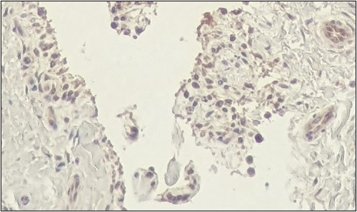
Application: Immunohistochemistry-ParaffinSample Tested: equine synovial tissue, Lymph node tissue and synovial tissueSpecies: EquineVerified Customer | Posted 05/09/2022Primary antibody was diluted 1:20-1:100. Secondary detection was performed with anti-mouse binding protein conjugated to HRP and subsequent incubation with DAB chromogen.

There are no reviews that match your criteria.
Protocols
Find general support by application which include: protocols, troubleshooting, illustrated assays, videos and webinars.
- 7-Amino Actinomycin D (7-AAD) Cell Viability Flow Cytometry Protocol
- Cellular Response to Hypoxia Protocols
- Extracellular Membrane Flow Cytometry Protocol
- Flow Cytometry Protocol for Cell Surface Markers
- Flow Cytometry Protocol for Staining Membrane Associated Proteins
- Flow Cytometry Staining Protocols
- Flow Cytometry Troubleshooting Guide
- Intracellular Flow Cytometry Protocol Using Alcohol (Methanol)
- Intracellular Flow Cytometry Protocol Using Detergents
- Intracellular Nuclear Staining Flow Cytometry Protocol Using Detergents
- Intracellular Staining Flow Cytometry Protocol Using Alcohol Permeabilization
- Intracellular Staining Flow Cytometry Protocol Using Detergents to Permeabilize Cells
- Propidium Iodide Cell Viability Flow Cytometry Protocol
- Protocol for Liperfluo
- Protocol for the Characterization of Human Th22 Cells
- Protocol for the Characterization of Human Th9 Cells
- Protocol: Annexin V and PI Staining by Flow Cytometry
- Protocol: Annexin V and PI Staining for Apoptosis by Flow Cytometry
- R&D Systems Quality Control Western Blot Protocol
- Troubleshooting Guide: Fluorokine Flow Cytometry Kits
- Troubleshooting Guide: Western Blot Figures
- Western Blot Conditions
- Western Blot Protocol
- Western Blot Protocol for Cell Lysates
- Western Blot Troubleshooting
- Western Blot Troubleshooting Guide
- View all Protocols, Troubleshooting, Illustrated assays and Webinars
Loading...
Associated Pathways


