GM-CSF was initially characterized as a factor that can support the in vitro colony formation of granulocyte-macrophage progenitors. It is also a growth factor for erythroid, megakaryocyte, and eosinophil progenitors. GM-CSF is produced by a number of different cell types (including T cells, B cells, macrophages, mast cells, endothelial cells, fibroblasts, and adipocytes) in response to cytokine or inflammatory stimuli. On mature hematopoietic cells, GM-CSF is a survival factor for and activates the effector functions of granulocytes, monocytes/macrophages, and eosinophils (1, 2). GM-CSF promotes a Th1 biased immune response, angiogenesis, allergic inflammation, and the development of autoimmunity (3‑5). It shows clinical effectiveness in ameliorating chemotherapy-induced neutropenia, and GM-CSF transfected tumor cells are utilized as cancer vaccines (6, 7). The 22 kDa glycosylated GM-CSF, similar to IL‑3 and IL‑5, is a cytokine with a core of four bundled alpha ‑helices (8‑10). Mature rat GM-CSF shares 56%‑69% amino acid sequence identity with canine, feline, human, mouse, and porcine GM‑CSF. GM‑CSF exerts its biological effects through a heterodimeric receptor complex composed of GM‑CSF R alpha /CD116 and the signal transducing common beta chain (CD131) which is also a component of the high-affinity receptors for IL-3 and IL-5 (11, 12). In addition, GM-CSF binds a naturally occurring soluble form of GM‑CSF R alpha (13). Rat GM‑CSF is active on mouse cells, although mouse GM‑CSF is only weakly active on rat cells (14, 15).

Key Product Details
Species Reactivity
Validated:
Rat
Cited:
Rat
Applications
Validated:
Western Blot, Neutralization, Intracellular Staining by Flow Cytometry, Immunocytochemistry, CyTOF-ready
Cited:
Immunohistochemistry-Paraffin, Flow Cytometry
Label
Unconjugated
Antibody Source
Monoclonal Mouse IgG2B Clone # 83308
Loading...
Product Specifications
Immunogen
E. coli-derived recombinant rat GM-CSF
Ala1-Lys127
Accession # P48750
Ala1-Lys127
Accession # P48750
Specificity
Detects rat GM-CSF in direct ELISAs and Western blots. In direct ELISAs and Western blots, no cross-reactivity with recombinant GM‑CSF from mouse, human, or pig is observed.
Clonality
Monoclonal
Host
Mouse
Isotype
IgG2B
Endotoxin Level
<0.10 EU per 1 μg of the antibody by the LAL method.
Scientific Data Images for Rat GM-CSF Antibody (83308)
Cell Proliferation Induced by GM‑CSF and Neutralization by Rat GM‑CSF Antibody.
Recombinant Rat GM-CSF (Catalog # 518-GM) stimulates proliferation in the DA3 mouse myeloma cell line in a dose-dependent manner (orange line). Proliferation elicited by Recombinant Rat GM-CSF (0.5 ng/mL) is neutralized (green line) by increasing concentrations of Rat GM-CSF Monoclonal Antibody (Catalog # MAB5181). The ND50 is typically 1-4 µg/mL.
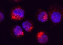
GM‑CSF in Rat Splenocytes.
GM-CSF was detected in immersion fixed rat splenocytes using Rat GM-CSF Monoclonal Antibody (Catalog # MAB5181) at 25 µg/mL for 3 hours at room temperature. Cells were stained using the NorthernLights™ 557-conjugated Anti-Mouse IgG Secondary Antibody (red; Catalog # NL007) and counterstained with DAPI (blue). Specific staining was localized to cytoplasm. View our protocol for Fluorescent ICC Staining of Non-adherent Cells.Applications for Rat GM-CSF Antibody (83308)
Application
Recommended Usage
CyTOF-ready
Ready to be labeled using established conjugation methods. No BSA or other carrier proteins that could interfere with conjugation.
Immunocytochemistry
8-25 µg/mL
Sample: Immersion fixed rat splenocytes
Sample: Immersion fixed rat splenocytes
Intracellular Staining by Flow Cytometry
2.5 µg/106 cells
Sample: Rat splenocytes fixed with paraformaldehyde and permeabilized with saponin
Sample: Rat splenocytes fixed with paraformaldehyde and permeabilized with saponin
Western Blot
1 µg/mL
Sample: Recombinant Rat GM-CSF (Catalog # 518-GM) under non-reducing conditions only. Catalog # AF518 is recommended to detect rat GM-CSF in Western blots.
Sample: Recombinant Rat GM-CSF (Catalog # 518-GM) under non-reducing conditions only. Catalog # AF518 is recommended to detect rat GM-CSF in Western blots.
Neutralization
Measured by its ability to neutralize GM‑CSF-induced proliferation in the DA3 mouse myeloma cell line. Ihle, J. N. et al. (1984) Advances in Viral Oncology. In G. Klein (eds): Raven Press, New York, NY. 4:95. The Neutralization Dose (ND50) is typically 1-4 µg/mL in the presence of 0.5 ng/mL Recombinant Rat GM‑CSF.
Reviewed Applications
Read 1 review rated 5 using MAB5181 in the following applications:
Flow Cytometry Panel Builder
Bio-Techne Knows Flow Cytometry
Save time and reduce costly mistakes by quickly finding compatible reagents using the Panel Builder Tool.
Advanced Features
- Spectra Viewer - Custom analysis of spectra from multiple fluorochromes
- Spillover Popups - Visualize the spectra of individual fluorochromes
- Antigen Density Selector - Match fluorochrome brightness with antigen density

Formulation, Preparation, and Storage
Purification
Protein A or G purified from hybridoma culture supernatant
Reconstitution
Reconstitute at 0.5 mg/mL in sterile PBS. For liquid material, refer to CoA for concentration.
Loading...
Formulation
Lyophilized from a 0.2 μm filtered solution in PBS with Trehalose. *Small pack size (SP) is supplied either lyophilized or as a 0.2 µm filtered solution in PBS.
Shipping
Lyophilized product is shipped at ambient temperature. Liquid small pack size (-SP) is shipped with polar packs. Upon receipt, store immediately at the temperature recommended below.
Stability & Storage
Use a manual defrost freezer and avoid repeated freeze-thaw cycles.
- 12 months from date of receipt, -20 to -70 °C as supplied.
- 1 month, 2 to 8 °C under sterile conditions after reconstitution.
- 6 months, -20 to -70 °C under sterile conditions after reconstitution.
Calculators
Background: GM-CSF
References
- Martinez-Moczygemba, M. and D.P. Huston (2003) J. Allergy Clin. Immunol. 112:653.
- Barreda, D.R. et al. (2004) Dev. Comp. Immunol. 28:509.
- Eksioglu, E.A. et al. (2007) Exp. Hematol. 35:1163.
- Cao, Y. (2007) J. Clin. Invest. 117:2362.
- Fleetwood, A.J. et al. (2005) Crit. Rev. Immunol. 25:405.
- Heuser, M. et al. (2007) Semin. Hematol. 44:148.
- Hege, K.M. et al. (2006) Int. Rev. Immunol. 25:321.
- Kaushansky, K. et al. (1992) Biochemistry 31:1881.
- Diederichs, K. et al. (1991) Science 254:1779.
- Smith, L.R. et al. (1994) Immunogenetics 39:80.
- Onetto-Pothier, N. et al. (1990) Blood 75:59.
- Hayashida, K. et al. (1990) Proc. Natl. Acad. Sci. 87:9655.
- Pelley, J.L. et al. (2007) Exp. Hematol. 35:1483.
- Oaks, M.K. et al. (1995) J. Interferon Cytokine Res. 15:1095.
- Vandenabeele, P. et al. (1990) Lymphokine Res. 9:381.
Long Name
Granulocyte Macrophage Growth Factor
Alternate Names
CSF-2, CSF2, GMCSF, Molgramostim, Sargramostim
Entrez Gene IDs
Gene Symbol
CSF2
UniProt
Additional GM-CSF Products
Product Documents for Rat GM-CSF Antibody (83308)
Certificate of Analysis
To download a Certificate of Analysis, please enter a lot or batch number in the search box below.
Note: Certificate of Analysis not available for kit components.
Product Specific Notices for Rat GM-CSF Antibody (83308)
For research use only
Related Research Areas
Citations for Rat GM-CSF Antibody (83308)
 Powered by Bioz
Customer Reviews for Rat GM-CSF Antibody (83308) (1)
5 out of 5
1 Customer Rating
Have you used Rat GM-CSF Antibody (83308)?
Submit a review and receive an Amazon gift card!
$25/€18/£15/$25CAN/¥2500 Yen for a review with an image
$10/€7/£6/$10CAN/¥1110 Yen for a review without an image
Submit a review
Customer Images
Showing
1
-
1 of
1 review
Showing All
Filter By:
-
Application: Immunocytochemistry/ImmunofluorescenceSample Tested: SplenocytesSpecies: RatVerified Customer | Posted 03/15/2022

There are no reviews that match your criteria.
Protocols
Find general support by application which include: protocols, troubleshooting, illustrated assays, videos and webinars.
- 7-Amino Actinomycin D (7-AAD) Cell Viability Flow Cytometry Protocol
- Appropriate Fixation of IHC/ICC Samples
- Cellular Response to Hypoxia Protocols
- ClariTSA™ Fluorophore Kits
- Detection & Visualization of Antibody Binding
- Extracellular Membrane Flow Cytometry Protocol
- Flow Cytometry Protocol for Cell Surface Markers
- Flow Cytometry Protocol for Staining Membrane Associated Proteins
- Flow Cytometry Staining Protocols
- Flow Cytometry Troubleshooting Guide
- ICC Cell Smear Protocol for Suspension Cells
- ICC Immunocytochemistry Protocol Videos
- ICC for Adherent Cells
- Immunocytochemistry (ICC) Protocol
- Immunocytochemistry Troubleshooting
- Immunofluorescence of Organoids Embedded in Cultrex Basement Membrane Extract
- Immunohistochemistry (IHC) and Immunocytochemistry (ICC) Protocols
- Intracellular Flow Cytometry Protocol Using Alcohol (Methanol)
- Intracellular Flow Cytometry Protocol Using Detergents
- Intracellular Nuclear Staining Flow Cytometry Protocol Using Detergents
- Intracellular Staining Flow Cytometry Protocol Using Alcohol Permeabilization
- Intracellular Staining Flow Cytometry Protocol Using Detergents to Permeabilize Cells
- Preparing Samples for IHC/ICC Experiments
- Preventing Non-Specific Staining (Non-Specific Binding)
- Primary Antibody Selection & Optimization
- Propidium Iodide Cell Viability Flow Cytometry Protocol
- Protocol for Liperfluo
- Protocol for VisUCyte™ HRP Polymer Detection Reagent
- Protocol for the Characterization of Human Th22 Cells
- Protocol for the Characterization of Human Th9 Cells
- Protocol for the Fluorescent ICC Staining of Cell Smears - Graphic
- Protocol for the Fluorescent ICC Staining of Cultured Cells on Coverslips - Graphic
- Protocol for the Preparation and Fluorescent ICC Staining of Cells on Coverslips
- Protocol for the Preparation and Fluorescent ICC Staining of Non-adherent Cells
- Protocol for the Preparation and Fluorescent ICC Staining of Stem Cells on Coverslips
- Protocol for the Preparation of a Cell Smear for Non-adherent Cell ICC - Graphic
- Protocol: Annexin V and PI Staining by Flow Cytometry
- Protocol: Annexin V and PI Staining for Apoptosis by Flow Cytometry
- R&D Systems Quality Control Western Blot Protocol
- TUNEL and Active Caspase-3 Detection by IHC/ICC Protocol
- The Importance of IHC/ICC Controls
- Troubleshooting Guide: Fluorokine Flow Cytometry Kits
- Troubleshooting Guide: Western Blot Figures
- Western Blot Conditions
- Western Blot Protocol
- Western Blot Protocol for Cell Lysates
- Western Blot Troubleshooting
- Western Blot Troubleshooting Guide
- View all Protocols, Troubleshooting, Illustrated assays and Webinars


