
Key Product Details
Species Reactivity
Validated:
Multi-Species
Cited:
Human, Mouse, Avian - Chicken, Avian - Coturnix japonica (Quail), Canine
Applications
Validated:
Immunohistochemistry, Western Blot, Neutralization
Cited:
Immunohistochemistry, Western Blot, Neutralization, ELISA Development
Label
Unconjugated
Antibody Source
Monoclonal Mouse IgG1 Clone # 20724
Loading...
Product Specifications
Immunogen
Spodoptera frugiperda, Sf 21 (baculovirus) derived recombinant human TGF-beta 3
Ala301-Ser412 (Tyr340Phe)
Accession # P10600
Ala301-Ser412 (Tyr340Phe)
Accession # P10600
Specificity
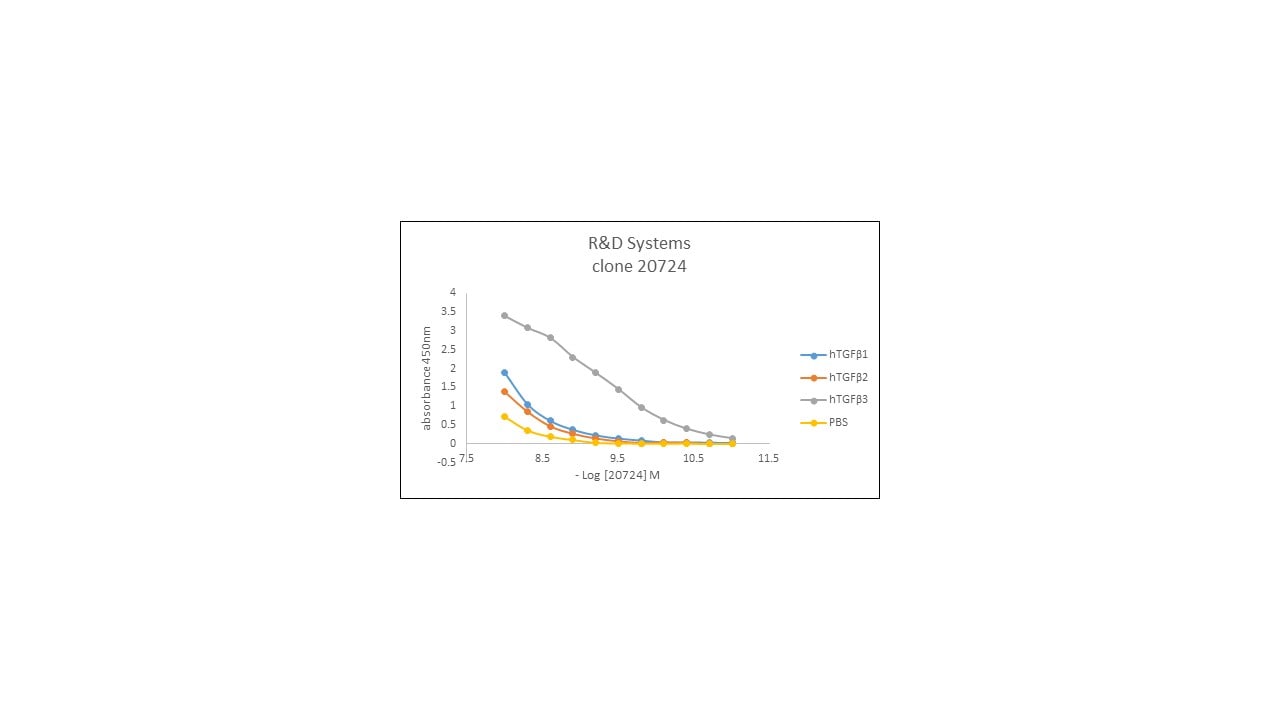
Detects TGF-beta 3 from multiple species in direct ELISAs and Western blots. In Western blots, less than 25% cross-reactivity with recombinant human (rh) TGF‑ beta 1.2 and rhTGF-beta 2 is observed, and less than 2% cross‑reactivity with recombinant amphibian TGF-beta 5 and recombinant human TGF-beta 1 is observed. Neutralizes the biological activity of TGF-beta 3 but not TGF-beta 1, TGF-beta 2, or TGF-beta 5.
Clonality
Monoclonal
Host
Mouse
Isotype
IgG1
Endotoxin Level
<0.10 EU per 1 μg of the antibody by the LAL method.
Scientific Data Images for TGF-beta 3 Antibody (20724)
TGF‑ beta 3 Inhibition of IL‑4-dependent Cell Proliferation and Neutralization by TGF‑ beta 3 Antibody.
TGF beta 3 Antibody (Catalog # MAB243) neutralizes Recombinant Human TGF-beta 3 (243-B3) inhibition of Recombinant Mouse IL-4 (404-ML) induced proliferation in the HT-2 mouse T cell line. The Neutralization Dose (ND50) is typically 0.100‑1.00 µg/mL.
TGF‑ beta 3 in Human Breast Cancer Tissue.
TGF-beta 3 was detected in immersion fixed paraffin-embedded sections of human breast cancer tissue using Mouse Anti-TGF-beta 3 Monoclonal Antibody (Catalog # MAB243) at 5 µg/mL overnight at 4 °C. Tissue was stained using the Anti-Mouse HRP-DAB Cell & Tissue Staining Kit (brown; CTS002) and counterstained with hematoxylin (blue). Specific staining was localized to cytoplasm in cancer cells. View our protocol for Chromogenic IHC Staining of Paraffin-embedded Tissue Sections.
Detection of Coturnix japonica TGF-beta 3 by Western Blot
TGF-beta signaling controls commitment into glial differentiation.(A) QNR/v-srcts cells were treated at 37°C with increasing concentrations (0.2–2 ng/ml) of recombinant TGF-beta 3 protein during 7 days. Pax6 and Glutamine Synthetase (GS) were detected by western-blot. Protein loading was normalized using Erk antiserum. (B) Pax6 expression was analyzed by immunofluorescence after treatment of QNR/v-Srcts/ICN cells at 37°C with DMSO (control) or 10 µM SB431542 during 7 days. The majority of control QNR/v-Srcts/ICN cells did not express nuclear Pax6 but we observed a weak peri-nuclear labeling. Magnification x40. Image collected and cropped by CiteAb from the following open publication (https://pubmed.ncbi.nlm.nih.gov/21042581), licensed under a CC-BY license. Not internally tested by R&D Systems.Applications for TGF-beta 3 Antibody (20724)
Application
Recommended Usage
Immunohistochemistry
8-25 µg/mL
Sample: Immersion fixed paraffin-embedded sections of human breast cancer tissue
Sample: Immersion fixed paraffin-embedded sections of human breast cancer tissue
Western Blot
1 µg/mL
Sample: Recombinant Human TGF-beta 3 (Catalog # 243-B3) under non-reducing conditions only
Sample: Recombinant Human TGF-beta 3 (Catalog # 243-B3) under non-reducing conditions only
Neutralization
Measured by its ability to neutralize TGF‑ beta 3 inhibition of IL‑4-dependent proliferation in the HT‑2 mouse T cell line. Tsang, M. et al. (1995) Cytokine 7:389. The Neutralization Dose (ND50) is typically 0.100-1.00 µg/mL in the presence of 0.1 ng/mL Recombinant Human TGF‑ beta 3 and 7.5 ng/mL Recombinant Mouse IL‑4.
Formulation, Preparation, and Storage
Purification
Protein A or G purified from ascites
Reconstitution
Reconstitute at 0.5 mg/mL in sterile PBS. For liquid material, refer to CoA for concentration.
Loading...
Formulation
Lyophilized from a 0.2 μm filtered solution in PBS with Trehalose. See Certificate of Analysis for details.
*Small pack size (-SP) is supplied either lyophilized or as a 0.2 µm filtered solution in PBS.
*Small pack size (-SP) is supplied either lyophilized or as a 0.2 µm filtered solution in PBS.
Shipping
Lyophilized product is shipped at ambient temperature. Liquid small pack size (-SP) is shipped with polar packs. Upon receipt, store immediately at the temperature recommended below.
Stability & Storage
Use a manual defrost freezer and avoid repeated freeze-thaw cycles.
- 12 months from date of receipt, -20 to -70 °C as supplied.
- 1 month, 2 to 8 °C under sterile conditions after reconstitution.
- 6 months, -20 to -70 °C under sterile conditions after reconstitution.
Calculators
Background: TGF-beta 3
References
- Sporn, M.B. (2006) Cytokine Growth Factor Rev. 17:3.
- Dunker, N. and K. Krieglstein (2000) Eur. J. Biochem. 267:6982.
- Wahl, S.M. (2006) Immunol. Rev. 213:213.
- Chang, H. et al. (2002) Endocr. Rev. 23:787.
- Lin, J.S. et al. (2006) Reproduction 132:179.
- Hinck, A.P. et al. (1996) Biochemistry 35:8517.
- Mittl, P.R.E. et al. (1996) Protein Sci. 5:1261.
- Derynck, R. et al. (1988) EMBO J. 7:3737.
- Miyazono, K. et al. (1988) J. Biol. Chem. 263:6407.
- Oklu, R. and R. Hesketh (2000) Biochem. J. 352:601.
- de Caestecker, M. et al. (2004) Cytokine Growth Factor Rev. 15:1.
- Zuniga, J.E. et al. (2005) J. Mol. Biol. 354:1052.
Long Name
Transforming Growth Factor beta 3
Alternate Names
ARVD1, LDS5, RNHF, TGFB3, TGFbeta 3
Gene Symbol
TGFB3
UniProt
Additional TGF-beta 3 Products
Product Documents for TGF-beta 3 Antibody (20724)
Certificate of Analysis
To download a Certificate of Analysis, please enter a lot or batch number in the search box below.
Note: Certificate of Analysis not available for kit components.
Product Specific Notices for TGF-beta 3 Antibody (20724)
For research use only
Citations for TGF-beta 3 Antibody (20724)
 Powered by Bioz
Customer Reviews for TGF-beta 3 Antibody (20724) (1)
5 out of 5
1 Customer Rating
Have you used TGF-beta 3 Antibody (20724)?
Submit a review and receive an Amazon gift card!
$25/€18/£15/$25CAN/¥2500 Yen for a review with an image
$10/€7/£6/$10CAN/¥1110 Yen for a review without an image
Submit a review
Customer Images
Showing
1
-
1 of
1 review
Showing All
Filter By:
-
Sample Tested: Recombinant proteinSpecies: HumanVerified Customer | Posted 03/28/2019direct ELISA to check specificity using in-house produced recombinant proteins

There are no reviews that match your criteria.
Protocols
Find general support by application which include: protocols, troubleshooting, illustrated assays, videos and webinars.
- Antigen Retrieval Protocol (PIER)
- Antigen Retrieval for Frozen Sections Protocol
- Appropriate Fixation of IHC/ICC Samples
- Cellular Response to Hypoxia Protocols
- Chromogenic IHC Staining of Formalin-Fixed Paraffin-Embedded (FFPE) Tissue Protocol
- Chromogenic Immunohistochemistry Staining of Frozen Tissue
- ClariTSA™ Fluorophore Kits
- Detection & Visualization of Antibody Binding
- Fluorescent IHC Staining of Frozen Tissue Protocol
- Graphic Protocol for Heat-induced Epitope Retrieval
- Graphic Protocol for the Preparation and Fluorescent IHC Staining of Frozen Tissue Sections
- Graphic Protocol for the Preparation and Fluorescent IHC Staining of Paraffin-embedded Tissue Sections
- Graphic Protocol for the Preparation of Gelatin-coated Slides for Histological Tissue Sections
- IHC Sample Preparation (Frozen sections vs Paraffin)
- Immunofluorescent IHC Staining of Formalin-Fixed Paraffin-Embedded (FFPE) Tissue Protocol
- Immunohistochemistry (IHC) and Immunocytochemistry (ICC) Protocols
- Immunohistochemistry Frozen Troubleshooting
- Immunohistochemistry Paraffin Troubleshooting
- Preparing Samples for IHC/ICC Experiments
- Preventing Non-Specific Staining (Non-Specific Binding)
- Primary Antibody Selection & Optimization
- Protocol for Heat-Induced Epitope Retrieval (HIER)
- Protocol for Making a 4% Formaldehyde Solution in PBS
- Protocol for VisUCyte™ HRP Polymer Detection Reagent
- Protocol for the Preparation & Fixation of Cells on Coverslips
- Protocol for the Preparation and Chromogenic IHC Staining of Frozen Tissue Sections
- Protocol for the Preparation and Chromogenic IHC Staining of Frozen Tissue Sections - Graphic
- Protocol for the Preparation and Chromogenic IHC Staining of Paraffin-embedded Tissue Sections
- Protocol for the Preparation and Chromogenic IHC Staining of Paraffin-embedded Tissue Sections - Graphic
- Protocol for the Preparation and Fluorescent IHC Staining of Frozen Tissue Sections
- Protocol for the Preparation and Fluorescent IHC Staining of Paraffin-embedded Tissue Sections
- Protocol for the Preparation of Gelatin-coated Slides for Histological Tissue Sections
- R&D Systems Quality Control Western Blot Protocol
- TUNEL and Active Caspase-3 Detection by IHC/ICC Protocol
- The Importance of IHC/ICC Controls
- Troubleshooting Guide: Immunohistochemistry
- Troubleshooting Guide: Western Blot Figures
- Western Blot Conditions
- Western Blot Protocol
- Western Blot Protocol for Cell Lysates
- Western Blot Troubleshooting
- Western Blot Troubleshooting Guide
- View all Protocols, Troubleshooting, Illustrated assays and Webinars
Loading...
Associated Pathways


