Best Seller
Human CXCL13/BLC/BCA-1 Antibody
R&D Systems | Catalog # AF801

Key Product Details
Species Reactivity
Validated:
Human
Cited:
Human, Mouse, Primate - Macaca mulatta (Rhesus Macaque), Rabbit, Transgenic Mouse, Xenograft
Applications
Validated:
Immunohistochemistry, Western Blot, Neutralization, Dual RNAscope ISH-IHC Compatible
Cited:
Immunohistochemistry, Immunohistochemistry-Paraffin, Immunohistochemistry-Frozen, Neutralization, Immunocytochemistry, Bioassay
Label
Unconjugated
Antibody Source
Polyclonal Goat IgG
Loading...
Product Specifications
Immunogen
E. coli-derived recombinant human CXCL13/BCA-1
Val23-Arg94
Accession # Q53X90
Val23-Arg94
Accession # Q53X90
Specificity
Detects CXCL13/BLC/BCA‑1 in direct ELISAs and Western blots.
Clonality
Polyclonal
Host
Goat
Isotype
IgG
Endotoxin Level
<0.10 EU per 1 μg of the antibody by the LAL method.
Scientific Data Images for Human CXCL13/BLC/BCA-1 Antibody
Chemotaxis Induced by CXCL13/BLC/BCA‑1 and Neutralization by Human CXCL13/BLC/BCA‑1 Antibody.
Recombinant Human CXCL13/BLC/BCA-1 (Catalog # 801-CX) chemoattracts the BaF3 mouse pro-B cell line transfected with human CXCR5 in a dose-dependent manner (orange line). The amount of cells that migrated through to the lower chemotaxis chamber was measured by Resazurin. Chemotaxis elicited by Recombinant Human CXCL13/BLC/BCA-1 (50 ng/mL) is neutralized (green line) by increasing concentrations of Goat Anti-Human CXCL13/BLC/BCA-1 Antigen Affinity-purified Polyclonal Antibody (Catalog # AF801). The ND50 is typically 1-4 µg/mL.
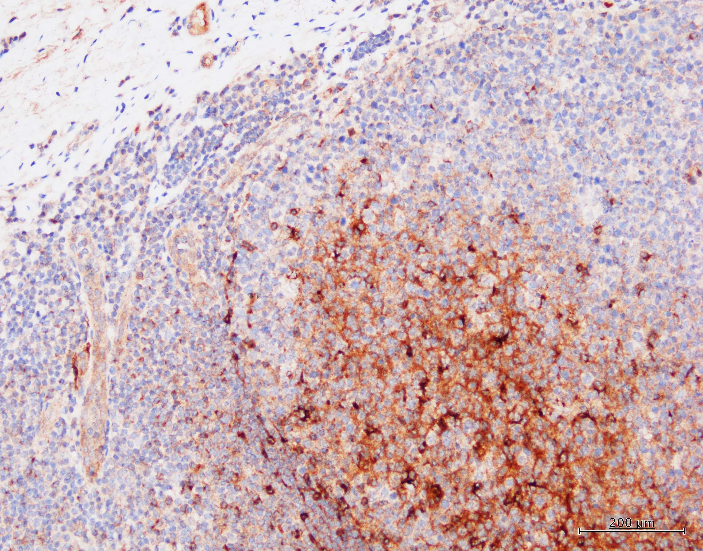
CXCL13/BLC/BCA‑1 in Human Lymphoma.
CXCL13/BLC/BCA-1 was detected in immersion fixed paraffin-embedded sections of human lymphoma using Goat Anti-Human CXCL13/BLC/BCA-1 Antigen Affinity-purified Polyclonal Antibody (Catalog # AF801) at 10 µg/mL overnight at 4 °C. Before incubation with the primary antibody, tissue was subjected to heat-induced epitope retrieval using Antigen Retrieval Reagent-Basic (Catalog # CTS013). Tissue was stained using the Anti-Goat HRP-DAB Cell & Tissue Staining Kit (brown; Catalog # CTS008) and counterstained with hematoxylin (blue). Specific staining was localized to cytoplasm. View our protocol for Chromogenic IHC Staining of Paraffin-embedded Tissue Sections.
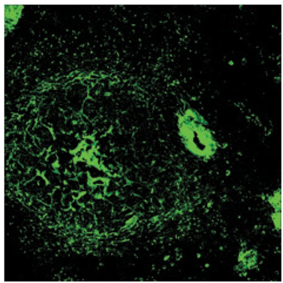
Detection of CXCL13/BLC/BCA‑1 in Human Lymphoma.
Formalin-fixed paraffin-embedded tissue sections of Hodgkin’s Lymphoma were probed for CXCL13 mRNA (ACD RNAScope Probe, catalog # 311321; Fast Red chromogen, ACD catalog # 322360). Adjacent tissue section was processed for immunohistochemistry using goat anti-human CXCL13 polyclonal antibody (R&D Systems catalog # AF801) at 5ug/mL with 1-hour incubation at room temperature followed by incubation with anti-goat IgG VisUCyte HRP Polymer Antibody (Catalog # VC004) and DAB chromogen (yellow-brown). Tissue was counterstained with hematoxylin (blue). Specific staining was localized to cytoplasm.
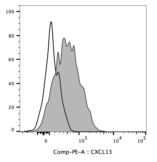
Detection of CXCL13/BLC/BCA-1 by Flow Cytometry
The anti-tumor function of Tfh cells was impaired in PDAC. (A) Representative flow cytometry data and the proportion of functional cTfh cells (CXCL13+ IL-21+) from the peripheral blood of healthy donors (n = 4) and patients with PDAC of different stages (nI = 5, nII = 4, nIII = 4). (B) Representative flow cytometry data and (C) the proportion of CD8+ T cells and B cells recruited to the bottom of the Transwell chamber by cTfh cells treated with or without rhCXCL13 or anti-CXCL13 neutralizing antibodies. (D) Representative flow cytometry data and the proportion of plasma cells (CD27+ CD38+) in B cells (CD19+ CD20+) from the peripheral blood of healthy donors (n = 4) and patients with PDAC at different stages (nI = 5, nII = 4, nIII = 4). (E) Representative flow cytometry data and the proportion of IgG-secreting plasma cells from cells in (D). (F) Representative flow cytometry data and the proportion of IgM-secreting plasma cells from cells in (D). (G) Representative flow cytometry data and the proportion of plasma cells (CD27+ CD38+) in B cells (CD19+ CD20+) co-cultured with cTfh cells sorted from the peripheral blood of healthy donors (n = 3) and patients with PDAC (n = 3). (H) Representative flow cytometry data and the proportion of IgG-secreting plasma cells from cells in (G). (I) Representative flow cytometry data and the proportion of IgM-secreting plasma cells from cells in (G). Asterisks indicated the significance level of the p-value (* p < 0.05 and ** p < 0.01). Image collected and cropped by CiteAb from the following open publication (https://pubmed.ncbi.nlm.nih.gov/34359579), licensed under a CC-BY license. Not internally tested by R&D Systems.Applications for Human CXCL13/BLC/BCA-1 Antibody
Application
Recommended Usage
Dual RNAscope ISH-IHC Compatible
5-15 µg/mL
Sample: Immersion fixed paraffin-embedded sections of human lymphoma
Sample: Immersion fixed paraffin-embedded sections of human lymphoma
Immunohistochemistry
5-15 µg/mL
Sample: Immersion fixed paraffin-embedded sections of human tonsil and human lymphoma
Sample: Immersion fixed paraffin-embedded sections of human tonsil and human lymphoma
Western Blot
0.1 µg/mL
Sample: Recombinant Human CXCL13/BLC/BCA‑1 (Catalog # 801-CX)
Sample: Recombinant Human CXCL13/BLC/BCA‑1 (Catalog # 801-CX)
Neutralization
Measured by its ability to neutralize CXCL13/BLC/BCA‑1-induced chemotaxis in the BaF3 mouse pro‑B cell line transfected with human CXCR5. The Neutralization Dose (ND50) is typically 1-4 µg/mL in the presence of 50 ng/mL Recombinant Human CXCL13/BLC/BCA‑1.
Reviewed Applications
Read 7 reviews rated 4.6 using AF801 in the following applications:
Formulation, Preparation, and Storage
Purification
Antigen Affinity-purified
Reconstitution
Reconstitute at 0.2 mg/mL in sterile PBS. For liquid material, refer to CoA for concentration.
Loading...
Formulation
Lyophilized from a 0.2 μm filtered solution in PBS with Trehalose. See Certificate of Analysis for details.
*Small pack size (-SP) is supplied either lyophilized or as a 0.2 µm filtered solution in PBS.
*Small pack size (-SP) is supplied either lyophilized or as a 0.2 µm filtered solution in PBS.
Shipping
Lyophilized product is shipped at ambient temperature. Liquid small pack size (-SP) is shipped with polar packs. Upon receipt, store immediately at the temperature recommended below.
Stability & Storage
Use a manual defrost freezer and avoid repeated freeze-thaw cycles.
- 12 months from date of receipt, -20 to -70 °C as supplied.
- 1 month, 2 to 8 °C under sterile conditions after reconstitution.
- 6 months, -20 to -70 °C under sterile conditions after reconstitution.
Calculators
Background: CXCL13/BLC/BCA-1
References
- Gunn, M.D. et al. (1998) Nature, 391:799.
- Legler, D.F. et al. (1998) J. Exp. Med. 187:655.
- Forster, R. et al. (1996) Cell 87:1037.
Alternate Names
ANGIE2, BCA-1, BCA1, BLC, BLR1L, SCYB13
Gene Symbol
CXCL13
UniProt
Additional CXCL13/BLC/BCA-1 Products
Product Documents for Human CXCL13/BLC/BCA-1 Antibody
Certificate of Analysis
To download a Certificate of Analysis, please enter a lot or batch number in the search box below.
Note: Certificate of Analysis not available for kit components.
Product Specific Notices for Human CXCL13/BLC/BCA-1 Antibody
For research use only
Citations for Human CXCL13/BLC/BCA-1 Antibody
 Powered by Bioz
Customer Reviews for Human CXCL13/BLC/BCA-1 Antibody (7)
4.6 out of 5
7 Customer Ratings
Have you used Human CXCL13/BLC/BCA-1 Antibody?
Submit a review and receive an Amazon gift card!
$25/€18/£15/$25CAN/¥2500 Yen for a review with an image
$10/€7/£6/$10CAN/¥1110 Yen for a review without an image
Submit a review
Customer Images
Showing
1
-
5 of
7 reviews
Showing All
Filter By:
-
Application: Flow CytometrySample Tested: SplenocytesSpecies: HumanVerified Customer | Posted 07/06/2022CXCL13 and negative control.

-
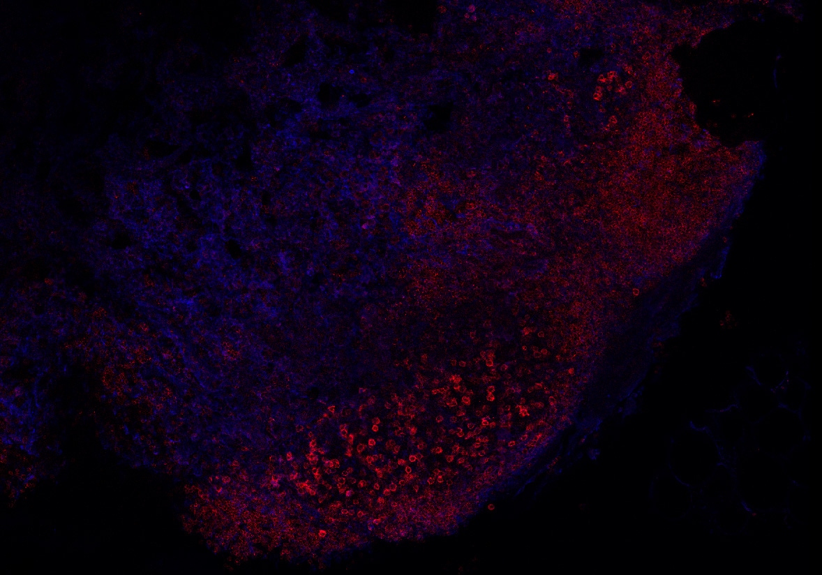
Application: Immunocytochemistry/ImmunofluorescenceSample Tested: Spleen tissueSpecies: HumanVerified Customer | Posted 07/06/2022Human spleens fixed in 4% PFA
-
Application: ImmunohistochemistrySample Tested: Tonsil tissueSpecies: HumanVerified Customer | Posted 12/10/2021Tonsil FFPE section. Primary Ab: 2ug/mL Detected using BOND Refine kit Rabbit anti-goat was used as Post Primary ER2 retrieval
-
Application: Immunofluorescence - paraffinSample Tested: Diffuse large B cell lymphomaSpecies: HumanVerified Customer | Posted 01/16/2020anti-CXCL13 on DLBCL, 1:200 dilutionpH 9 antigen retrieval
-
Application: ImmunohistochemistrySample Tested: Lymph node tissueSpecies: Rhesus MacaqueVerified Customer | Posted 04/23/2018The CXCL13 can be observed in blue while CXCR5+ cells are observed in red
-
Application: ImmunohistochemistrySample Tested: Spleen tissueSpecies: Rhesus MacaqueVerified Customer | Posted 03/21/2018
-
Application: Immunohistochemistry-ParaffinSample Tested: See PMID 23778140Species: HumanVerified Customer | Posted 01/09/2015
There are no reviews that match your criteria.
Protocols
Find general support by application which include: protocols, troubleshooting, illustrated assays, videos and webinars.
- Antigen Retrieval Protocol (PIER)
- Antigen Retrieval for Frozen Sections Protocol
- Appropriate Fixation of IHC/ICC Samples
- Cellular Response to Hypoxia Protocols
- Chromogenic IHC Staining of Formalin-Fixed Paraffin-Embedded (FFPE) Tissue Protocol
- Chromogenic Immunohistochemistry Staining of Frozen Tissue
- ClariTSA™ Fluorophore Kits
- Detection & Visualization of Antibody Binding
- Fluorescent IHC Staining of Frozen Tissue Protocol
- Graphic Protocol for Heat-induced Epitope Retrieval
- Graphic Protocol for the Preparation and Fluorescent IHC Staining of Frozen Tissue Sections
- Graphic Protocol for the Preparation and Fluorescent IHC Staining of Paraffin-embedded Tissue Sections
- Graphic Protocol for the Preparation of Gelatin-coated Slides for Histological Tissue Sections
- IHC Sample Preparation (Frozen sections vs Paraffin)
- ISH-IHC Protocol for Chromogenic Detection on Formalin Fixed Paraffin Embedded (FFPE) Tissue
- Immunofluorescent IHC Staining of Formalin-Fixed Paraffin-Embedded (FFPE) Tissue Protocol
- Immunohistochemistry (IHC) and Immunocytochemistry (ICC) Protocols
- Immunohistochemistry Frozen Troubleshooting
- Immunohistochemistry Paraffin Troubleshooting
- Preparing Samples for IHC/ICC Experiments
- Preventing Non-Specific Staining (Non-Specific Binding)
- Primary Antibody Selection & Optimization
- Protocol for Heat-Induced Epitope Retrieval (HIER)
- Protocol for Making a 4% Formaldehyde Solution in PBS
- Protocol for VisUCyte™ HRP Polymer Detection Reagent
- Protocol for the Preparation & Fixation of Cells on Coverslips
- Protocol for the Preparation and Chromogenic IHC Staining of Frozen Tissue Sections
- Protocol for the Preparation and Chromogenic IHC Staining of Frozen Tissue Sections - Graphic
- Protocol for the Preparation and Chromogenic IHC Staining of Paraffin-embedded Tissue Sections
- Protocol for the Preparation and Chromogenic IHC Staining of Paraffin-embedded Tissue Sections - Graphic
- Protocol for the Preparation and Fluorescent IHC Staining of Frozen Tissue Sections
- Protocol for the Preparation and Fluorescent IHC Staining of Paraffin-embedded Tissue Sections
- Protocol for the Preparation of Gelatin-coated Slides for Histological Tissue Sections
- R&D Systems Quality Control Western Blot Protocol
- TUNEL and Active Caspase-3 Detection by IHC/ICC Protocol
- The Importance of IHC/ICC Controls
- Troubleshooting Guide: Immunohistochemistry
- Troubleshooting Guide: Western Blot Figures
- Western Blot Conditions
- Western Blot Protocol
- Western Blot Protocol for Cell Lysates
- Western Blot Troubleshooting
- Western Blot Troubleshooting Guide
- View all Protocols, Troubleshooting, Illustrated assays and Webinars
Loading...
Associated Pathways
