
CD30 ligand (CD30L)/TNFSF8 is a type II membrane protein belonging to the TNF superfamily. CD30L is expressed on the cell surface of activated T cells, B cells, and monocytes. The protein is also constitutively expressed on granulocytes and medullary thymic epithelial cells. The specific receptor for CD30L is CD30/TNFRSF8, a type I transmembrane glycoprotein belonging to the TNF receptor superfamily. CD30 was originally identified as a cell surface antigen of Hodgkin's and Reed-Sternberg cells using the monoclonal antibody Ki-1. CD30 is also expressed on different non-Hodgkin's lymphomas, virus-infected T and B cells, and on normal T and B cells after activation. Among T cells, CD30 is preferentially expressed on a subset of T cells producing Th2-type cytokines and on CD4+/CD8+ thymocytes that coexpress CD45RO and IL-4 receptor. CD30 ligation by CD30L mediates pleiotropic effects including cell proliferation, activation, differentiation and cell death by apoptosis. CD30 can act as a costimulatory molecule in thymic negative selection and may also play a critical role in the pathophysiology of Hodgkin's disease and other CD30+ lymphomas. Human and mouse CD30 ligand cDNAs share 70% sequence homology.
Human CD30 Ligand/TNFSF8 Antibody (116614)
R&D Systems | Catalog # MAB1028


Scientific images may be resized, cropped, or adjusted for brightness or contrast for presentation purposes. These updates are limited to presentation and do not affect the underlying scientific interpretation of the data. Where available, additional source data may be provided upon request.
Key Product Details
Validated by
Species Reactivity
Validated:
Cited:
Applications
Validated:
Cited:
Label
Antibody Source
Product Specifications
Immunogen
Gln63-Asp234
Accession # P32971
Specificity
Clonality
Host
Isotype
Endotoxin Level
Scientific Data Images for Human CD30 Ligand/TNFSF8 Antibody (116614)
Detection of CD30 Ligand/TNFSF8 in Human PBMCs by Flow Cytometry.
Human peripheral blood mononuclear cells (PBMCs) treated with 50 ng/mL PMA and 200 ng/mL Calcium Ionomycin overnight were stained with Mouse Anti-Human CD3e APC-conjugated Monoclonal Antibody (Catalog # FAB100A) and either (A) Mouse Anti-Human CD30 Ligand/TNFSF8 Monoclonal Antibody (Catalog # MAB1028) or (B) Mouse IgG2BIsotype Control (Catalog # MAB004) followed by Allophycocyanin-conjugated Anti-Mouse IgG Secondary Antibody (Catalog # F0101B). View our protocol for Staining Membrane-associated Proteins.
IL‑6 Secretion Induced by CD30 Ligand/TNFSF8 and Neutralization by Human CD30 Ligand/TNFSF8 Antibody.
In the presence of a cross-linking antibody, Mouse polyHistidine Monoclonal Antibody (10 µg/mL, Catalog # MAB050), Recombinant Human CD30 Ligand/TNFSF8 (Catalog # 1028-CL) stimulates IL-6 secretion in the HDLM human Hodgkin’s lymphoma cell line in a dose-dependent manner (orange line), as measured by the Human IL-6 Quantikine ELISA Kit (Catalog # D6050). Under these conditions, IL-6 secretion elicited by Recombinant Human CD30 Ligand/TNFSF8 (1 µg/mL) is neutralized (green line) by increasing concentrations of Mouse Anti-Human CD30 Ligand/TNFSF8 Monoclonal Antibody (Catalog # MAB1028). The ND50 is typically 1-4 µg/mL.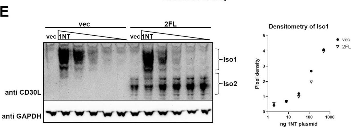
Detection of CD30 Ligand/TNFSF8 by Western Blot
Iso2 partially blocks Iso1-mediated stimulation of K299 cells. (A) HEK cells were transfected with either vector (vec) control or untagged Iso1 (1NT) with or without untagged Iso2 (2NT) or FLAG-tagged Iso2 (2FL). Transfected HEK cells were co-cultured with a K299-NFkB-Luc reporter clonal line. Two asterisks indicate p ≤ 0.01, three indicate p ≤ 0.001, and four indicate p ≤ 0.0001. (B,C) HEK cells were transfected with increasing quantities of 1NT plasmid with either vec or 2NT at a constant quantity, and then co-cultured with the K299-NFkB-Luc reporter line. Reporter activity (B) and IL-6 production (C) were measured. (D) HEK cells were transfected with either 1NT and vec or Iso2 and assessed for 1NT expression by flow cytometry. (E) Cells were transfected with increasing quantities of 1NT and either vec or 2FL, and the lysates were subject to SDS-PAGE and Western blot using a pan-CD30L antibody. Densitometry was used to quantify Iso1 bands, normalizing to GAPDH. Error bars for all are from standard deviations between technical replicates; representative experiment of ≥3 shown. Image collected and cropped by CiteAb from the following open publication (https://www.mdpi.com/1467-3045/46/3/172), licensed under a CC-BY license. Not internally tested by R&D Systems.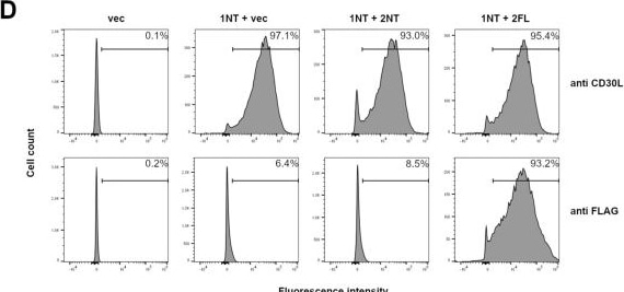
Detection of CD30 Ligand/TNFSF8 by Flow Cytometry
Iso2 partially blocks Iso1-mediated stimulation of K299 cells. (A) HEK cells were transfected with either vector (vec) control or untagged Iso1 (1NT) with or without untagged Iso2 (2NT) or FLAG-tagged Iso2 (2FL). Transfected HEK cells were co-cultured with a K299-NFkB-Luc reporter clonal line. Two asterisks indicate p ≤ 0.01, three indicate p ≤ 0.001, and four indicate p ≤ 0.0001. (B,C) HEK cells were transfected with increasing quantities of 1NT plasmid with either vec or 2NT at a constant quantity, and then co-cultured with the K299-NFkB-Luc reporter line. Reporter activity (B) and IL-6 production (C) were measured. (D) HEK cells were transfected with either 1NT and vec or Iso2 and assessed for 1NT expression by flow cytometry. (E) Cells were transfected with increasing quantities of 1NT and either vec or 2FL, and the lysates were subject to SDS-PAGE and Western blot using a pan-CD30L antibody. Densitometry was used to quantify Iso1 bands, normalizing to GAPDH. Error bars for all are from standard deviations between technical replicates; representative experiment of ≥3 shown. Image collected and cropped by CiteAb from the following open publication (https://www.mdpi.com/1467-3045/46/3/172), licensed under a CC-BY license. Not internally tested by R&D Systems.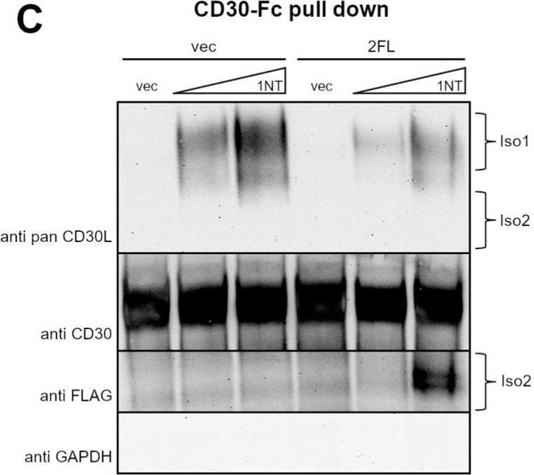
Detection of CD30 Ligand/TNFSF8 by Western Blot
Iso2 can block Iso1/CD30 interaction and anti-Iso1 antibody binding. (A) HEK cells were transfected with increasing quantities of untagged Iso1 (1NT) plasmid with a constant amount of either vector (vec) control or FLAG-tagged Iso2 (2FL). Lysates were used in a plate-based binding assay using CD30 recombinant protein as the capture and an Iso1-specific anti-CD30L antibody was used for detection. (B) HEK cells were transfected with 1NT with or without 2FL, and 2FL alone. The lysates were used in a plate-based binding assay using an anti-pan-CD30L antibody for capture and an Iso1-specific anti-CD30L antibody for detection. (C,D) HEK cells were transfected with two quantities of 1NT and a constant quantity of 2FL or vector (vec) control in 10 cm plates. Additionally, 10% of the lysates were set aside as the input whole-cell lysate fraction (C), and the remainder was incubated with CD30-Fc recombinant protein and protein A/G beads (D). Both were subject to SDS-PAGE and Western blot. Error bars for all are from standard deviations between technical replicates; representative experiment of ≥3 shown. Image collected and cropped by CiteAb from the following open publication (https://www.mdpi.com/1467-3045/46/3/172), licensed under a CC-BY license. Not internally tested by R&D Systems.Applications for Human CD30 Ligand/TNFSF8 Antibody (116614)
CyTOF-ready
Flow Cytometry
Sample: Human peripheral blood mononuclear cells (PBMCs) treated with PMA and Calcium Ionomycin
Neutralization
Reviewed Applications
Read 1 review rated 3 using MAB1028 in the following applications:
Flow Cytometry Panel Builder
Bio-Techne Knows Flow Cytometry
Save time and reduce costly mistakes by quickly finding compatible reagents using the Panel Builder Tool.
Advanced Features
- Spectra Viewer - Custom analysis of spectra from multiple fluorochromes
- Spillover Popups - Visualize the spectra of individual fluorochromes
- Antigen Density Selector - Match fluorochrome brightness with antigen density

Formulation, Preparation, and Storage
Purification
Reconstitution
Reconstitute at 0.5 mg/mL in sterile PBS. For liquid material, refer to CoA for concentration.
Formulation
Shipping
Stability & Storage
- 12 months from date of receipt, -20 to -70 °C as supplied.
- 1 month, 2 to 8 °C under sterile conditions after reconstitution.
- 6 months, -20 to -70 °C under sterile conditions after reconstitution.
Calculators
Background: CD30 Ligand/TNFSF8
References
- Brunangelo, F. et al. (1995) Blood 85:1.
- Gruss, H.-J. and F. Herrmann (1996) Leukemia and Lymphoma 20:397.
- Chiarle, R. et al. (1999) J. Immunol. 163:194.
Alternate Names
Gene Symbol
UniProt
Additional CD30 Ligand/TNFSF8 Products
Product Documents for Human CD30 Ligand/TNFSF8 Antibody (116614)
Certificate of Analysis
To download a Certificate of Analysis, please enter a lot or batch number in the search box below.
Note: Certificate of Analysis not available for kit components.
Product Specific Notices for Human CD30 Ligand/TNFSF8 Antibody (116614)
For research use only
Citations for Human CD30 Ligand/TNFSF8 Antibody (116614)
 Powered by Bioz
Customer Reviews for Human CD30 Ligand/TNFSF8 Antibody (116614) (1)
Have you used Human CD30 Ligand/TNFSF8 Antibody (116614)?
Submit a review and receive an Amazon gift card!
$25/€18/£15/$25CAN/¥2500 for a review with an image
$10/€7/£6/$10CAN/¥1110 for a review without an image
Submit a review
-
Application: ELISASample Tested: 293T human embryonic kidney cell lineSpecies: HumanVerified Customer | Posted 06/04/2019
There are no reviews that match your criteria.
Protocols
Find general support by application which include: protocols, troubleshooting, illustrated assays, videos and webinars.
- 7-Amino Actinomycin D (7-AAD) Cell Viability Flow Cytometry Protocol
- Extracellular Membrane Flow Cytometry Protocol
- Flow Cytometry Protocol for Cell Surface Markers
- Flow Cytometry Protocol for Staining Membrane Associated Proteins
- Flow Cytometry Staining Protocols
- Flow Cytometry Troubleshooting Guide
- Intracellular Flow Cytometry Protocol Using Alcohol (Methanol)
- Intracellular Flow Cytometry Protocol Using Detergents
- Intracellular Nuclear Staining Flow Cytometry Protocol Using Detergents
- Intracellular Staining Flow Cytometry Protocol Using Alcohol Permeabilization
- Intracellular Staining Flow Cytometry Protocol Using Detergents to Permeabilize Cells
- Propidium Iodide Cell Viability Flow Cytometry Protocol
- Protocol for Liperfluo
- Protocol for the Characterization of Human Th22 Cells
- Protocol for the Characterization of Human Th9 Cells
- Protocol: Annexin V and PI Staining by Flow Cytometry
- Protocol: Annexin V and PI Staining for Apoptosis by Flow Cytometry
- Troubleshooting Guide: Fluorokine Flow Cytometry Kits
- View all Protocols, Troubleshooting, Illustrated assays and Webinars
Associated Pathways
