Human CD31/PECAM-1 Antibody (9G11)
R&D Systems | Catalog # BBA7

Key Product Details
Validated by
Knockout/Knockdown
Species Reactivity
Validated:
Human
Cited:
Human, Mouse, Rat, Porcine, Primate - Macaca mulatta (Rhesus Macaque)
Applications
Validated:
Knockout Validated, Immunohistochemistry, Western Blot, Immunocytochemistry, Immunoprecipitation
Cited:
Immunohistochemistry, Immunohistochemistry-Paraffin, Immunohistochemistry-Frozen, Western Blot, Neutralization, Flow Cytometry, Immunofluorescence, Immunocytochemistry, Immunocytochemistry/ Immunofluorescence, Bioassay, Functional Assay
Label
Unconjugated
Antibody Source
Monoclonal Mouse IgG1 Clone # 9G11
Loading...
Product Specifications
Immunogen
Activated HUVEC human umbilical vein endothelial cells
Specificity
Detects human CD31/PEACAM-1. In Western blots, no cross-reactivity with recombinant human (rh) E-Selectin, rhICAM-1, -2, -3, rhVCAM‑1, or recombinant mouse VCAM‑1 was observed.
Clonality
Monoclonal
Host
Mouse
Isotype
IgG1
Scientific Data Images for Human CD31/PECAM-1 Antibody (9G11)
Detection of Human CD31/PECAM‑1 by Western Blot.
Western blot shows lysates of HUVEC human umbilical vein endothelial cells. PVDF membrane was probed with 1 µg/mL of Mouse Anti-Human CD31/PECAM-1 Monoclonal Antibody (Catalog # BBA7) followed by HRP-conjugated Anti-Mouse IgG Secondary Antibody (HAF018). A specific band was detected for CD31/PECAM-1 at approximately 130 kDa (as indicated). This experiment was conducted under reducing conditions and using Immunoblot Buffer Group 1.
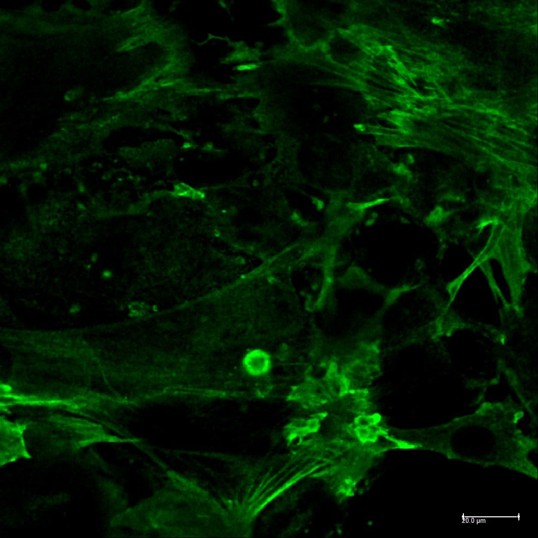
CD31/PECAM‑1 in HUVEC Human Cells.
CD31/PECAM-1 was detected in immersion fixed HUVEC human umbilical vein endothelial cells using 10 µg/mL Mouse Anti-Human CD31/PECAM-1 Monoclonal Antibody (Catalog # BBA7) for 3 hours at room temperature. Cells were stained with the NorthernLights™ 557-conjugated Anti-Mouse IgG Secondary Antibody (red; NL007) and counterstained with DAPI (blue). View our protocol for Fluorescent ICC Staining of Cells on Coverslips.
CD31/PECAM‑1 Specificity is Shown by Immunocytochemistry in Knockout Cell Line.
CD31/PECAM‑1 was detected in immersion fixed THP‑1 human acute monocytic leukemia cell line but is not detected in CD31/PECAM‑1 knockout (KO) THP‑1 human cell line using Mouse Anti-Human CD31/PECAM‑1 Monoclonal Antibody (Catalog # BBA7) at 8 µg/mL for 3 hours at room temperature. Cells were stained using the NorthernLights™ 557-conjugated Anti-Mouse IgG Secondary Antibody (red; NL007) and counterstained with DAPI (blue). Specific staining was localized to cell membrane. Staining was performed using our protocol for Fluorescent ICC Staining of Non-adherent Cells.
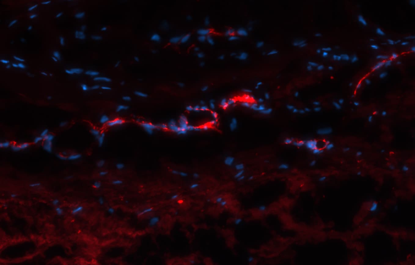
Detection of CD31/PECAM‑1 in Artery in Human Liver.
CD31/PECAM‑1 was detected in immersion fixed paraffin-embedded sections of Artery in Human Liver using Mouse Anti-Human CD31/PECAM‑1 Monoclonal Antibody (Catalog # BBA7) at 10 µg/mL for 1 hour at room temperature followed by incubation with the Anti-Mouse IgG VisUCyte™ HRP Polymer Antibody (Catalog # VC001). Before incubation with the primary antibody, tissue was subjected to heat-induced epitope retrieval using VisUCyte Antigen Retrieval Reagent-Basic (Catalog # VCTS021). Tissue was stained using DAB (brown) and counterstained with hematoxylin (blue). Specific staining was localized to endothelial cells. View our protocol for IHC Staining with VisUCyte HRP Polymer Detection Reagents.
Detection of Human CD31/PECAM-1 by Immunocytochemistry/ Immunofluorescence
Characterization&functional analysis of expanded AEPCs compared with HUVECs. All the experiments performed using AEPCs&HUVECs at passage number 5. (A) Fluorescent immunocytochemistry of the expression profiles of endothelial cell-specific markers, CD31, vWF,&the binding capacity of isolectin-B4, in AEPCs&HUVECs. Nuclear staining was assessed by 4′,6-diamidino-2-phenylindole (DAPI). Photographs taken using a confocal microscope (OLYMPUS, FV1000 with a CCD camera DP71&an objective lens UPlanFL N 40×/1.30 oil; 400× magnification) as a plane image. Bars represent 100 µm. The graphs show the %age of endothelial marker-positive cells among DAPI-positive cells. The data represent two independent experiments (2 donors),&%ages determined from cultivations performed in triplicate. The bars represent means ± SD (n = 6, **P < 0.01, two-tailed unpaired t-test). Image collected & cropped by CiteAb from the following open publication (https://pubmed.ncbi.nlm.nih.gov/35110646), licensed under a CC-BY license. Not internally tested by R&D Systems.
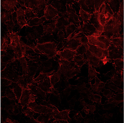
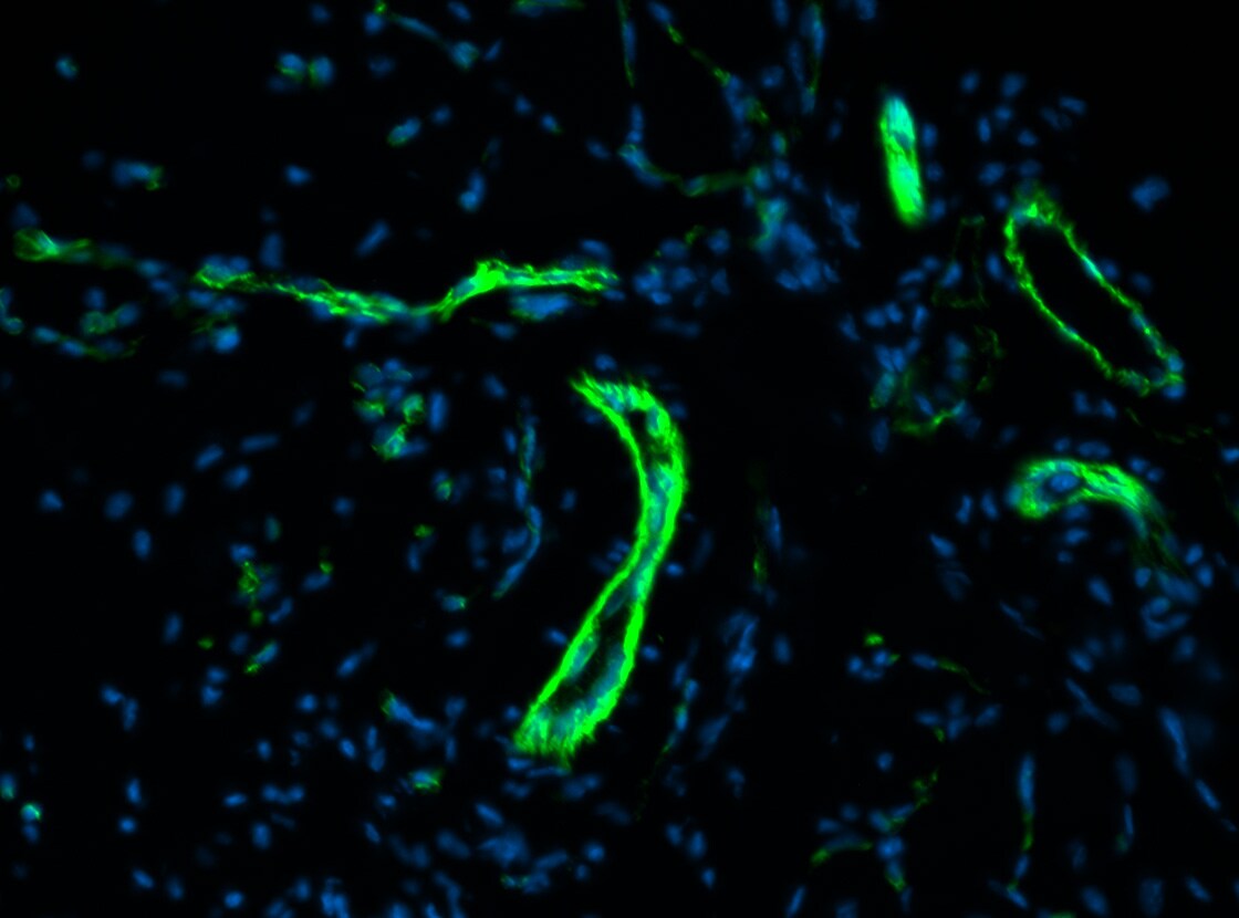
Detection of CD31/PECAM-1 in Human Lung Tissue.
IF staining of CD31/PECAM-1 in human lung tissue. Image from a verified customer review.Applications for Human CD31/PECAM-1 Antibody (9G11)
Application
Recommended Usage
Immunocytochemistry
8-25 µg/mL
Sample: Immersion fixed HUVEC human umbilical vein endothelial cells
Sample: Immersion fixed HUVEC human umbilical vein endothelial cells
Immunohistochemistry
5-25 µg/mL
Sample: Immersion fixed paraffin-embedded sections of Artery in Human Liver
Sample: Immersion fixed paraffin-embedded sections of Artery in Human Liver
Immunoprecipitation
Lampugnani, M.G. et al. (1992) J. Cell Biol. 118:1511.
Knockout Validated
CD31/PECAM‑1 was detected in immersion fixed THP‑1 human acute monocytic leukemia cell line but is not detected in CD31/PECAM‑1 knockout (KO) THP‑1 human cell line.
Western Blot
1 µg/mL
Sample: HUVEC human umbilical vein endothelial cells
Sample: HUVEC human umbilical vein endothelial cells
Reviewed Applications
Read 8 reviews rated 4.3 using BBA7 in the following applications:
Formulation, Preparation, and Storage
Purification
Protein A or G purified from hybridoma culture supernatant
Reconstitution
Sterile PBS to a final concentration of 0.5 mg/mL.
Loading...
Formulation
Lyophilized from a 0.2 μm filtered solution in PBS with Trehalose.
Shipping
The product is shipped at ambient temperature. Upon receipt, store it immediately at the temperature recommended below.
Stability & Storage
Use a manual defrost freezer and avoid repeated freeze-thaw cycles.
- 12 months from date of receipt, -20 to -70 °C as supplied.
- 1 month, 2 to 8 °C under sterile conditions after reconstitution.
- 6 months, -20 to -70 °C under sterile conditions after reconstitution.
Calculators
Background: CD31/PECAM-1
Long Name
Platelet Endothelial Cell Adhesion Molecule 1
Alternate Names
CD31, EndoCAM, PECA1, PECAM-1, PECAM1
Gene Symbol
PECAM1
Additional CD31/PECAM-1 Products
Product Documents for Human CD31/PECAM-1 Antibody (9G11)
Certificate of Analysis
To download a Certificate of Analysis, please enter a lot or batch number in the search box below.
Note: Certificate of Analysis not available for kit components.
Product Specific Notices for Human CD31/PECAM-1 Antibody (9G11)
For research use only
Related Research Areas
Citations for Human CD31/PECAM-1 Antibody (9G11)
 Powered by Bioz
Customer Reviews for Human CD31/PECAM-1 Antibody (9G11) (8)
4.3 out of 5
8 Customer Ratings
Have you used Human CD31/PECAM-1 Antibody (9G11)?
Submit a review and receive an Amazon gift card!
$25/€18/£15/$25CAN/¥2500 Yen for a review with an image
$10/€7/£6/$10CAN/¥1110 Yen for a review without an image
Submit a review
Customer Images

Showing
1
-
5 of
8 reviews
Showing All
Filter By:
-
Application: ImmunofluorescenceSample Tested: Adult lungSpecies: HumanVerified Customer | Posted 09/10/2025IF staining of pecam 1 in human lung tissueIF staining of pecam1 in human lung tissue

-
Application: Western BlotSample Tested: HUVEC human umbilical vein endothelial cellsSpecies: HumanVerified Customer | Posted 03/09/2022
-
Application: MicroarraysSample Tested: EDTA PlasmaSpecies: HumanVerified Customer | Posted 01/14/2021
-
Application: MicroarraysSample Tested: EDTA PlasmaSpecies: HumanVerified Customer | Posted 11/14/2018
-
Application: MicroarraySample Tested: EDTA PlasmaSpecies: HumanVerified Customer | Posted 11/02/2018
-
Application: Immunocytochemistry/ImmunofluorescenceSample Tested: HUVEC human umbilical vein endothelial cellsSpecies: HumanVerified Customer | Posted 02/28/2017Antibody was reconstituted at 0.5mg/ml and used at 1:50 dilution overnight in 4ºC.
-
Application: ImmunohistochemistrySample Tested: arterySpecies: HumanVerified Customer | Posted 08/29/2016Frozen human artery. Permeabilized with 80% acetone. Primary antibody at 1:100 incubate at 4oC overnight Secondary antibody: CY3 donkey anti-mouse secondary antibody. 1 hour at room temperature.
-
Application: ImmunohistochemistrySample Tested: Muscle tissueSpecies: HumanVerified Customer | Posted 08/29/2016Antigen retrieval: 10mM Citrate pH6. Primary antibody at 1:100 incubate at 4oC overnight Secondary antibody: Alexa 647 donkey anti-mouse secondary antibody. 1 hour at room temperature.
There are no reviews that match your criteria.
Protocols
Find general support by application which include: protocols, troubleshooting, illustrated assays, videos and webinars.
- Antigen Retrieval Protocol (PIER)
- Antigen Retrieval for Frozen Sections Protocol
- Appropriate Fixation of IHC/ICC Samples
- Cellular Response to Hypoxia Protocols
- Chromogenic IHC Staining of Formalin-Fixed Paraffin-Embedded (FFPE) Tissue Protocol
- Chromogenic Immunohistochemistry Staining of Frozen Tissue
- ClariTSA™ Fluorophore Kits
- Detection & Visualization of Antibody Binding
- Fluorescent IHC Staining of Frozen Tissue Protocol
- Graphic Protocol for Heat-induced Epitope Retrieval
- Graphic Protocol for the Preparation and Fluorescent IHC Staining of Frozen Tissue Sections
- Graphic Protocol for the Preparation and Fluorescent IHC Staining of Paraffin-embedded Tissue Sections
- Graphic Protocol for the Preparation of Gelatin-coated Slides for Histological Tissue Sections
- ICC Cell Smear Protocol for Suspension Cells
- ICC Immunocytochemistry Protocol Videos
- ICC for Adherent Cells
- IHC Sample Preparation (Frozen sections vs Paraffin)
- Immunocytochemistry (ICC) Protocol
- Immunocytochemistry Troubleshooting
- Immunofluorescence of Organoids Embedded in Cultrex Basement Membrane Extract
- Immunofluorescent IHC Staining of Formalin-Fixed Paraffin-Embedded (FFPE) Tissue Protocol
- Immunohistochemistry (IHC) and Immunocytochemistry (ICC) Protocols
- Immunohistochemistry Frozen Troubleshooting
- Immunohistochemistry Paraffin Troubleshooting
- Immunoprecipitation Protocol
- Preparing Samples for IHC/ICC Experiments
- Preventing Non-Specific Staining (Non-Specific Binding)
- Primary Antibody Selection & Optimization
- Protocol for Heat-Induced Epitope Retrieval (HIER)
- Protocol for Making a 4% Formaldehyde Solution in PBS
- Protocol for VisUCyte™ HRP Polymer Detection Reagent
- Protocol for the Fluorescent ICC Staining of Cell Smears - Graphic
- Protocol for the Fluorescent ICC Staining of Cultured Cells on Coverslips - Graphic
- Protocol for the Preparation & Fixation of Cells on Coverslips
- Protocol for the Preparation and Chromogenic IHC Staining of Frozen Tissue Sections
- Protocol for the Preparation and Chromogenic IHC Staining of Frozen Tissue Sections - Graphic
- Protocol for the Preparation and Chromogenic IHC Staining of Paraffin-embedded Tissue Sections
- Protocol for the Preparation and Chromogenic IHC Staining of Paraffin-embedded Tissue Sections - Graphic
- Protocol for the Preparation and Fluorescent ICC Staining of Cells on Coverslips
- Protocol for the Preparation and Fluorescent ICC Staining of Non-adherent Cells
- Protocol for the Preparation and Fluorescent ICC Staining of Stem Cells on Coverslips
- Protocol for the Preparation and Fluorescent IHC Staining of Frozen Tissue Sections
- Protocol for the Preparation and Fluorescent IHC Staining of Paraffin-embedded Tissue Sections
- Protocol for the Preparation of Gelatin-coated Slides for Histological Tissue Sections
- Protocol for the Preparation of a Cell Smear for Non-adherent Cell ICC - Graphic
- R&D Systems Quality Control Western Blot Protocol
- TUNEL and Active Caspase-3 Detection by IHC/ICC Protocol
- The Importance of IHC/ICC Controls
- Troubleshooting Guide: Immunohistochemistry
- Troubleshooting Guide: Western Blot Figures
- Western Blot Conditions
- Western Blot Protocol
- Western Blot Protocol for Cell Lysates
- Western Blot Troubleshooting
- Western Blot Troubleshooting Guide
- View all Protocols, Troubleshooting, Illustrated assays and Webinars
Loading...