CD40 is a type I transmembrane glycoprotein belonging to the TNF receptor superfamily. The mature hCD40 consists of a 172 amino acid (aa) extracellular domain, a 22 aa transmembrane region and a 62 aa cytoplasmic domain (1). Human and mouse CD40 share 62% aa identity. CD40 is expressed in B cells, follicular dendritic cells, dendritic cells, activated monocytes, macrophages, endothelial cells, vascular smooth muscle cells, and several tumor cell lines (2). The extracellular domain has the cysteine-rich repeat regions, which are characteristic for many of the receptors of the TNF superfamily. Interaction of CD40 with its ligand, CD40L, leads to aggregation of CD40 molecules, which in turn interact with cytoplasmic components to initiate signaling pathways. Early studies on the CD40-CD40L system revealed its role in humoral immunity. Interaction between CD40L on T cells and CD40 on B cells stimulated B cell proliferation and provided the signal for immunoglobulin isotype switching (3). Mutations in the CD40L gene, which resulted in a CD40L molecule unable to interact with CD40, are responsible for the hyper-IgM syndrome (4). Cross-linking of CD40 with antibodies or by CD40 binding to CD40L produces cell type-specific responses which include costimulation and induction of proliferation, induction of cytokine production, rescue from apoptosis, and upregulation of adhesion molecules (5). Some of the early events of intracellular signaling by the
CD40‑CD40L system include the association of the CD40 with TRAFs and the activation of various kinases (6‑8).

Key Product Details
Species Reactivity
Validated:
Human
Cited:
Human, Mouse
Applications
Validated:
Flow Cytometry, Immunocytochemistry, Agonist Activity, CyTOF-ready
Cited:
Immunohistochemistry-Paraffin, Neutralization, Flow Cytometry, Bioassay, Functional Assay, Stimulation
Label
Unconjugated
Antibody Source
Monoclonal Mouse IgG2B Clone # 82111
Loading...
Product Specifications
Immunogen
Mouse myeloma cell line NS0-derived recombinant human CD40/TNFRSF5
Glu21-Arg193
Accession # P25942
Glu21-Arg193
Accession # P25942
Specificity
Detects human CD40/TNFRSF5 in direct ELISAs and Western blots. In direct ELISAs, does not cross-react with recombinant human (rh) 4‑1BB, rhCD27, rhCD30, recombinant mouse CD40, rhDR3, rhDR6, rhEDAR, rhFas, rhGITR, rhHVEM, rhLTR beta, rhNGF R, rhOPG, rhRANK, rhTAJ, rhTNF RI, or rhTNF RII.
Clonality
Monoclonal
Host
Mouse
Isotype
IgG2B
Endotoxin Level
<0.10 EU per 1 μg of the antibody by the LAL method.
Scientific Data Images for Human CD40/TNFRSF5 Antibody
CD40/TNFRSF5 in Human PBMCs.
CD40/TNFRSF5 was detected in immersion fixed human peripheral blood mononuclear cells (PBMCs) using Mouse Anti-Human CD40/TNFRSF5 Monoclonal Antibody (Catalog # MAB6321) at 10 µg/mL for 3 hours at room temperature. Cells were stained using the NorthernLights™ 557-conjugated Anti-Mouse IgG Secondary Antibody (red; Catalog # NL007) and counterstained with DAPI (blue). Specific staining was localized to cell surfaces and cytoplasm. View our protocol for Fluorescent ICC Staining of Non-adherent Cells.
Human CD40/TNFRSF5 Antibody Stimulates Cell Proliferation in Human B Cells.
Mouse Anti-Human CD40/TNFRSF5 Monoclonal Antibody (Catalog # MAB6321) stimulates human B cell proliferation in the presence of Recombinant Human IL-4 (Catalog # 204-IL) in a dose-dependent manner, as measured by Resazurin (Catalog # AR002). The ED50 for this effect is typically 0.035-0.175 µg/mL
Detection of CD40/TNFRSF5 in PBMC lymphocytes by Flow Cytometry
PBMC lymphocytes were stained with Mouse Anti-Human CD19 APC‑conjugated Monoclonal Antibody (Catalog # FAB4867A) and either (A) Mouse Anti-Human CD40/TNFRSF5 Monoclonal Antibody (Catalog # MAB6321) or (B) isotype control antibody (Catalog # MAB0041) followed by Phycoerythrin-conjugated Anti-Mouse IgG Secondary Antibody (Catalog # F0102B). View our protocol for Staining Membrane-associated Proteins.Applications for Human CD40/TNFRSF5 Antibody
Application
Recommended Usage
Agonist Activity
Measured in a cell proliferation assay using B cell enriched human peripheral blood lymphocytes in the presence of IL-4. Banchereau, J. et al. (1991) Science 251:70. The ED50 for this effect is typically 0.035-0.175 μg/mL.
CyTOF-ready
Ready to be labeled using established conjugation methods. No BSA or other carrier proteins that could interfere with conjugation.
Flow Cytometry
2.5 µg/106 cells
Sample: Human whole blood CD19+ B cells
Sample: Human whole blood CD19+ B cells
Immunocytochemistry
8-25 µg/mL
Sample: Immersion fixed human peripheral blood mononuclear cells (PBMCs)
Sample: Immersion fixed human peripheral blood mononuclear cells (PBMCs)
Reviewed Applications
Read 1 review rated 4 using MAB6321 in the following applications:
Flow Cytometry Panel Builder
Bio-Techne Knows Flow Cytometry
Save time and reduce costly mistakes by quickly finding compatible reagents using the Panel Builder Tool.
Advanced Features
- Spectra Viewer - Custom analysis of spectra from multiple fluorochromes
- Spillover Popups - Visualize the spectra of individual fluorochromes
- Antigen Density Selector - Match fluorochrome brightness with antigen density

Formulation, Preparation, and Storage
Purification
Protein A or G purified from hybridoma culture supernatant
Reconstitution
Reconstitute at 0.5 mg/mL in sterile PBS. For liquid material, refer to CoA for concentration.
Loading...
Formulation
Lyophilized from a 0.2 μm filtered solution in PBS with Trehalose. *Small pack size (SP) is supplied either lyophilized or as a 0.2 µm filtered solution in PBS.
Shipping
Lyophilized product is shipped at ambient temperature. Liquid small pack size (-SP) is shipped with polar packs. Upon receipt, store immediately at the temperature recommended below.
Stability & Storage
Use a manual defrost freezer and avoid repeated freeze-thaw cycles.
- 12 months from date of receipt, -20 to -70 °C as supplied.
- 1 month, 2 to 8 °C under sterile conditions after reconstitution.
- 6 months, -20 to -70 °C under sterile conditions after reconstitution.
Calculators
Background: CD40/TNFRSF5
References
- Torres, R.M. and E.A. Clark (1992) J. Immunol. 148:620.
- Schonbeck, U. et al. (1997) J. Biol. Chem. 272:19569.
- Armitage, R.J. et al. (1993) J. Immunol. 150:3671.
- Callard, R.E. et al. (1993) Immunol. Today 14:559.
- Stout, R.D. and J. Suttles (1996) Immunol. Today 17:487.
- Pullen, S.S. et al. (1999) Biochemistry 38:10168.
- Faris, M. et al. (1994) J. Exp. Med. 179:1923.
- Hanissian, S.H. and R.S Geha (1997) Immunity 6:379.
Alternate Names
CD40, TNFRSF5
Gene Symbol
CD40
UniProt
Additional CD40/TNFRSF5 Products
Product Documents for Human CD40/TNFRSF5 Antibody
Certificate of Analysis
To download a Certificate of Analysis, please enter a lot or batch number in the search box below.
Note: Certificate of Analysis not available for kit components.
Product Specific Notices for Human CD40/TNFRSF5 Antibody
For research use only
Related Research Areas
Citations for Human CD40/TNFRSF5 Antibody
 Powered by Bioz
Customer Reviews for Human CD40/TNFRSF5 Antibody (1)
4 out of 5
1 Customer Rating
Have you used Human CD40/TNFRSF5 Antibody?
Submit a review and receive an Amazon gift card!
$25/€18/£15/$25CAN/¥2500 Yen for a review with an image
$10/€7/£6/$10CAN/¥1110 Yen for a review without an image
Submit a review
Customer Images
Showing
1
-
1 of
1 review
Showing All
Filter By:
-
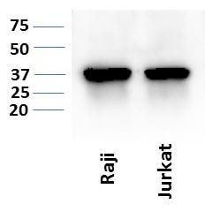
Application: Western BlotSample Tested: Min6 mouse insulinoma whole cell lysate and Cancer cell lysatesSpecies: HumanVerified Customer | Posted 06/02/2019

There are no reviews that match your criteria.
Protocols
Find general support by application which include: protocols, troubleshooting, illustrated assays, videos and webinars.
- 7-Amino Actinomycin D (7-AAD) Cell Viability Flow Cytometry Protocol
- Appropriate Fixation of IHC/ICC Samples
- Cellular Response to Hypoxia Protocols
- ClariTSA™ Fluorophore Kits
- Detection & Visualization of Antibody Binding
- Extracellular Membrane Flow Cytometry Protocol
- Flow Cytometry Protocol for Cell Surface Markers
- Flow Cytometry Protocol for Staining Membrane Associated Proteins
- Flow Cytometry Staining Protocols
- Flow Cytometry Troubleshooting Guide
- ICC Cell Smear Protocol for Suspension Cells
- ICC Immunocytochemistry Protocol Videos
- ICC for Adherent Cells
- Immunocytochemistry (ICC) Protocol
- Immunocytochemistry Troubleshooting
- Immunofluorescence of Organoids Embedded in Cultrex Basement Membrane Extract
- Immunohistochemistry (IHC) and Immunocytochemistry (ICC) Protocols
- Intracellular Flow Cytometry Protocol Using Alcohol (Methanol)
- Intracellular Flow Cytometry Protocol Using Detergents
- Intracellular Nuclear Staining Flow Cytometry Protocol Using Detergents
- Intracellular Staining Flow Cytometry Protocol Using Alcohol Permeabilization
- Intracellular Staining Flow Cytometry Protocol Using Detergents to Permeabilize Cells
- Preparing Samples for IHC/ICC Experiments
- Preventing Non-Specific Staining (Non-Specific Binding)
- Primary Antibody Selection & Optimization
- Propidium Iodide Cell Viability Flow Cytometry Protocol
- Protocol for Liperfluo
- Protocol for VisUCyte™ HRP Polymer Detection Reagent
- Protocol for the Characterization of Human Th22 Cells
- Protocol for the Characterization of Human Th9 Cells
- Protocol for the Fluorescent ICC Staining of Cell Smears - Graphic
- Protocol for the Fluorescent ICC Staining of Cultured Cells on Coverslips - Graphic
- Protocol for the Preparation and Fluorescent ICC Staining of Cells on Coverslips
- Protocol for the Preparation and Fluorescent ICC Staining of Non-adherent Cells
- Protocol for the Preparation and Fluorescent ICC Staining of Stem Cells on Coverslips
- Protocol for the Preparation of a Cell Smear for Non-adherent Cell ICC - Graphic
- Protocol: Annexin V and PI Staining by Flow Cytometry
- Protocol: Annexin V and PI Staining for Apoptosis by Flow Cytometry
- TUNEL and Active Caspase-3 Detection by IHC/ICC Protocol
- The Importance of IHC/ICC Controls
- Troubleshooting Guide: Fluorokine Flow Cytometry Kits
- View all Protocols, Troubleshooting, Illustrated assays and Webinars
Loading...
Associated Pathways

