CD44 is a ubiquitously expressed protein that is the major receptor for hyaluronan and exerts control over cell growth and migration (1‑3). Human CD44 has a 20 amino acid (aa) signal sequence, an extracellular domain (ECD) with a 100 aa hyaluronan-binding disulfide-stabilized link region and a 325‑530 aa stem region, a 21 aa transmembrane domain, and a 72 aa cytoplasmic domain. Within the stem, ten variably spliced exons (v1‑10, exons 6‑15) produce multiple protein isoforms (1‑3). The standard or hematopoietic form, CD44s or CD44H, does not include the variable segments (1‑3). Cancer aggressiveness and T cell activation have been correlated with expression of specific isoforms (1, 3). With variable N- and O-glycosylation and splicing within the stalk, CD44 can range from 80 to 200 kDa (1). Within the N‑terminal invariant portion of the ECD (aa 21‑220), human CD44 shares 76%, 76%, 86%, 83% and 79% identity with corresponding mouse, rat, equine, canine and bovine CD44, respectively. The many reported functions of CD44 fall within three categories (1). First, CD44 binds hyaluronan and other ligands within the extracellular matrix and can function as a “platform” for growth factors and metalloproteinases. Second, CD44 can function as a co‑receptor that modifies activity of receptors including MET and the ERBB family of tyrosine kinases. Third, the CD44 intracellular domain links the plasma membrane to the actin cytoskeleton via the ERM proteins, ezrin, radixin and moesin. CD44 can be synthesized in a soluble form (4) or may be cleaved at multiple sites by either membrane-type matrix metalloproteinases, or ADAM proteases to produce soluble ectodomains (5, 6). The cellular portion may then undergo gamma secretase-dependent intramembrane cleavage to form an A beta -like transmembrane portion and a cytoplasmic signaling portion that affects gene expression (7, 8). These cleavage events are thought to promote metastasis by enhancing tumor cell motility and growth (1, 5).
Human CD44 s Pan Specific Antibody (691534)
R&D Systems | Catalog # MAB7045

Key Product Details
Species Reactivity
Validated:
Human
Cited:
Human, Mouse
Applications
Validated:
Multiplex Immunofluorescence, Immunohistochemistry, Western Blot, Flow Cytometry, Immunocytochemistry, COMET, CyTOF-reported
Cited:
Immunohistochemistry, Immunohistochemistry-Paraffin, Western Blot, ELISA Detection
Label
Unconjugated
Antibody Source
Monoclonal Mouse IgG2A Clone # 691534
Loading...
Product Specifications
Immunogen
Mouse myeloma cell line NS0-derived recombinant human CD44s
Gln21-Pro220
Accession # P16070
Gln21-Pro220
Accession # P16070
Specificity
Detects human CD44 in direct ELISAs and Western blots. In direct ELISAs, no cross-reactivity with recombinant CD44 from mouse, rat, or pig is observed.
Clonality
Monoclonal
Host
Mouse
Isotype
IgG2A
Scientific Data Images for Human CD44 s Pan Specific Antibody (691534)
Detection of CD44 in Human Lymph Node via seqIF™ staining on COMET™
CD44 was detected in immersion fixed paraffin-embedded sections of human Lymph Node using Mouse Anti-Human CD44, Monoclonal Antibody (MAB7045) at 1 μg/mL at 37 ° Celsius for 4 minutes. Before incubation with the primary antibody, tissue underwent an all-in-one dewaxing and antigen retrieval preprocessing using PreTreatment Module (PT Module) and Dewax and HIER Buffer H (pH 9; Epredia Catalog # TA-999-DHBH). Tissue was stained using the Alexa Fluor™ 647 Goat anti-Mouse IgG Secondary Antibody at 1:200 at 37 ° Celsius for 2 minutes. (Yellow; Lunaphore Catalog # DR647MS) and counterstained with DAPI (blue; Lunaphore Catalog # DR100). Specific staining was localized to the membrane. Protocol available in COMET™ Panel Builder.
Detection of Human CD44 by Western Blot.
Western blot shows lysates of HeLa human cervical epithelial carcinoma cell line, HUVEC human umbilical vein endothelial cells, and PC-3 human prostate cancer cell line. PVDF membrane was probed with 0.2 µg/mL of Mouse Anti-Human CD44s Pan Specific Monoclonal Antibody (Catalog # MAB7045) followed by HRP-conjugated Anti-Mouse IgG Secondary Antibody (HAF007). Specific bands were detected for CD44 at approximately 80 to 100 kDa (as indicated). This experiment was conducted under reducing conditions and using Immunoblot Buffer Group 1.
Detection of CD44 in Human Blood Lymphocytes by Flow Cytometry.
Human peripheral blood lymphocytes were stained with Mouse Anti-Human CD44s Pan Specific Monoclonal Antibody (Catalog # MAB7045, filled histogram) or isotype control antibody (MAB003, open histogram), followed by Allophycocyanin-conjugated Anti-Mouse IgG Secondary Antibody (F0101B).
CD44 in Human PBMCs.
CD44 was detected in immersion fixed human peripheral blood mononuclear cells (PBMCs) using Mouse Anti-Human CD44s Pan Specific Monoclonal Antibody (Catalog # MAB7045) at 10 µg/mL for 3 hours at room temperature. Cells were stained using the NorthernLights™ 557-conjugated Anti-Mouse IgG Secondary Antibody (red; NL007) and counterstained with DAPI (blue). Specific staining was localized to cell surfaces. View our protocol for Fluorescent ICC Staining of Non-adherent Cells.
CD44 in Human Tonsil.
CD44 was detected in immersion fixed paraffin-embedded sections of human tonsil using Mouse Anti-Human CD44s Pan Specific Monoclonal Antibody (Catalog # MAB7045) at 15 µg/mL overnight at 4 °C. Tissue was stained using the Anti-Mouse HRP-DAB Cell & Tissue Staining Kit (brown; CTS002) and counterstained with hematoxylin (blue). View our protocol for Chromogenic IHC Staining of Paraffin-embedded Tissue Sections. & Daudi (Negative).")
Detection of CD44 in PC‑3 (Positive) & Daudi (Negative).
CD44 was detected in immersion fixed PC‑3 human prostate cancer cells (Positive) & absent in Daudi human Burkitt's lymphoma cells (Negative) using Mouse Anti-Human CD44 s Pan Specific Monoclonal Antibody (Catalog # MAB7045) at 8 µg/mL for 3 hours at room temperature. Cells were stained using the NorthernLights™ 557-conjugated Anti-Mouse IgG Secondary Antibody (red; Catalog # NL007) and counterstained with DAPI (blue). Specific staining was localized to Nuclear and cytoplasmic. View our protocol for Fluorescent ICC Staining of Cells on Coverslips.
Detection of CD44 by Flow Cytometry.
MDA‑MB‑231 human breast cancer parental cell line WT and CD44 KO cells were labelled with a green or violet, fluorescent dye, respectively. WT and KO cells were mixed in a 1:1 ratio, fixed in 4% PFA and permeabilized in 0.1% saponin. 400,000 cells were stained with Mouse Anti-Human CD44 s Pan Specific Monoclonal Antibody (Catalog # MAB7045) and a secondary antibody. Antibody staining was quantified, and representative images showing the staining intensity in the KO population (pink histogram, dashed line) compared to the WT cells (green histogram, solid line) are presented. Histograms with dotted lines represent secondary antibody-only controls in both WT and KO cells. Primary antibody concentration: 1 μg/mL. Image, protocol and testing courtesy of YCharOS Inc. (ycharos.com).Applications for Human CD44 s Pan Specific Antibody (691534)
Application
Recommended Usage
COMET
Optimal dilutions of this antibody should be experimentally determined.
CyTOF-reported
This clone has been commercially reported for use in CyTOF®. Ready to be labeled using established conjugation methods. No BSA or other carrier proteins that could interfere with conjugation.
Flow Cytometry
2.5 µg/106 cells
Sample: Human peripheral blood lymphocytes & MDA-MB-231 human breast cancer parental cell line
Sample: Human peripheral blood lymphocytes & MDA-MB-231 human breast cancer parental cell line
Immunocytochemistry
8-25 µg/mL
Sample: Immersion fixed human peripheral blood mononuclear cells (PBMCs), PC‑3 human prostate cancer cells (Positive) & Daudi human Burkitt's lymphoma cells (Negative)
Sample: Immersion fixed human peripheral blood mononuclear cells (PBMCs), PC‑3 human prostate cancer cells (Positive) & Daudi human Burkitt's lymphoma cells (Negative)
Immunohistochemistry
8-25 µg/mL
Sample: Immersion fixed paraffin-embedded sections of human tonsil
Sample: Immersion fixed paraffin-embedded sections of human tonsil
Multiplex Immunofluorescence
1 µg/mL
Sample: Immersion fixed paraffin-embedded sections of Human Lymph Node
Sample: Immersion fixed paraffin-embedded sections of Human Lymph Node
Western Blot
0.2 µg/mL
Sample: HeLa human cervical epithelial carcinoma cell line, HUVEC human umbilical vein endothelial cells, and PC‑3 human prostate cancer cell line
Sample: HeLa human cervical epithelial carcinoma cell line, HUVEC human umbilical vein endothelial cells, and PC‑3 human prostate cancer cell line
Reviewed Applications
Read 2 reviews rated 4.5 using MAB7045 in the following applications:
Flow Cytometry Panel Builder
Bio-Techne Knows Flow Cytometry
Save time and reduce costly mistakes by quickly finding compatible reagents using the Panel Builder Tool.
Advanced Features
- Spectra Viewer - Custom analysis of spectra from multiple fluorochromes
- Spillover Popups - Visualize the spectra of individual fluorochromes
- Antigen Density Selector - Match fluorochrome brightness with antigen density

Formulation, Preparation, and Storage
Purification
Protein A or G purified from hybridoma culture supernatant
Reconstitution
Sterile PBS to a final concentration of 0.5 mg/mL. For liquid material, refer to CoA for concentration.
Loading...
Formulation
Lyophilized from a 0.2 μm filtered solution in PBS with Trehalose. See Certificate of Analysis for details.
*Small pack size (-SP) is supplied either lyophilized or as a 0.2 µm filtered solution in PBS.
*Small pack size (-SP) is supplied either lyophilized or as a 0.2 µm filtered solution in PBS.
Shipping
Lyophilized product is shipped at ambient temperature. Liquid small pack size (-SP) is shipped with polar packs. Upon receipt, store immediately at the temperature recommended below.
Stability & Storage
Use a manual defrost freezer and avoid repeated freeze-thaw cycles.
- 12 months from date of receipt, -20 to -70 °C as supplied.
- 1 month, 2 to 8 °C under sterile conditions after reconstitution.
- 6 months, -20 to -70 °C under sterile conditions after reconstitution.
Calculators
Background: CD44
References
- Ponta, H. et al. (2003) Nat. Rev. Mol. Cell Biol. 4:33.
- Screaton, G.R. et al. (1992) Proc. Natl. Acad. Sci. USA 89:12160.
- Lynch, K.W. (2004) Nat. Rev. Immunol. 4:931.
- Yu, Q. and B.P. Toole (1996) J. Biol. Chem. 271:20603.
- Nagano, O. and H. Saya (2004) Cancer Sci. 95:930.
- Nakamura, H. et al. (2004) Cancer Res. 64:876.
- Murakami, D. et al. (2003) Oncogene 22:1511.
- Lammich, S. et al. (2002) J. Biol. Chem. 277:44754.
Alternate Names
CD44, ECMR-III, HCAM, HCELL, LHR, MDU2, MDU3, MIC4, MUTCH-I, Pgp1
Gene Symbol
CD44
UniProt
Additional CD44 Products
Product Documents for Human CD44 s Pan Specific Antibody (691534)
Certificate of Analysis
To download a Certificate of Analysis, please enter a lot or batch number in the search box below.
Note: Certificate of Analysis not available for kit components.
Product Specific Notices for Human CD44 s Pan Specific Antibody (691534)
For research use only
Related Research Areas
Citations for Human CD44 s Pan Specific Antibody (691534)
 Powered by Bioz
Customer Reviews for Human CD44 s Pan Specific Antibody (691534) (2)
4.5 out of 5
2 Customer Ratings
Have you used Human CD44 s Pan Specific Antibody (691534)?
Submit a review and receive an Amazon gift card!
$25/€18/£15/$25CAN/¥2500 Yen for a review with an image
$10/€7/£6/$10CAN/¥1110 Yen for a review without an image
Submit a review
Customer Images
Showing
1
-
2 of
2 reviews
Showing All
Filter By:
-
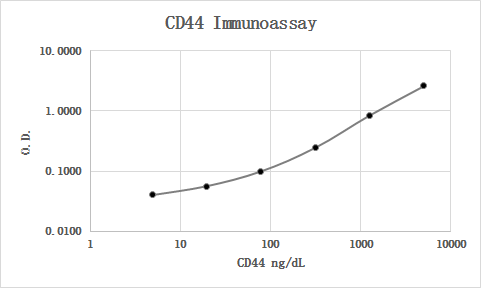
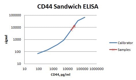
Application: ELISASample Tested: Serum and PlasmaSpecies: HumanVerified Customer | Posted 06/12/2019

-
Application: ELISASample Tested: Serum and PlasmaSpecies: HumanVerified Customer | Posted 01/30/2019MAB7045 was used as the capture antibody along with BBA10 as the detection antibody. Recombinant human CD44 Fc Chimera (3660-CD) was used as the calibrator material. Human serum and plasma samples were diluted 1:5 and all were quantifiable. Parallelism looked good.
There are no reviews that match your criteria.
Protocols
Find general support by application which include: protocols, troubleshooting, illustrated assays, videos and webinars.
- 7-Amino Actinomycin D (7-AAD) Cell Viability Flow Cytometry Protocol
- Antigen Retrieval Protocol (PIER)
- Antigen Retrieval for Frozen Sections Protocol
- Appropriate Fixation of IHC/ICC Samples
- Cellular Response to Hypoxia Protocols
- Chromogenic IHC Staining of Formalin-Fixed Paraffin-Embedded (FFPE) Tissue Protocol
- Chromogenic Immunohistochemistry Staining of Frozen Tissue
- ClariTSA™ Fluorophore Kits
- Detection & Visualization of Antibody Binding
- Extracellular Membrane Flow Cytometry Protocol
- Flow Cytometry Protocol for Cell Surface Markers
- Flow Cytometry Protocol for Staining Membrane Associated Proteins
- Flow Cytometry Staining Protocols
- Flow Cytometry Troubleshooting Guide
- Fluorescent IHC Staining of Frozen Tissue Protocol
- Graphic Protocol for Heat-induced Epitope Retrieval
- Graphic Protocol for the Preparation and Fluorescent IHC Staining of Frozen Tissue Sections
- Graphic Protocol for the Preparation and Fluorescent IHC Staining of Paraffin-embedded Tissue Sections
- Graphic Protocol for the Preparation of Gelatin-coated Slides for Histological Tissue Sections
- ICC Cell Smear Protocol for Suspension Cells
- ICC Immunocytochemistry Protocol Videos
- ICC for Adherent Cells
- IHC Sample Preparation (Frozen sections vs Paraffin)
- Immunocytochemistry (ICC) Protocol
- Immunocytochemistry Troubleshooting
- Immunofluorescence of Organoids Embedded in Cultrex Basement Membrane Extract
- Immunofluorescent IHC Staining of Formalin-Fixed Paraffin-Embedded (FFPE) Tissue Protocol
- Immunohistochemistry (IHC) and Immunocytochemistry (ICC) Protocols
- Immunohistochemistry Frozen Troubleshooting
- Immunohistochemistry Paraffin Troubleshooting
- Intracellular Flow Cytometry Protocol Using Alcohol (Methanol)
- Intracellular Flow Cytometry Protocol Using Detergents
- Intracellular Nuclear Staining Flow Cytometry Protocol Using Detergents
- Intracellular Staining Flow Cytometry Protocol Using Alcohol Permeabilization
- Intracellular Staining Flow Cytometry Protocol Using Detergents to Permeabilize Cells
- Preparing Samples for IHC/ICC Experiments
- Preventing Non-Specific Staining (Non-Specific Binding)
- Primary Antibody Selection & Optimization
- Propidium Iodide Cell Viability Flow Cytometry Protocol
- Protocol for Heat-Induced Epitope Retrieval (HIER)
- Protocol for Liperfluo
- Protocol for Making a 4% Formaldehyde Solution in PBS
- Protocol for VisUCyte™ HRP Polymer Detection Reagent
- Protocol for the Characterization of Human Th22 Cells
- Protocol for the Characterization of Human Th9 Cells
- Protocol for the Fluorescent ICC Staining of Cell Smears - Graphic
- Protocol for the Fluorescent ICC Staining of Cultured Cells on Coverslips - Graphic
- Protocol for the Preparation & Fixation of Cells on Coverslips
- Protocol for the Preparation and Chromogenic IHC Staining of Frozen Tissue Sections
- Protocol for the Preparation and Chromogenic IHC Staining of Frozen Tissue Sections - Graphic
- Protocol for the Preparation and Chromogenic IHC Staining of Paraffin-embedded Tissue Sections
- Protocol for the Preparation and Chromogenic IHC Staining of Paraffin-embedded Tissue Sections - Graphic
- Protocol for the Preparation and Fluorescent ICC Staining of Cells on Coverslips
- Protocol for the Preparation and Fluorescent ICC Staining of Non-adherent Cells
- Protocol for the Preparation and Fluorescent ICC Staining of Stem Cells on Coverslips
- Protocol for the Preparation and Fluorescent IHC Staining of Frozen Tissue Sections
- Protocol for the Preparation and Fluorescent IHC Staining of Paraffin-embedded Tissue Sections
- Protocol for the Preparation of Gelatin-coated Slides for Histological Tissue Sections
- Protocol for the Preparation of a Cell Smear for Non-adherent Cell ICC - Graphic
- Protocol: Annexin V and PI Staining by Flow Cytometry
- Protocol: Annexin V and PI Staining for Apoptosis by Flow Cytometry
- R&D Systems Quality Control Western Blot Protocol
- TUNEL and Active Caspase-3 Detection by IHC/ICC Protocol
- The Importance of IHC/ICC Controls
- Troubleshooting Guide: Fluorokine Flow Cytometry Kits
- Troubleshooting Guide: Immunohistochemistry
- Troubleshooting Guide: Western Blot Figures
- Western Blot Conditions
- Western Blot Protocol
- Western Blot Protocol for Cell Lysates
- Western Blot Troubleshooting
- Western Blot Troubleshooting Guide
- View all Protocols, Troubleshooting, Illustrated assays and Webinars
Loading...
Associated Pathways


