Human Cytokeratin 14 Antibody (LL001)
R&D Systems | Catalog # MAB3164

Key Product Details
Species Reactivity
Validated:
Human
Cited:
Human, Mouse
Applications
Validated:
Immunohistochemistry, Western Blot, Immunocytochemistry
Cited:
Immunohistochemistry, Immunohistochemistry-Paraffin
Label
Unconjugated
Antibody Source
Monoclonal Mouse IgG2A Clone # LL001
Loading...
Product Specifications
Immunogen
Human Cytokeratin 14 synthetic peptide
DGKVVSTHEQVLRTKN
DGKVVSTHEQVLRTKN
Specificity
Detects human Cytokeratin 14 in Western blots.
Clonality
Monoclonal
Host
Mouse
Isotype
IgG2A
Scientific Data Images for Human Cytokeratin 14 Antibody (LL001)
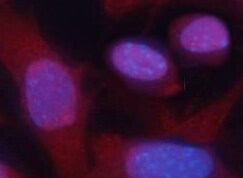
Cytokeratin 14 in A431 Human Cell Line.
Cytokeratin 14 was detected in immersion fixed A431 human epithelial carcinoma cell line using Mouse Anti-Human Cytokeratin 14 Monoclonal Antibody (Catalog # MAB3164) at 10 µg/mL for 3 hours at room temperature. Cells were stained using the NorthernLights™ 557-conjugated Anti-Mouse IgG Secondary Antibody (red; NL007) and counterstained with DAPI (blue). Specific staining was localized to cytoplasm. Staining was performed using our protocol for Fluorescent ICC Staining of Non-adherent Cells.
Cytokeratin 14 in NHEK Human Normal Epidermal Keratinocytes.
Cytokeratin 14 was detected in immersion fixed NHEK human normal epidermal keratinocytes using Mouse Anti-Human Cytokeratin 14 Monoclonal Antibody (Catalog # MAB3164) at 10 µg/mL for 3 hours at room temperature. Cells were stained using the NorthernLights™ 557-conjugated Anti-Mouse IgG Secondary Antibody (red; Catalog # NL007). CKAP4/p63 was also detected using Sheep Anti-Human CKAP4/p63 Affinity-purified Polyclonal Antibody (Catalog # AF7355) and stained using the NorthernLights™ 493-conjugated Anti-Sheep IgG Secondary Antibody (green; Catalog # NL012). Cells were counterstained with DAPI (blue). Specific staining of Cytokeratin 14 was localized to intermediate filaments. View our protocol for Fluorescent ICC Staining of Cells on Coverslips.
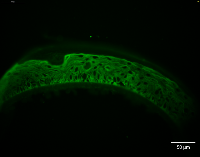
Detection of Cytokeratin 14 in human skin.
Cytokeratin 14 was detected in immersion fixed paraffin-embedded sections of human skin using Mouse Anti-Human Cytokeratin 14 Monoclonal Antibody (Catalog # MAB3164) at 3 µg/mL for 1 hour at room temperature followed by incubation with the Anti-Mouse IgG VisUCyte™ HRP Polymer Antibody (Catalog # VC001). Before incubation with the primary antibody, tissue was subjected to heat-induced epitope retrieval using VisUCyte Antigen Retrieval Reagent-Basic (Catalog # VCTS021). Tissue was stained using DAB (brown) and counterstained with hematoxylin (blue). Specific staining was localized to keratinocytes. View our protocol for IHC Staining with VisUCyte HRP Polymer Detection Reagents.Applications for Human Cytokeratin 14 Antibody (LL001)
Application
Recommended Usage
Immunocytochemistry
8-25 µg/mL
Sample: Immersion fixed A431 human epithelial carcinoma cell line and NHEK human normal epidermal keratinocytes
Sample: Immersion fixed A431 human epithelial carcinoma cell line and NHEK human normal epidermal keratinocytes
Immunohistochemistry
3-25 µg/mL
Sample: Immersion fixed paraffin-embedded sections of human skin
Sample: Immersion fixed paraffin-embedded sections of human skin
Western Blot
Purkis, P. et al. (1990) J. Cell Sci. 97:39. This application was not tested by R&D Systems.
Reviewed Applications
Read 3 reviews rated 4 using MAB3164 in the following applications:
Formulation, Preparation, and Storage
Purification
Protein A or G purified from hybridoma culture supernatant
Reconstitution
Reconstitute at 0.5 mg/mL in sterile PBS. For liquid material, refer to CoA for concentration.
Loading...
Formulation
Lyophilized from a 0.2 μm filtered solution in PBS with Trehalose. *Small pack size (SP) is supplied either lyophilized or as a 0.2 µm filtered solution in PBS.
Shipping
Lyophilized product is shipped at ambient temperature. Liquid small pack size (-SP) is shipped with polar packs. Upon receipt, store immediately at the temperature recommended below.
Stability & Storage
Use a manual defrost freezer and avoid repeated freeze-thaw cycles.
- 12 months from date of receipt, -20 to -70 °C as supplied.
- 1 month, 2 to 8 °C under sterile conditions after reconstitution.
- 6 months, -20 to -70 °C under sterile conditions after reconstitution.
Calculators
Background: Cytokeratin 14
Additional Cytokeratin 14 Products
Product Documents for Human Cytokeratin 14 Antibody (LL001)
Certificate of Analysis
To download a Certificate of Analysis, please enter a lot or batch number in the search box below.
Note: Certificate of Analysis not available for kit components.
Product Specific Notices for Human Cytokeratin 14 Antibody (LL001)
For research use only
Related Research Areas
Citations for Human Cytokeratin 14 Antibody (LL001)
 Powered by Bioz
Customer Reviews for Human Cytokeratin 14 Antibody (LL001) (3)
4 out of 5
3 Customer Ratings
Have you used Human Cytokeratin 14 Antibody (LL001)?
Submit a review and receive an Amazon gift card!
$25/€18/£15/$25CAN/¥2500 Yen for a review with an image
$10/€7/£6/$10CAN/¥1110 Yen for a review without an image
Submit a review
Customer Images
Showing
1
-
3 of
3 reviews
Showing All
Filter By:
-
Application: Immunocytochemistry/ImmunofluorescenceSample Tested: fibroblastsSpecies: HumanVerified Customer | Posted 01/14/2022

-
Application: Immunocytochemistry/ImmunofluorescenceSample Tested: Skin tissueSpecies: HumanVerified Customer | Posted 02/27/2020Anti-keratin 14 antibody was tested at 1:50 and 1:100 dilutions and the protocol includes citrate antigen retrieval. Pepsin and pronase were also used as alternative methods for antigen retrieval, without success. The antibody is not specific of the basal layer of the skin and targets all the layers.
-
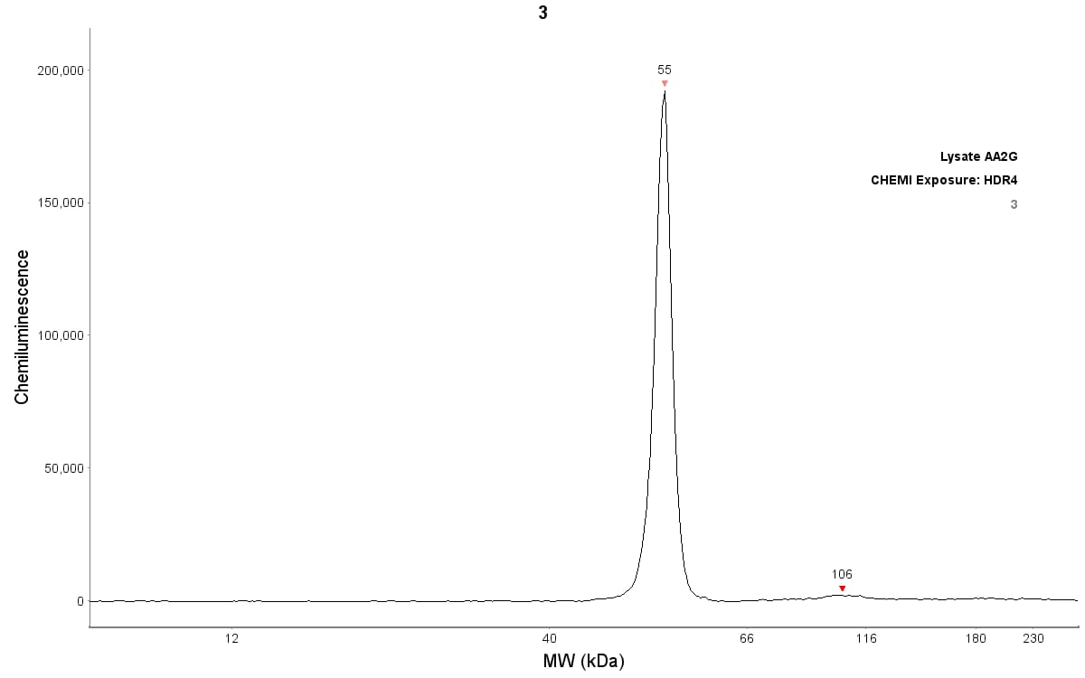
Application: Simple WesternSample Tested: Skin tissueSpecies: HumanVerified Customer | Posted 06/17/2019Detection of keratin 14 by Simple Western (JESS) in reconstructed human epidermis lysate (about 200 µg/mL protein concentration). Antibody dilution: 1/50
There are no reviews that match your criteria.
Protocols
Find general support by application which include: protocols, troubleshooting, illustrated assays, videos and webinars.
- Antigen Retrieval Protocol (PIER)
- Antigen Retrieval for Frozen Sections Protocol
- Appropriate Fixation of IHC/ICC Samples
- Cellular Response to Hypoxia Protocols
- Chromogenic IHC Staining of Formalin-Fixed Paraffin-Embedded (FFPE) Tissue Protocol
- Chromogenic Immunohistochemistry Staining of Frozen Tissue
- ClariTSA™ Fluorophore Kits
- Detection & Visualization of Antibody Binding
- Fluorescent IHC Staining of Frozen Tissue Protocol
- Graphic Protocol for Heat-induced Epitope Retrieval
- Graphic Protocol for the Preparation and Fluorescent IHC Staining of Frozen Tissue Sections
- Graphic Protocol for the Preparation and Fluorescent IHC Staining of Paraffin-embedded Tissue Sections
- Graphic Protocol for the Preparation of Gelatin-coated Slides for Histological Tissue Sections
- ICC Cell Smear Protocol for Suspension Cells
- ICC Immunocytochemistry Protocol Videos
- ICC for Adherent Cells
- IHC Sample Preparation (Frozen sections vs Paraffin)
- Immunocytochemistry (ICC) Protocol
- Immunocytochemistry Troubleshooting
- Immunofluorescence of Organoids Embedded in Cultrex Basement Membrane Extract
- Immunofluorescent IHC Staining of Formalin-Fixed Paraffin-Embedded (FFPE) Tissue Protocol
- Immunohistochemistry (IHC) and Immunocytochemistry (ICC) Protocols
- Immunohistochemistry Frozen Troubleshooting
- Immunohistochemistry Paraffin Troubleshooting
- Preparing Samples for IHC/ICC Experiments
- Preventing Non-Specific Staining (Non-Specific Binding)
- Primary Antibody Selection & Optimization
- Protocol for Heat-Induced Epitope Retrieval (HIER)
- Protocol for Making a 4% Formaldehyde Solution in PBS
- Protocol for VisUCyte™ HRP Polymer Detection Reagent
- Protocol for the Fluorescent ICC Staining of Cell Smears - Graphic
- Protocol for the Fluorescent ICC Staining of Cultured Cells on Coverslips - Graphic
- Protocol for the Preparation & Fixation of Cells on Coverslips
- Protocol for the Preparation and Chromogenic IHC Staining of Frozen Tissue Sections
- Protocol for the Preparation and Chromogenic IHC Staining of Frozen Tissue Sections - Graphic
- Protocol for the Preparation and Chromogenic IHC Staining of Paraffin-embedded Tissue Sections
- Protocol for the Preparation and Chromogenic IHC Staining of Paraffin-embedded Tissue Sections - Graphic
- Protocol for the Preparation and Fluorescent ICC Staining of Cells on Coverslips
- Protocol for the Preparation and Fluorescent ICC Staining of Non-adherent Cells
- Protocol for the Preparation and Fluorescent ICC Staining of Stem Cells on Coverslips
- Protocol for the Preparation and Fluorescent IHC Staining of Frozen Tissue Sections
- Protocol for the Preparation and Fluorescent IHC Staining of Paraffin-embedded Tissue Sections
- Protocol for the Preparation of Gelatin-coated Slides for Histological Tissue Sections
- Protocol for the Preparation of a Cell Smear for Non-adherent Cell ICC - Graphic
- R&D Systems Quality Control Western Blot Protocol
- TUNEL and Active Caspase-3 Detection by IHC/ICC Protocol
- The Importance of IHC/ICC Controls
- Troubleshooting Guide: Immunohistochemistry
- Troubleshooting Guide: Western Blot Figures
- Western Blot Conditions
- Western Blot Protocol
- Western Blot Protocol for Cell Lysates
- Western Blot Troubleshooting
- Western Blot Troubleshooting Guide
- View all Protocols, Troubleshooting, Illustrated assays and Webinars
Loading...