
Key Product Details
Species Reactivity
Validated:
Human
Cited:
Human, Monkey
Applications
Validated:
Immunohistochemistry, Western Blot, Intracellular Staining by Flow Cytometry, Simple Western, CyTOF-ready
Cited:
Immunohistochemistry, Immunohistochemistry-Paraffin, Western Blot, Flow Cytometry, Immunocytochemistry, ELISA Capture, ELISA Development (Detection)
Label
Unconjugated
Antibody Source
Polyclonal Goat IgG
Loading...
Product Specifications
Immunogen
E. coli-derived recombinant human Galectin‑3
Ala2-Ile250
Accession # P17931.5
Ala2-Ile250
Accession # P17931.5
Specificity
Detects human Galectin-3 in direct ELISAs and Western blots.
Clonality
Polyclonal
Host
Goat
Isotype
IgG
Scientific Data Images for Human Galectin-3 Antibody
Detection of Human Galectin‑3 by Western Blot.
Western blot shows lysates of COLO 205 human colorectal adenocarcinoma cell line, MCF-7 human breast cancer cell line, and U-118-MG human glioblastoma/astrocytoma cell line. PVDF membrane was probed with 0.1 µg/mL of Goat Anti-Human Galectin-3 Antigen Affinity-purified Polyclonal Antibody (Catalog # AF1154) followed by HRP-conjugated Anti-Goat IgG Secondary Antibody (HAF017). A specific band was detected for Galectin-3 at approximately 28 kDa (as indicated). This experiment was conducted under reducing conditions and using Immunoblot Buffer Group 1.
Detection of Human Galectin‑3 by Simple WesternTM.
Simple Western lane view shows lysates of Exosome Standards (Human Urine) (NBP2-49840) and MCF‑7 human breast cancer cell line, loaded at 0.5 mg/ml. A specific band was detected for Galectin‑3 at approximately 37 kDa (as indicated) using 10 µg/ml of Goat Anti-Human Galectin‑3 Antigen Affinity-purified Polyclonal Antibody (Catalog # AF1154) followed by HRP-conjugated Donkey Anti-Goat Secondary Antibody (Catalog # 042-206). This experiment was conducted under reducing conditions and using the 12-230kDa separation system.
Detection of Human Galectin‑3 by Simple WesternTM.
Simple Western lane view shows lysates of COLO 205 human colorectal adenocarcinoma cell line, MCF-7 human breast cancer cell line, and U-118-MG human glioblastoma/astrocytoma cell line, loaded at 0.2 mg/mL. A specific band was detected for Galectin-3 at approximately 37-38 kDa (as indicated) using 10 µg/mL of Goat Anti-Human Galectin-3 Antigen Affinity-purified Polyclonal Antibody (Catalog # AF1154) followed by 1:50 dilution of HRP-conjugated Anti-Goat IgG Secondary Antibody (Catalog # HAF109). This experiment was conducted under reducing conditions and using the 12-230 kDa separation system.
Galectin‑3 in Human Colon.
Galectin-3 was detected in formalin fixed paraffin-embedded sections of human colon using Goat Anti-Human Galectin-3 Antigen Affinity-purified Polyclonal Antibody (Catalog # AF1154) at 1.7 µg/mL overnight at 4 °C. Tissue was stained using the Anti-Goat HRP-DAB Cell & Tissue Staining Kit (brown; (CTS008) and counterstained with hematoxylin (blue). View our protocol for Chromogenic IHC Staining of immersion fixed paraffin-embedded Tissue Sections.
Detection of Human Galectin-3 by Western Blot
GPNMB and galectin-3 levels are elevated in FTD-GRN brains. a, b GPNMB and galectin-3 levels (ng/mg protein) were measured in frontal lobe tissue lysates generated from cognitively normal controls (CTL; n = 27) and FTD-GRN patients (n = 25). Data analyzed using unpaired t-test. c Representative immunoblots for GPNMB and galectin-3 in frontal lobe lysates from cognitively normal controls (n = 8) and FTD-GRN (n = 8) patients. d GPNMB levels (ng/mL) in CSF samples form cognitively normal controls (n = 14), FTD-GRN (n = 9), FTD-C9orf72 (n = 12) and FTD-MAPT (n = 12) samples quantified by ELISA. Data analyzed using one-way ANOVA. e, f GPNMB immunostaining was performed on frontal lobe tissue sections from cognitively normal controls (n = 5) (e) and FTD-GRN (n = 5) (f) patients. g, h Immunostaining for p-TDP 43 was stained on adjacent sections from identical samples in e, f as marker of FTLD pathology. i GPNMB staining intensity in human brain sections (e, f) were measured and presented as fold change. Representative immunofluorescence staining for cell markers (green) (j, n, r), GPNMB (red) (k, o, s), DAPI (blue) (i, p, t) in paraffin sections of brains from FTD-GRN cases. Iba-1, GFAP, NeuN used for markers of human microglia, astrocytes, and neurons respectively. GPNMB and Iba-1 signals overlap (arrow) (m) whereas, no overlapping signal was observed in co-staining with GFAP or NeuN (q, u). Scale bars were labeled in the images. Data analyzed by unpaired t-test. Scale bars (20 µm) labeled in images and quantitative data are shown as mean ± SEM, *p < 0.05; **p < 0.01; ***p < 0.001; ****p < 0.0001 Image collected and cropped by CiteAb from the following open publication (https://pubmed.ncbi.nlm.nih.gov/33028409), licensed under a CC-BY license. Not internally tested by R&D Systems.
Human Galectin-3 ELISA Standard Curve
Recombinant Human Galectin‑3 (Catalog # 1154-GA) was serially diluted and captured by Mouse Anti-Human Galectin‑3 Monoclonal Antibody (Catalog # MAB11541) coated on a Clear Polystyrene Microplate (Catalog # DY990). Goat Anti-Human Galectin‑3 Antigen Affinity-purified Polyclonal Antibody (Catalog # AF1154) was biotinylated and incubated with the protein captured on the plate. Detection of the standard curve was achieved by incubating Streptavidin-HRP (Catalog # DY998)Applications for Human Galectin-3 Antibody
Application
Recommended Usage
CyTOF-ready
Ready to be labeled using established conjugation methods. No BSA or other carrier proteins that could interfere with conjugation.
Immunohistochemistry
5-15 µg/mL
Sample: Immersion fixed paraffin-embedded sections of human colon and lung cancer tissue
Sample: Immersion fixed paraffin-embedded sections of human colon and lung cancer tissue
Intracellular Staining by Flow Cytometry
2.5 µg/106 cells
Sample: Human PBMCs fixed with paraformaldehyde and permeabilized with saponin
Sample: Human PBMCs fixed with paraformaldehyde and permeabilized with saponin
Simple Western
10 µg/mL
Sample: Exosome Standards (Human Urine) (Catalog # NBP2-49840), COLO 205 human colorectal adenocarcinoma cell line, MCF‑7 human breast cancer cell line, and U‑118‑MG human glioblastoma/astrocytoma cell line
Sample: Exosome Standards (Human Urine) (Catalog # NBP2-49840), COLO 205 human colorectal adenocarcinoma cell line, MCF‑7 human breast cancer cell line, and U‑118‑MG human glioblastoma/astrocytoma cell line
Western Blot
0.1 µg/mL
Sample: COLO 205 human colorectal adenocarcinoma cell line, MCF‑7 human breast cancer cell line, and U‑118‑MG human glioblastoma/astrocytoma cell line
Sample: COLO 205 human colorectal adenocarcinoma cell line, MCF‑7 human breast cancer cell line, and U‑118‑MG human glioblastoma/astrocytoma cell line
Reviewed Applications
Read 3 reviews rated 4.7 using AF1154 in the following applications:
Flow Cytometry Panel Builder
Bio-Techne Knows Flow Cytometry
Save time and reduce costly mistakes by quickly finding compatible reagents using the Panel Builder Tool.
Advanced Features
- Spectra Viewer - Custom analysis of spectra from multiple fluorochromes
- Spillover Popups - Visualize the spectra of individual fluorochromes
- Antigen Density Selector - Match fluorochrome brightness with antigen density

Formulation, Preparation, and Storage
Purification
Antigen Affinity-purified
Reconstitution
Reconstitute at 0.2 mg/mL in sterile PBS. For liquid material, refer to CoA for concentration.
Loading...
Formulation
Lyophilized from a 0.2 μm filtered solution in PBS with Trehalose. See Certificate of Analysis for details.
*Small pack size (-SP) is supplied either lyophilized or as a 0.2 µm filtered solution in PBS.
*Small pack size (-SP) is supplied either lyophilized or as a 0.2 µm filtered solution in PBS.
Shipping
Lyophilized product is shipped at ambient temperature. Liquid small pack size (-SP) is shipped with polar packs. Upon receipt, store immediately at the temperature recommended below.
Stability & Storage
Use a manual defrost freezer and avoid repeated freeze-thaw cycles.
- 12 months from date of receipt, -20 to -70 °C as supplied.
- 1 month, 2 to 8 °C under sterile conditions after reconstitution.
- 6 months, -20 to -70 °C under sterile conditions after reconstitution.
Calculators
Background: Galectin-3
References
- Rabinovich, A. et al. (2002) Trends in Immunol. 23:313.
- Rabinovich, A. et al. (2002) J. Leukocyte Biology 71:741.
- Hughes, R.C. (2001) Biochimie 83:667.
- R&D Systems Cytokine Bulletin; Summer 2002.
- Gorski, J.P. et al. (2002) J. Biol. Chem. 277:18840.
Alternate Names
AGE-R3, CBP35, GAL3, Galectin3, L29, LGALS3, Mac-2
Gene Symbol
LGALS3
UniProt
Additional Galectin-3 Products
Product Documents for Human Galectin-3 Antibody
Certificate of Analysis
To download a Certificate of Analysis, please enter a lot or batch number in the search box below.
Note: Certificate of Analysis not available for kit components.
Product Specific Notices for Human Galectin-3 Antibody
For research use only
Citations for Human Galectin-3 Antibody
 Powered by Bioz
Customer Reviews for Human Galectin-3 Antibody (3)
4.7 out of 5
3 Customer Ratings
Have you used Human Galectin-3 Antibody?
Submit a review and receive an Amazon gift card!
$25/€18/£15/$25CAN/¥2500 Yen for a review with an image
$10/€7/£6/$10CAN/¥1110 Yen for a review without an image
Submit a review
Customer Images
Showing
1
-
3 of
3 reviews
Showing All
Filter By:
-
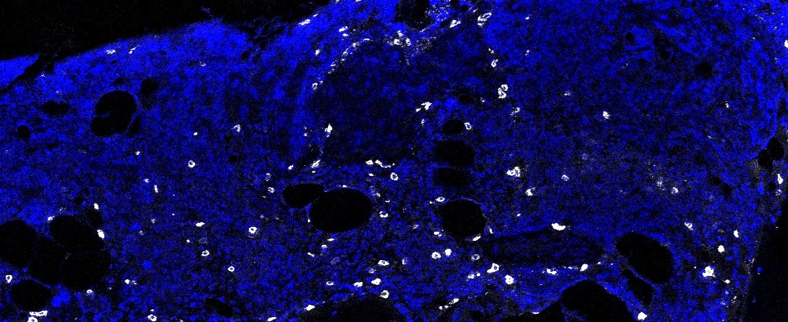
Application: Imaging Mass CytometrySample Tested: bone marrowSpecies: HumanVerified Customer | Posted 11/12/2021IMC of human bone marrow FFPE samples DNA-blue Lgals3- white
 Bio-Techne ResponseThis review was submitted through the legacy Novus Innovators Program, reflecting a new species or application tested on a primary antibody.
 -
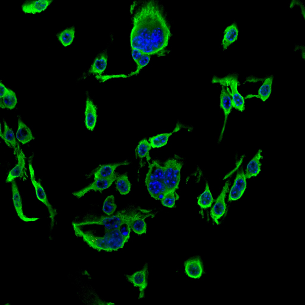
Application: Immunocytochemistry/ImmunofluorescenceSample Tested: RAW 264.7 mouse monocyte/macrophage cell lineSpecies: MouseVerified Customer | Posted 11/18/2016
-
Application: ImmunofluorescenceSample Tested: See PMID 23121677Species: HumanVerified Customer | Posted 01/05/2015
There are no reviews that match your criteria.
Protocols
Find general support by application which include: protocols, troubleshooting, illustrated assays, videos and webinars.
- 7-Amino Actinomycin D (7-AAD) Cell Viability Flow Cytometry Protocol
- Antigen Retrieval Protocol (PIER)
- Antigen Retrieval for Frozen Sections Protocol
- Appropriate Fixation of IHC/ICC Samples
- Cellular Response to Hypoxia Protocols
- Chromogenic IHC Staining of Formalin-Fixed Paraffin-Embedded (FFPE) Tissue Protocol
- Chromogenic Immunohistochemistry Staining of Frozen Tissue
- ClariTSA™ Fluorophore Kits
- Detection & Visualization of Antibody Binding
- Extracellular Membrane Flow Cytometry Protocol
- Flow Cytometry Protocol for Cell Surface Markers
- Flow Cytometry Protocol for Staining Membrane Associated Proteins
- Flow Cytometry Staining Protocols
- Flow Cytometry Troubleshooting Guide
- Fluorescent IHC Staining of Frozen Tissue Protocol
- Graphic Protocol for Heat-induced Epitope Retrieval
- Graphic Protocol for the Preparation and Fluorescent IHC Staining of Frozen Tissue Sections
- Graphic Protocol for the Preparation and Fluorescent IHC Staining of Paraffin-embedded Tissue Sections
- Graphic Protocol for the Preparation of Gelatin-coated Slides for Histological Tissue Sections
- IHC Sample Preparation (Frozen sections vs Paraffin)
- Immunofluorescent IHC Staining of Formalin-Fixed Paraffin-Embedded (FFPE) Tissue Protocol
- Immunohistochemistry (IHC) and Immunocytochemistry (ICC) Protocols
- Immunohistochemistry Frozen Troubleshooting
- Immunohistochemistry Paraffin Troubleshooting
- Intracellular Flow Cytometry Protocol Using Alcohol (Methanol)
- Intracellular Flow Cytometry Protocol Using Detergents
- Intracellular Nuclear Staining Flow Cytometry Protocol Using Detergents
- Intracellular Staining Flow Cytometry Protocol Using Alcohol Permeabilization
- Intracellular Staining Flow Cytometry Protocol Using Detergents to Permeabilize Cells
- Preparing Samples for IHC/ICC Experiments
- Preventing Non-Specific Staining (Non-Specific Binding)
- Primary Antibody Selection & Optimization
- Propidium Iodide Cell Viability Flow Cytometry Protocol
- Protocol for Heat-Induced Epitope Retrieval (HIER)
- Protocol for Liperfluo
- Protocol for Making a 4% Formaldehyde Solution in PBS
- Protocol for VisUCyte™ HRP Polymer Detection Reagent
- Protocol for the Characterization of Human Th22 Cells
- Protocol for the Characterization of Human Th9 Cells
- Protocol for the Preparation & Fixation of Cells on Coverslips
- Protocol for the Preparation and Chromogenic IHC Staining of Frozen Tissue Sections
- Protocol for the Preparation and Chromogenic IHC Staining of Frozen Tissue Sections - Graphic
- Protocol for the Preparation and Chromogenic IHC Staining of Paraffin-embedded Tissue Sections
- Protocol for the Preparation and Chromogenic IHC Staining of Paraffin-embedded Tissue Sections - Graphic
- Protocol for the Preparation and Fluorescent IHC Staining of Frozen Tissue Sections
- Protocol for the Preparation and Fluorescent IHC Staining of Paraffin-embedded Tissue Sections
- Protocol for the Preparation of Gelatin-coated Slides for Histological Tissue Sections
- Protocol: Annexin V and PI Staining by Flow Cytometry
- Protocol: Annexin V and PI Staining for Apoptosis by Flow Cytometry
- R&D Systems Quality Control Western Blot Protocol
- TUNEL and Active Caspase-3 Detection by IHC/ICC Protocol
- The Importance of IHC/ICC Controls
- Troubleshooting Guide: Fluorokine Flow Cytometry Kits
- Troubleshooting Guide: Immunohistochemistry
- Troubleshooting Guide: Western Blot Figures
- Western Blot Conditions
- Western Blot Protocol
- Western Blot Protocol for Cell Lysates
- Western Blot Troubleshooting
- Western Blot Troubleshooting Guide
- View all Protocols, Troubleshooting, Illustrated assays and Webinars
Loading...