
Key Product Details
Species Reactivity
Validated:
Human
Cited:
Human, Mouse, Rat, Porcine, Bovine, Primate - Chlorocebus pygerythrus (Vervet Monkey), Primate - Macaca fascicularis (Crab-eating Monkey or Cynomolgus Macaque), Xenograft
Applications
Validated:
Immunohistochemistry, Western Blot, Immunocytochemistry, Simple Western
Cited:
Immunohistochemistry, Immunohistochemistry-Paraffin, Western Blot, Flow Cytometry, Immunocytochemistry, Chromatin Immunoprecipitation (ChIP)
Label
Unconjugated
Antibody Source
Polyclonal Goat IgG
Loading...
Product Specifications
Immunogen
E. coli-derived recombinant human GATA-3
Pro135-Ser258
Accession # P23771
Pro135-Ser258
Accession # P23771
Specificity
Detects human and mouse GATA-3 in direct ELISAs and Western blots.
Clonality
Polyclonal
Host
Goat
Isotype
IgG
Scientific Data Images for Human GATA-3 Antibody
Detection of Human and Mouse GATA‑3 by Western Blot.
Western blot shows lysates of MCF-7 human breast cancer cell line, Jurkat human acute T cell leukemia cell line, and EL-4 mouse lymphoblast cell line. PVDF membrane was probed with 0.25 µg/mL of Goat Anti-Human GATA-3 Antigen Affinity-purified Polyclonal Antibody (Catalog # AF2605) followed by HRP-conjugated Anti-Goat IgG Secondary Antibody (Catalog # HAF109). Specific bands were detected for GATA-3 full length (FL) at approximately 55 kDa and the splice form (SF) at approximately 40 kDa (as indicated). This experiment was conducted under reducing conditions and using Immunoblot Buffer Group 1.
GATA‑3 in Human Breast Cancer Tissue.
GATA-3 was detected in immersion fixed paraffin-embedded sections of human breast cancer tissue using Goat Anti-Human GATA-3 Antigen Affinity-purified Polyclonal Antibody (Catalog # AF2605) at 3 µg/mL overnight at 4 °C. Before incubation with the primary antibody tissue was subjected to heat-induced epitope retrieval using Antigen Retrieval Reagent-Basic (Catalog # CTS013). Tissue was stained using the Anti-Goat HRP-DAB Cell & Tissue Staining Kit (brown; Catalog # CTS008) and counterstained with hematoxylin (blue). View our protocol for Chromogenic IHC Staining of Paraffin-embedded Tissue Sections.
GATA‑3 in MCF-7 Human Cell Line.
GATA-3 was detected in immersion fixed MCF-7 human breast cancer cell line using 10 µg/mL Goat Anti-Human GATA-3 Antigen Affinity-purified Polyclonal Antibody (Catalog # AF2605) for 3 hours at room temperature. Cells were stained with the NorthernLights™ 557-conjugated Anti-Goat IgG Secondary Antibody (red, upper panel; Catalog # NL001) and counterstained with DAPI (blue, lower panel). View our protocol for Fluorescent ICC Staining of Cells on Coverslips.
GATA‑3 in Human Breast Cancer Tissue.
GATA-3 was detected in immersion fixed paraffin-embedded sections of human breast cancer tissue using Goat Anti-Human GATA-3 Antigen Affinity-purified Polyclonal Antibody (Catalog # AF2605) at 3 µg/mL overnight at 4 °C. Before incubation with the primary antibody tissue was subjected to heat-induced epitope retrieval using Antigen Retrieval Reagent-Basic (Catalog # CTS013). Tissue was stained using the Anti-Goat HRP-DAB Cell & Tissue Staining Kit (brown; Catalog # CTS008) and counterstained with hematoxylin (blue). View our protocol for Chromogenic IHC Staining of Paraffin-embedded Tissue Sections.
Detection of Human GATA‑3 by Simple WesternTM.
Simple Western lane view shows lysates of MCF-7 human breast cancer cell line and Jurkat human acute T cell leukemia cell line, loaded at 0.2 mg/mL. Specific bands were detected for GATA-3 full length (FL) at approximately 62 kDa and the splice form (SF) at approximately 52 kDa (as indicated) using 2.5 µg/mL of Goat Anti-Human GATA-3 Antigen Affinity-purified Polyclonal Antibody (Catalog # AF2605) followed by 1:50 dilution of HRP-conjugated Anti-Goat IgG Secondary Antibody (Catalog # HAF109). This experiment was conducted under reducing conditions and using the 12-230 kDa separation system.
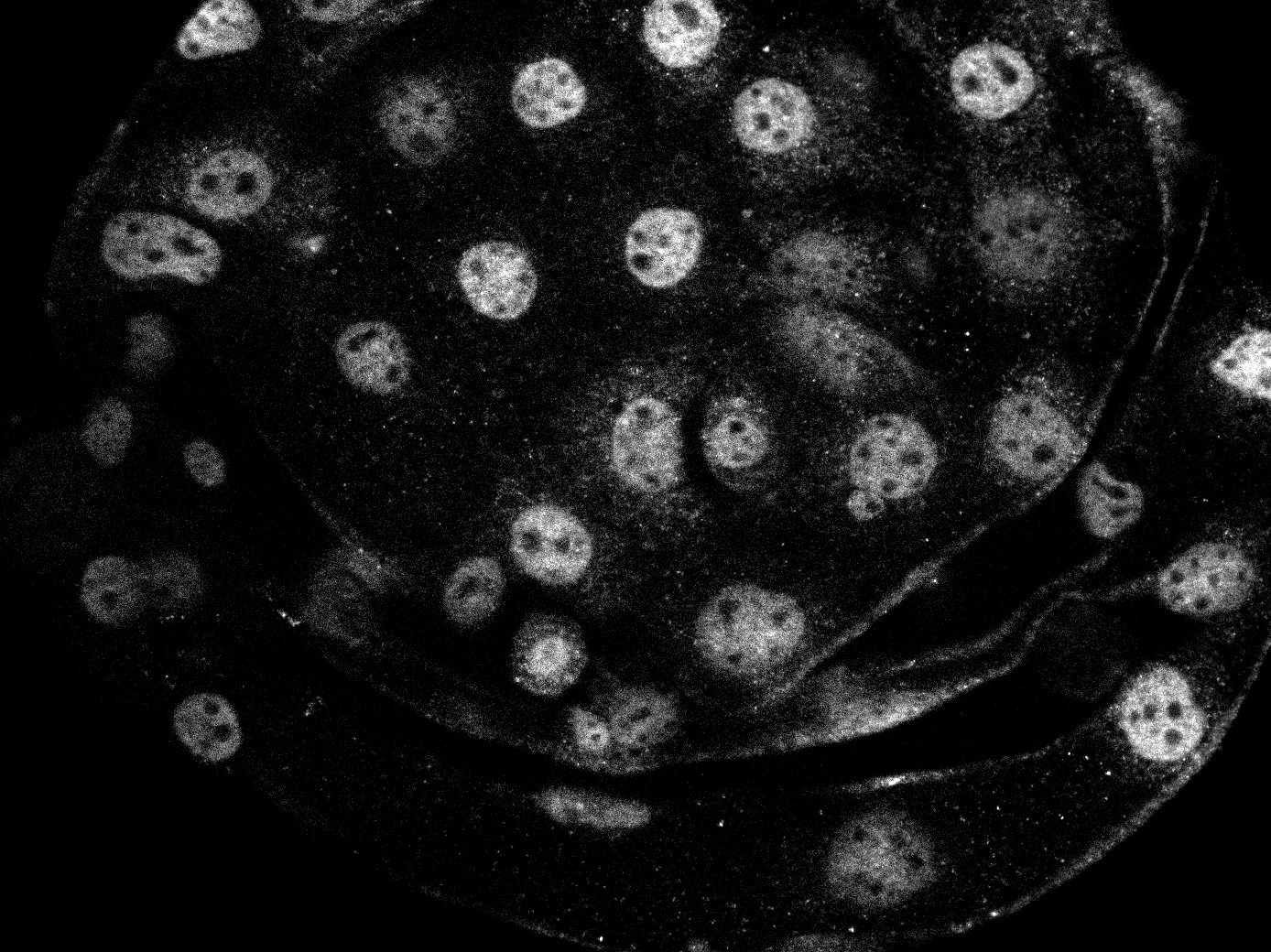
Detection of Mouse GATA-3 by Immunohistochemistry
Generation of a self‐organizing de novo nephron from the differentiated Sca1+ Oct4+ monolayer cultures. A) Bright field image of self‐organizing organoid‐like structure from monolayer culture of kidney stem cells. The photo shows glomerulus‐like structure (black arrow), tubule‐like structure (red arrows), and collecting duct‐like structure (green arrow). Scale bar, 100 µm. A1) Glomerulus‐like structure stained with nephrin (white arrow). Scale bar, 50 µm. A2) Glomerulus‐like structure stained with nephrin (white arrow) and proximal tubule‐like structure stained with LTL (red arrows). Scale bar, 50 µm. A3) Glomerulus‐like structure stained with podocin (white arrow). Scale bar, 50 µm. A4) Collecting duct‐like structure stained with GATA3 (white arrow). Scale bar, 50 µm.,C) Western blot analysis of CD31, nephrin, and podocin expression in undifferentiated and differentiated Sca1+ Oct4+ cells. GAPDH was used as a loading control. Data is representative of three independent experiments. Values represent the mean ± SD (n = 3). p < 0.05 versus control by 2‐tailed Student's t test. Image collected and cropped by CiteAb from the following open publication (https://pubmed.ncbi.nlm.nih.gov/35315252), licensed under a CC-BY license. Not internally tested by R&D Systems.Applications for Human GATA-3 Antibody
Application
Recommended Usage
Immunocytochemistry
5-15 µg/mL
Sample: Immersion fixed MCF-7 human breast cancer cell line
Sample: Immersion fixed MCF-7 human breast cancer cell line
Immunohistochemistry
5-15 µg/mL
Sample: Immersion fixed paraffin-embedded sections of human breast cancer tissue
Sample: Immersion fixed paraffin-embedded sections of human breast cancer tissue
Simple Western
2.5 µg/mL
Sample: MCF‑7 human breast cancer cell line and Jurkat human acute T cell leukemia cell line
Sample: MCF‑7 human breast cancer cell line and Jurkat human acute T cell leukemia cell line
Western Blot
0.25 µg/mL
Sample: MCF‑7 human breast cancer cell line, Jurkat human acute T cell leukemia cell line, and EL‑4 mouse lymphoblast cell line
Sample: MCF‑7 human breast cancer cell line, Jurkat human acute T cell leukemia cell line, and EL‑4 mouse lymphoblast cell line
Reviewed Applications
Read 1 review rated 5 using AF2605 in the following applications:
Formulation, Preparation, and Storage
Purification
Antigen Affinity-purified
Reconstitution
Reconstitute at 0.2 mg/mL in sterile PBS. For liquid material, refer to CoA for concentration.
Loading...
Formulation
Lyophilized from a 0.2 μm filtered solution in PBS with Trehalose. *Small pack size (SP) is supplied either lyophilized or as a 0.2 µm filtered solution in PBS.
Shipping
Lyophilized product is shipped at ambient temperature. Liquid small pack size (-SP) is shipped with polar packs. Upon receipt, store immediately at the temperature recommended below.
Stability & Storage
Use a manual defrost freezer and avoid repeated freeze-thaw cycles.
- 12 months from date of receipt, -20 to -70 °C as supplied.
- 1 month, 2 to 8 °C under sterile conditions after reconstitution.
- 6 months, -20 to -70 °C under sterile conditions after reconstitution.
Calculators
Background: GATA-3
Additional GATA-3 Products
Product Documents for Human GATA-3 Antibody
Certificate of Analysis
To download a Certificate of Analysis, please enter a lot or batch number in the search box below.
Note: Certificate of Analysis not available for kit components.
Product Specific Notices for Human GATA-3 Antibody
For research use only
Related Research Areas
Citations for Human GATA-3 Antibody
 Powered by Bioz
Customer Reviews for Human GATA-3 Antibody (1)
5 out of 5
1 Customer Rating
Have you used Human GATA-3 Antibody?
Submit a review and receive an Amazon gift card!
$25/€18/£15/$25CAN/¥2500 Yen for a review with an image
$10/€7/£6/$10CAN/¥1110 Yen for a review without an image
Submit a review
Customer Images
Showing
1
-
1 of
1 review
Showing All
Filter By:
-
Application: Immunocytochemistry/ImmunofluorescenceSample Tested: blastocystSpecies: BovineVerified Customer | Posted 08/09/2022use at 10 µg/ml

There are no reviews that match your criteria.
Protocols
Find general support by application which include: protocols, troubleshooting, illustrated assays, videos and webinars.
- Antigen Retrieval Protocol (PIER)
- Antigen Retrieval for Frozen Sections Protocol
- Appropriate Fixation of IHC/ICC Samples
- Cellular Response to Hypoxia Protocols
- Chromogenic IHC Staining of Formalin-Fixed Paraffin-Embedded (FFPE) Tissue Protocol
- Chromogenic Immunohistochemistry Staining of Frozen Tissue
- ClariTSA™ Fluorophore Kits
- Detection & Visualization of Antibody Binding
- Fluorescent IHC Staining of Frozen Tissue Protocol
- Graphic Protocol for Heat-induced Epitope Retrieval
- Graphic Protocol for the Preparation and Fluorescent IHC Staining of Frozen Tissue Sections
- Graphic Protocol for the Preparation and Fluorescent IHC Staining of Paraffin-embedded Tissue Sections
- Graphic Protocol for the Preparation of Gelatin-coated Slides for Histological Tissue Sections
- ICC Cell Smear Protocol for Suspension Cells
- ICC Immunocytochemistry Protocol Videos
- ICC for Adherent Cells
- IHC Sample Preparation (Frozen sections vs Paraffin)
- Immunocytochemistry (ICC) Protocol
- Immunocytochemistry Troubleshooting
- Immunofluorescence of Organoids Embedded in Cultrex Basement Membrane Extract
- Immunofluorescent IHC Staining of Formalin-Fixed Paraffin-Embedded (FFPE) Tissue Protocol
- Immunohistochemistry (IHC) and Immunocytochemistry (ICC) Protocols
- Immunohistochemistry Frozen Troubleshooting
- Immunohistochemistry Paraffin Troubleshooting
- Preparing Samples for IHC/ICC Experiments
- Preventing Non-Specific Staining (Non-Specific Binding)
- Primary Antibody Selection & Optimization
- Protocol for Heat-Induced Epitope Retrieval (HIER)
- Protocol for Making a 4% Formaldehyde Solution in PBS
- Protocol for VisUCyte™ HRP Polymer Detection Reagent
- Protocol for the Fluorescent ICC Staining of Cell Smears - Graphic
- Protocol for the Fluorescent ICC Staining of Cultured Cells on Coverslips - Graphic
- Protocol for the Preparation & Fixation of Cells on Coverslips
- Protocol for the Preparation and Chromogenic IHC Staining of Frozen Tissue Sections
- Protocol for the Preparation and Chromogenic IHC Staining of Frozen Tissue Sections - Graphic
- Protocol for the Preparation and Chromogenic IHC Staining of Paraffin-embedded Tissue Sections
- Protocol for the Preparation and Chromogenic IHC Staining of Paraffin-embedded Tissue Sections - Graphic
- Protocol for the Preparation and Fluorescent ICC Staining of Cells on Coverslips
- Protocol for the Preparation and Fluorescent ICC Staining of Non-adherent Cells
- Protocol for the Preparation and Fluorescent ICC Staining of Stem Cells on Coverslips
- Protocol for the Preparation and Fluorescent IHC Staining of Frozen Tissue Sections
- Protocol for the Preparation and Fluorescent IHC Staining of Paraffin-embedded Tissue Sections
- Protocol for the Preparation of Gelatin-coated Slides for Histological Tissue Sections
- Protocol for the Preparation of a Cell Smear for Non-adherent Cell ICC - Graphic
- R&D Systems Quality Control Western Blot Protocol
- TUNEL and Active Caspase-3 Detection by IHC/ICC Protocol
- The Importance of IHC/ICC Controls
- Troubleshooting Guide: Immunohistochemistry
- Troubleshooting Guide: Western Blot Figures
- Western Blot Conditions
- Western Blot Protocol
- Western Blot Protocol for Cell Lysates
- Western Blot Troubleshooting
- Western Blot Troubleshooting Guide
- View all Protocols, Troubleshooting, Illustrated assays and Webinars


