Interleukin-17 (IL-17) is a pro-inflammatory cytokine secreted by activated T cells. It is the prototype member of the IL-17 family that also includes IL-17B, C, D, E, and F.
Human IL-17/IL-17A Antibody (41802)
R&D Systems | Catalog # MAB3171

Key Product Details
Validated by
Biological Validation
Species Reactivity
Validated:
Human
Cited:
Human, Mouse
Applications
Validated:
Immunohistochemistry, Western Blot, Intracellular Staining by Flow Cytometry, Immunoprecipitation, CyTOF-ready
Cited:
Immunohistochemistry, Immunohistochemistry-Paraffin, Western Blot, Immunocytochemistry, Confocal Imaging, ELISA Capture
Label
Unconjugated
Antibody Source
Monoclonal Mouse IgG1 Clone # 41802
Loading...
Product Specifications
Immunogen
E. coli-derived recombinant human IL-17
Ile20-Ala155
Accession # Q16552
Ile20-Ala155
Accession # Q16552
Specificity
Detects human IL-17 in direct ELISAs. In direct ELISAs, 100% cross-reactivity with recombinant human (rh) IL-17A/IL-17F heterodimer is observed. No cross-reactivity with recombinant mouse IL-17, recombinant canine Il-17, rhIL-17B, rhIL-17C, rhIL-17D, rhIL-17E, or rhIL-17F is observed.
Clonality
Monoclonal
Host
Mouse
Isotype
IgG1
Scientific Data Images for Human IL-17/IL-17A Antibody (41802)
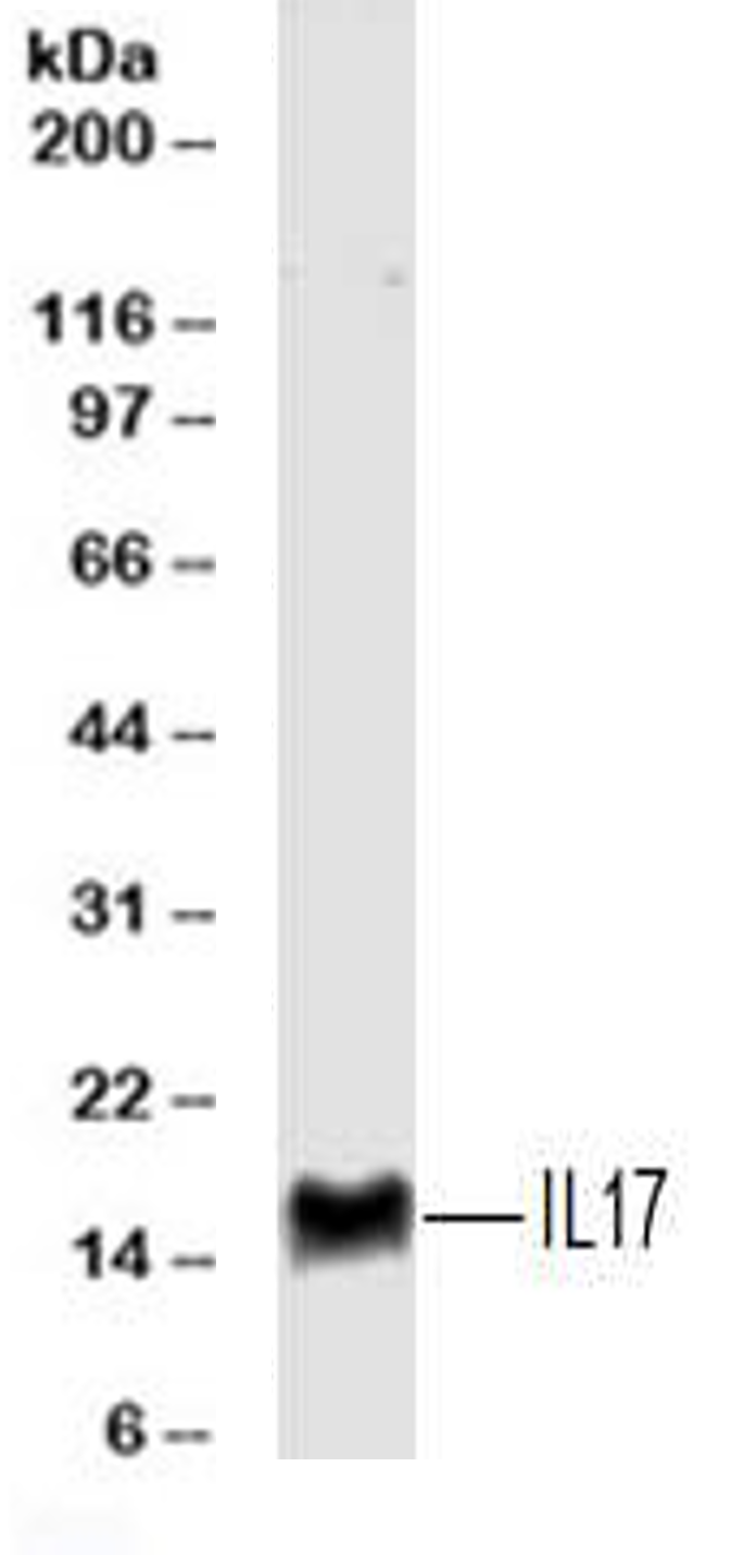
Detection of IL-17/IL-17A Mouse by Western Blot.
Western blot shows lysates of purified naïve human CD4+T cells orin vitrodifferentiated human Th17 CD4+ T cells. PVDF membrane was probed with 2 µg/mL of Mouse Anti-Human IL-17 Monoclonal Antibody (Catalog # MAB3171) followed by HRP-conjugated Anti-Mouse IgG Secondary Antibody (Catalog # HAF007). A specific band was detected for IL-17 at approximately 17 kDa (as indicated). This experiment was conducted under reducing conditions and using Immunoblot Buffer Group 1.
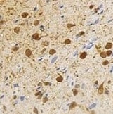
IL‑17/IL‑17A in Human Crohn's Disease Intestine.
IL-17/IL-17A was detected in immersion fixed paraffin-embedded sections of human Crohn's disease intestine using Mouse Anti-Human IL-17/IL-17A Monoclonal Antibody (Catalog # MAB3171) at 0.5 µg/mL for 1 hour at room temperature followed by incubation with the Anti-Mouse IgG VisUCyte™ HRP Polymer Antibody (Catalog # VC001). Before incubation with the primary antibody, tissue was subjected to heat-induced epitope retrieval using Antigen Retrieval Reagent-Basic (Catalog # CTS013). Tissue was stained using DAB (brown) and counterstained with hematoxylin (blue). Specific staining was localized to lymphocytes. View our protocol for IHC Staining with VisUCyte HRP Polymer Detection Reagents.
Immunoprecipitation of Human IL-17.
Human IL-17 was immunoprecipitated from 100 µg of humanin vitrodifferentiated Th17 cell lysate following incubation with 3 µg Mouse Anti-Human IL-17 Monoclonal Antibody (Catalog # MAB3171) or isotype control antibody (Catalog # MAB002) overnight at 4 °C. IL-17-antibody complexes were absorbed using anti-mouse agarose beads. Immunoprecipitated IL-17 was detected by Western blot using 1 µg/mL Goat Anti-Human IL-17 Antigen Affinity-purified Polyclonal Antibody (Catalog # AF-317-NA). View our recommended buffer recipes for immunoprecipitation.
Detection of IL‑17 in Human PBMCs by Flow Cytometry.
Human peripheral blood mononuclear cells were unstimulated (light orange filled histogram) or treated with 50 ng/mL PMA and 250 ng/mL Ca2+ionomycin for 16 hours, then stained with Mouse Anti-Human IL-17 Monoclonal Antibody (Catalog # MAB3171, dark orange filled histogram) or isotype control antibody (Catalog # MAB002, open histogram), followed by Allophycocyanin-conjugated Anti-Mouse IgG F(ab')2Secondary Antibody (Catalog # F0101B). To facilitate intracellular staining, cells were fixed with paraformaldehyde and permeabilized with saponin.Applications for Human IL-17/IL-17A Antibody (41802)
Application
Recommended Usage
CyTOF-ready
Ready to be labeled using established conjugation methods. No BSA or other carrier proteins that could interfere with conjugation.
Immunohistochemistry
0.5-25 µg/mL
Sample: Immersion fixed paraffin-embedded sections of human Crohn's disease intestine
Sample: Immersion fixed paraffin-embedded sections of human Crohn's disease intestine
Immunoprecipitation
3 µg/100 µg cell lysate
Sample: Human in vitro differentiated Th17 cells
Sample: Human in vitro differentiated Th17 cells
Intracellular Staining by Flow Cytometry
2.5 µg/106 cells
Sample: Human peripheral blood mononuclear cells treated with PMA and Ca2+ ionomycin, fixed with paraformaldehyde, and permeabilized with saponin
Sample: Human peripheral blood mononuclear cells treated with PMA and Ca2+ ionomycin, fixed with paraformaldehyde, and permeabilized with saponin
Western Blot
2 µg/mL
Sample: Purified naïve human CD4+ T cells or in vitro differentiated human Th17 CD4+ T cells
Sample: Purified naïve human CD4+ T cells or in vitro differentiated human Th17 CD4+ T cells
Reviewed Applications
Read 3 reviews rated 4.3 using MAB3171 in the following applications:
Flow Cytometry Panel Builder
Bio-Techne Knows Flow Cytometry
Save time and reduce costly mistakes by quickly finding compatible reagents using the Panel Builder Tool.
Advanced Features
- Spectra Viewer - Custom analysis of spectra from multiple fluorochromes
- Spillover Popups - Visualize the spectra of individual fluorochromes
- Antigen Density Selector - Match fluorochrome brightness with antigen density

Formulation, Preparation, and Storage
Purification
Protein A or G purified from hybridoma culture supernatant
Reconstitution
Reconstitute at 0.5 mg/mL in sterile PBS. For liquid material, refer to CoA for concentration.
Loading...
Formulation
Lyophilized from a 0.2 μm filtered solution in PBS with Trehalose. *Small pack size (SP) is supplied either lyophilized or as a 0.2 µm filtered solution in PBS.
Shipping
Lyophilized product is shipped at ambient temperature. Liquid small pack size (-SP) is shipped with polar packs. Upon receipt, store immediately at the temperature recommended below.
Stability & Storage
Use a manual defrost freezer and avoid repeated freeze-thaw cycles.
- 12 months from date of receipt, -20 to -70 °C as supplied.
- 1 month, 2 to 8 °C under sterile conditions after reconstitution.
- 6 months, -20 to -70 °C under sterile conditions after reconstitution.
Calculators
Background: IL-17/IL-17A
Long Name
Interleukin 17
Alternate Names
CTLA-8, CTLA8, IL-17A, IL17, IL17A
Entrez Gene IDs
Gene Symbol
IL17A
UniProt
Additional IL-17/IL-17A Products
Product Documents for Human IL-17/IL-17A Antibody (41802)
Certificate of Analysis
To download a Certificate of Analysis, please enter a lot or batch number in the search box below.
Note: Certificate of Analysis not available for kit components.
Product Specific Notices for Human IL-17/IL-17A Antibody (41802)
For research use only
Citations for Human IL-17/IL-17A Antibody (41802)
 Powered by Bioz
Customer Reviews for Human IL-17/IL-17A Antibody (41802) (3)
4.3 out of 5
3 Customer Ratings
Have you used Human IL-17/IL-17A Antibody (41802)?
Submit a review and receive an Amazon gift card!
$25/€18/£15/$25CAN/¥2500 Yen for a review with an image
$10/€7/£6/$10CAN/¥1110 Yen for a review without an image
Submit a review
Customer Images
Showing
1
-
3 of
3 reviews
Showing All
Filter By:
-
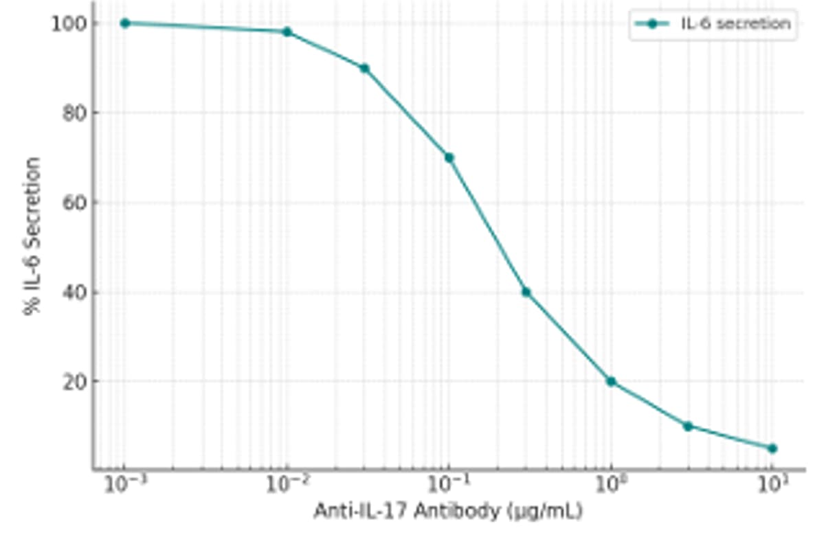
Application: Block/NeutralizeSample Tested: HT-29 human colon adenocarcinoma cell lineSpecies: HumanVerified Customer | Posted 06/02/2025we used it in a cell-based neutralization assay designed to measure IL-17A activity. We used HT-29 cells, which respond to IL-17 stimulation by secreting IL-6. IL-17 (10 ng/mL) was pre-incubated with increasing concentrations of the anti-IL-17 antibody, followed by stimulation of cells. IL-6 secretion in the supernatant was quantified via MSD.

-
Application: Western BlotSample Tested: Cell LysatesSpecies: HumanVerified Customer | Posted 04/03/2023The Western blot of cell lysates obtained from human CD4+T cells. The well was loaded with 50 micrograms of sample under reducing conditions.
-
Application: ImmunohistochemistrySample Tested: Ischemic brain tissueSpecies: HumanVerified Customer | Posted 10/14/2021
There are no reviews that match your criteria.
Protocols
Find general support by application which include: protocols, troubleshooting, illustrated assays, videos and webinars.
- 7-Amino Actinomycin D (7-AAD) Cell Viability Flow Cytometry Protocol
- Antigen Retrieval Protocol (PIER)
- Antigen Retrieval for Frozen Sections Protocol
- Appropriate Fixation of IHC/ICC Samples
- Cellular Response to Hypoxia Protocols
- Chromogenic IHC Staining of Formalin-Fixed Paraffin-Embedded (FFPE) Tissue Protocol
- Chromogenic Immunohistochemistry Staining of Frozen Tissue
- ClariTSA™ Fluorophore Kits
- Detection & Visualization of Antibody Binding
- Extracellular Membrane Flow Cytometry Protocol
- Flow Cytometry Protocol for Cell Surface Markers
- Flow Cytometry Protocol for Staining Membrane Associated Proteins
- Flow Cytometry Staining Protocols
- Flow Cytometry Troubleshooting Guide
- Fluorescent IHC Staining of Frozen Tissue Protocol
- Graphic Protocol for Heat-induced Epitope Retrieval
- Graphic Protocol for the Preparation and Fluorescent IHC Staining of Frozen Tissue Sections
- Graphic Protocol for the Preparation and Fluorescent IHC Staining of Paraffin-embedded Tissue Sections
- Graphic Protocol for the Preparation of Gelatin-coated Slides for Histological Tissue Sections
- IHC Sample Preparation (Frozen sections vs Paraffin)
- Immunofluorescent IHC Staining of Formalin-Fixed Paraffin-Embedded (FFPE) Tissue Protocol
- Immunohistochemistry (IHC) and Immunocytochemistry (ICC) Protocols
- Immunohistochemistry Frozen Troubleshooting
- Immunohistochemistry Paraffin Troubleshooting
- Immunoprecipitation Protocol
- Intracellular Flow Cytometry Protocol Using Alcohol (Methanol)
- Intracellular Flow Cytometry Protocol Using Detergents
- Intracellular Nuclear Staining Flow Cytometry Protocol Using Detergents
- Intracellular Staining Flow Cytometry Protocol Using Alcohol Permeabilization
- Intracellular Staining Flow Cytometry Protocol Using Detergents to Permeabilize Cells
- Preparing Samples for IHC/ICC Experiments
- Preventing Non-Specific Staining (Non-Specific Binding)
- Primary Antibody Selection & Optimization
- Propidium Iodide Cell Viability Flow Cytometry Protocol
- Protocol for Heat-Induced Epitope Retrieval (HIER)
- Protocol for Liperfluo
- Protocol for Making a 4% Formaldehyde Solution in PBS
- Protocol for VisUCyte™ HRP Polymer Detection Reagent
- Protocol for the Characterization of Human Th22 Cells
- Protocol for the Characterization of Human Th9 Cells
- Protocol for the Preparation & Fixation of Cells on Coverslips
- Protocol for the Preparation and Chromogenic IHC Staining of Frozen Tissue Sections
- Protocol for the Preparation and Chromogenic IHC Staining of Frozen Tissue Sections - Graphic
- Protocol for the Preparation and Chromogenic IHC Staining of Paraffin-embedded Tissue Sections
- Protocol for the Preparation and Chromogenic IHC Staining of Paraffin-embedded Tissue Sections - Graphic
- Protocol for the Preparation and Fluorescent IHC Staining of Frozen Tissue Sections
- Protocol for the Preparation and Fluorescent IHC Staining of Paraffin-embedded Tissue Sections
- Protocol for the Preparation of Gelatin-coated Slides for Histological Tissue Sections
- Protocol: Annexin V and PI Staining by Flow Cytometry
- Protocol: Annexin V and PI Staining for Apoptosis by Flow Cytometry
- R&D Systems Quality Control Western Blot Protocol
- TUNEL and Active Caspase-3 Detection by IHC/ICC Protocol
- The Importance of IHC/ICC Controls
- Troubleshooting Guide: Fluorokine Flow Cytometry Kits
- Troubleshooting Guide: Immunohistochemistry
- Troubleshooting Guide: Western Blot Figures
- Western Blot Conditions
- Western Blot Protocol
- Western Blot Protocol for Cell Lysates
- Western Blot Troubleshooting
- Western Blot Troubleshooting Guide
- View all Protocols, Troubleshooting, Illustrated assays and Webinars
Loading...
Associated Pathways


