
Key Product Details
Species Reactivity
Validated:
Human
Cited:
Human, Rat
Applications
Validated:
Immunohistochemistry, Western Blot, Neutralization, Simple Western
Cited:
Immunohistochemistry, Immunohistochemistry-Frozen, Western Blot, Neutralization, Immunocytochemistry, ELISA Capture
Label
Unconjugated
Antibody Source
Polyclonal Goat IgG
Loading...
Product Specifications
Immunogen
E. coli-derived recombinant human IL‑1ra/IL-1F3
Arg26-Glu177
Accession # P18510
Arg26-Glu177
Accession # P18510
Specificity
Detects human IL‑1ra/IL-1F3 in direct ELISAs and Western blots. In direct ELISAs and Western blots (non-reducing conditions), less than 15% cross-reactivity with recombinant mouse IL-1ra is observed.
Clonality
Polyclonal
Host
Goat
Isotype
IgG
Endotoxin Level
<0.10 EU per 1 μg of the antibody by the LAL method.
Scientific Data Images for Human IL-1ra/IL-1F3 Antibody
Detection of Human IL‑1ra/IL‑1F3 by Western Blot.
Western blot shows lysates of RT-4 human bladder carcinoma cell line and A431 human epithelial carcinoma cell line. PVDF membrane was probed with 0.5 µg/mL of Goat Anti-Human IL-1ra/IL-1F3 Antigen Affinity-purified Polyclonal Antibody (Catalog # AF-280-NA) followed by HRP-conjugated Anti-Goat IgG Secondary Antibody (Catalog # HAF017). A specific band was detected for IL-1ra/IL-1F3 at approximately 18-20 kDa (as indicated). This experiment was conducted under reducing conditions and using Immunoblot Buffer Group 1.
IL‑1ra/IL‑1F3 in Human Skin.
IL-1ra/IL-1F3 was detected in immersion fixed paraffin-embedded sections of human skin using Goat Anti-Human IL-1ra/IL-1F3 Antigen Affinity-purified Polyclonal Antibody (Catalog # AF-280-NA) at 5 µg/mL overnight at 4 °C. Tissue was stained using the Anti-Goat HRP-DAB Cell & Tissue Staining Kit (brown; Catalog # CTS008) and counterstained with hematoxylin (blue). Specific staining was localized to keratinocytes. View our protocol for Chromogenic IHC Staining of Paraffin-embedded Tissue Sections.
Detection of Human and Mouse IL‑1ra/IL‑1F3 by Simple WesternTM.
Simple Western lane view shows lysates of RT-4 human bladder carcinoma cell line, A431 human epithelial carcinoma cell line, human skin cancer tissue, and mouse skin cancer tissue, loaded at 0.2 mg/mL. A specific band was detected for IL-1ra/IL-1F3 at approximately 24-26 kDa (as indicated) using 20 µg/mL of Goat Anti-Human IL-1ra/IL-1F3 Antigen Affinity-purified Polyclonal Antibody (Catalog # AF-280-NA) followed by 1:50 dilution of HRP-conjugated Anti-Goat IgG Secondary Antibody (Catalog # HAF109). This experiment was conducted under reducing conditions and using the 12-230 kDa separation system.
IL‑1ra/IL‑1F3 Inhibition of IL‑1 alpha /IL‑1F1-dependent Cell Proliferation and Neutralization by Human IL‑1ra/IL‑1F3 Antibody.
Recombinant Human IL-1ra/IL-1F3 (Catalog # 280-RA) inhibits Recombinant Human IL-1a/IL-1F1 (Catalog # 200-LA) induced proliferation in the D10.G4.1 mouse helper T cell line in a dose-dependent manner (orange line), as measured by Resazurin (Catalog # AR002). Inhibition of Recombinant Human IL-1a/IL-1F1 (50 pg/mL) activity elicited by Recombinant Human IL-1ra/IL-1F3 (50 ng/mL) is neutralized (green line) by increasing concentrations of Goat Anti-Human IL-1ra/IL-1F3 Antigen Affinity-purified Polyclonal Antibody (Catalog # AF-280-NA). The ND50 is typically ≤ 4 µg/mL.
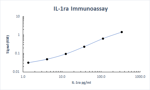
Human IL-1ra / IL-1F3 ELISA Standard Curve
Recombinant Human IL‑1ra/IL‑1F3 (Catalog # 280-RA) was serially diluted and captured by Mouse Anti-Human IL‑1ra/IL‑1F3 Monoclonal Antibody (Catalog # MAB280) coated on a Clear Polystyrene Microplate (Catalog # DY990). Goat Anti-Human IL‑1ra/IL‑1F3 Antigen Affinity-purified Polyclonal Antibody (Catalog # AF-280-NA) was biotinylated and incubated with the protein captured on the plate. Detection of the standard curve was achieved by incubating Streptavidin-HRP (Catalog # DY998)Applications for Human IL-1ra/IL-1F3 Antibody
Application
Recommended Usage
Immunohistochemistry
5-15 µg/mL
Sample: Immersion fixed paraffin-embedded sections of human skin
Sample: Immersion fixed paraffin-embedded sections of human skin
Simple Western
20 µg/mL
Sample: RT-4 human bladder carcinoma cell line, A431 human epithelial carcinoma cell line, human skin cancer tissue, and mouse skin cancer tissue
Sample: RT-4 human bladder carcinoma cell line, A431 human epithelial carcinoma cell line, human skin cancer tissue, and mouse skin cancer tissue
Western Blot
0.5 µg/mL
Sample:
Sample:
RT-4 human bladder carcinoma cell line and A431 human epithelial carcinoma cell line
Neutralization
Measured by its ability to neutralize IL‑1ra/IL‑1F3 inhibition of IL‑1 alpha /IL‑1F1-dependent proliferation in the D10.G4.1 mouse helper T cell line. Symons, J.A. et al. (1987) in Lymphokines and Interferons, a Practical Approach. Clemens, M.J. et al. (eds): IRL Press. 272. The Neutralization Dose (ND50) is typically ≤4 µg/mL in the presence of 50 ng/mL Recombinant Human IL‑1ra/IL‑1F3 and 50 pg/mL Recombinant Human IL‑1 alpha /IL‑1F1.
Reviewed Applications
Read 2 reviews rated 5 using AF-280-NA in the following applications:
Formulation, Preparation, and Storage
Purification
Antigen Affinity-purified
Reconstitution
Reconstitute at 0.2 mg/mL in sterile PBS. For liquid material, refer to CoA for concentration.
Loading...
Formulation
Lyophilized from a 0.2 μm filtered solution in PBS with Trehalose. *Small pack size (SP) is supplied either lyophilized or as a 0.2 µm filtered solution in PBS.
Shipping
Lyophilized product is shipped at ambient temperature. Liquid small pack size (-SP) is shipped with polar packs. Upon receipt, store immediately at the temperature recommended below.
Stability & Storage
Use a manual defrost freezer and avoid repeated freeze-thaw cycles.
- 12 months from date of receipt, -20 to -70 °C as supplied.
- 1 month, 2 to 8 °C under sterile conditions after reconstitution.
- 6 months, -20 to -70 °C under sterile conditions after reconstitution.
Calculators
Background: IL-1ra/IL-1F3
Long Name
Interleukin 1 Receptor Antagonist
Alternate Names
DIRA, ICIL-1ra, IL-1F3, IL-1ra3, IL-1RN, IL1ra, IL1RN, MVCD4
Gene Symbol
IL1RN
UniProt
Additional IL-1ra/IL-1F3 Products
Product Documents for Human IL-1ra/IL-1F3 Antibody
Certificate of Analysis
To download a Certificate of Analysis, please enter a lot or batch number in the search box below.
Note: Certificate of Analysis not available for kit components.
Product Specific Notices for Human IL-1ra/IL-1F3 Antibody
For research use only
Citations for Human IL-1ra/IL-1F3 Antibody
 Powered by Bioz
Customer Reviews for Human IL-1ra/IL-1F3 Antibody (2)
5 out of 5
2 Customer Ratings
Have you used Human IL-1ra/IL-1F3 Antibody?
Submit a review and receive an Amazon gift card!
$25/€18/£15/$25CAN/¥2500 Yen for a review with an image
$10/€7/£6/$10CAN/¥1110 Yen for a review without an image
Submit a review
Customer Images
Showing
1
-
2 of
2 reviews
Showing All
Filter By:
-
Application: ELISASample Tested: SerumSpecies: PrimateVerified Customer | Posted 11/28/2022Used as a capture antibody in our NHP ELISA

-
Application: ELISASample Tested: SerumSpecies: HumanVerified Customer | Posted 11/18/2019
There are no reviews that match your criteria.
Protocols
Find general support by application which include: protocols, troubleshooting, illustrated assays, videos and webinars.
- Antigen Retrieval Protocol (PIER)
- Antigen Retrieval for Frozen Sections Protocol
- Appropriate Fixation of IHC/ICC Samples
- Cellular Response to Hypoxia Protocols
- Chromogenic IHC Staining of Formalin-Fixed Paraffin-Embedded (FFPE) Tissue Protocol
- Chromogenic Immunohistochemistry Staining of Frozen Tissue
- ClariTSA™ Fluorophore Kits
- Detection & Visualization of Antibody Binding
- Fluorescent IHC Staining of Frozen Tissue Protocol
- Graphic Protocol for Heat-induced Epitope Retrieval
- Graphic Protocol for the Preparation and Fluorescent IHC Staining of Frozen Tissue Sections
- Graphic Protocol for the Preparation and Fluorescent IHC Staining of Paraffin-embedded Tissue Sections
- Graphic Protocol for the Preparation of Gelatin-coated Slides for Histological Tissue Sections
- IHC Sample Preparation (Frozen sections vs Paraffin)
- Immunofluorescent IHC Staining of Formalin-Fixed Paraffin-Embedded (FFPE) Tissue Protocol
- Immunohistochemistry (IHC) and Immunocytochemistry (ICC) Protocols
- Immunohistochemistry Frozen Troubleshooting
- Immunohistochemistry Paraffin Troubleshooting
- Preparing Samples for IHC/ICC Experiments
- Preventing Non-Specific Staining (Non-Specific Binding)
- Primary Antibody Selection & Optimization
- Protocol for Heat-Induced Epitope Retrieval (HIER)
- Protocol for Making a 4% Formaldehyde Solution in PBS
- Protocol for VisUCyte™ HRP Polymer Detection Reagent
- Protocol for the Preparation & Fixation of Cells on Coverslips
- Protocol for the Preparation and Chromogenic IHC Staining of Frozen Tissue Sections
- Protocol for the Preparation and Chromogenic IHC Staining of Frozen Tissue Sections - Graphic
- Protocol for the Preparation and Chromogenic IHC Staining of Paraffin-embedded Tissue Sections
- Protocol for the Preparation and Chromogenic IHC Staining of Paraffin-embedded Tissue Sections - Graphic
- Protocol for the Preparation and Fluorescent IHC Staining of Frozen Tissue Sections
- Protocol for the Preparation and Fluorescent IHC Staining of Paraffin-embedded Tissue Sections
- Protocol for the Preparation of Gelatin-coated Slides for Histological Tissue Sections
- R&D Systems Quality Control Western Blot Protocol
- TUNEL and Active Caspase-3 Detection by IHC/ICC Protocol
- The Importance of IHC/ICC Controls
- Troubleshooting Guide: Immunohistochemistry
- Troubleshooting Guide: Western Blot Figures
- Western Blot Conditions
- Western Blot Protocol
- Western Blot Protocol for Cell Lysates
- Western Blot Troubleshooting
- Western Blot Troubleshooting Guide
- View all Protocols, Troubleshooting, Illustrated assays and Webinars
Loading...
Associated Pathways
