
Key Product Details
Species Reactivity
Validated:
Human
Cited:
Human, Mouse, Avian - Chicken, Boa constrictor, Chicken, Snake, Snake - Aspidelaps lubricus (Cape Coral Snake), Snake - Naja annulifera (Banded or Ringed Water Cobra), Transgenic Mouse
Applications
Validated:
Immunohistochemistry, Western Blot, Immunocytochemistry, Simple Western
Cited:
Immunohistochemistry, Immunohistochemistry-Paraffin, Western Blot, Flow Cytometry, Immunocytochemistry, Chromatin Immunoprecipitation (ChIP)
Label
Unconjugated
Antibody Source
Polyclonal Goat IgG
Loading...
Product Specifications
Immunogen
E. coli-derived recombinant human Islet-1
Met4-Ala349
Accession # P61371
Met4-Ala349
Accession # P61371
Specificity
Detects human Islet-1 in direct ELISAs and Western blots. In direct ELISAs, approximately 10% cross-reactivity with recombinant human Islet-2 is observed.
Clonality
Polyclonal
Host
Goat
Isotype
IgG
Scientific Data Images for Human Islet-1 Antibody
Detection of Human Islet‑1 by Western Blot.
Western blot shows lysates of JOY6 human induced pluripotent stem cells undifferentiated and differentiatied into motor neurons. PVDF membrane was probed with 1 µg/mL of Goat Anti-Human Islet-1 Antigen Affinity-purified Polyclonal Antibody (Catalog # AF1837) followed by HRP-conjugated Anti-Goat IgG Secondary Antibody (Catalog # HAF017). A specific band was detected for Islet-1 at approximately 42 kDa (as indicated). This experiment was conducted under reducing conditions and using Immunoblot Buffer Group 1.
Islet‑1 in iPS2 Human Stem Cells.
Islet-1 was detected in immersion fixed iPS2 human induced pluripotent stem cells differentiated to endocrine progenitor cells using Goat Anti-Human Islet-1 Antigen Affinity-purified Polyclonal Antibody (Catalog # AF1837) at 10 µg/mL for 3 hours at room temperature. Cells were stained using the NorthernLights™ 557-conjugated Anti-Goat IgG Secondary Antibody (red; Catalog # NL001) and counterstained with DAPI(blue). Specific staining was localized to the nucleus. View our protocol for Fluorescent ICC Staining of Cells on Coverslips.
Islet‑1 in Human Pancreas.
Islet‑1 was detected in immersion fixed paraffin-embedded sections of human pancreas using Goat Anti-Human Islet‑1 Antigen Affinity-purified Polyclonal Antibody (Catalog # AF1837) at 1 µg/mL for 1 hour at room temperature followed by incubation with the Anti-Goat IgG VisUCyte™ HRP Polymer Antibody (VC004). Before incubation with the primary antibody, tissue was subjected to heat-induced epitope retrieval using Antigen Retrieval Reagent-Basic (CTS013). Tissue was stained using DAB (brown) and counterstained with hematoxylin (blue). Specific staining was localized to nuclei in islet cells. Staining was performed using our protocol for IHC Staining with VisUCyte HRP Polymer Detection Reagents.
Detection of Human Islet‑1 by Simple WesternTM.
Simple Western lane view shows lysates of JOY6 human induced pluripotent stem cells undifferentiated and differentiated into motor neurons, loaded at 0.2 mg/mL. A specific band was detected for Islet-1 at approximately 53 kDa (as indicated) using 10 µg/mL of Goat Anti-Human Islet-1 Antigen Affinity-purified Polyclonal Antibody (Catalog # AF1837) followed by 1:50 dilution of HRP-conjugated Anti-Goat IgG Secondary Antibody (Catalog # HAF109). This experiment was conducted under reducing conditions and using the 12-230 kDa separation system.
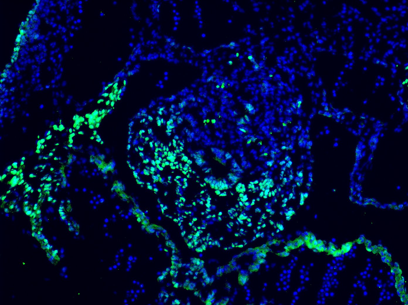
Detection of Mouse Islet-1 by Immunohistochemistry
Efficiency of Isl1 ablation in stomachs of Isl1MCM/Del mutant mouse stomachs at E18.5. (A) Tamoxifen-inducible Cre recombinase excised DNA sequences flanked by two loxP sites. (B)Isl1 RNA levels were ablated in Isl1MCM/Del mutant stomachs as seen by semi-quantitative PCR. Isl1F/+mice showed a 592 base pair product whereas Isl1MCM/Del mice generated a 303 base pair product. (C) Isl1 was significantly down-regulated at the protein levels in Isl1MCM/Del mutant stomachs as shown by western blot. Expression of embryos at E11.5 was used as positive control. (D) Isl1 protein expression in Isl1F/+and Isl1MCM/Del embryonic pylorus. Isl1 expression was significantly reduced in Isl1MCM/Del embryonic stomachs, as seen by immunofluorescence. Images in Isl1F/+and Isl1MCM/Del were processed on the same slide and photographed at the same exposure. Enlarged images of the boxed areas are shown on the right side of the merged pictures. Yellow arrowheads show representative Isl1-positive cells, and white arrowheads show representative Isl1-negative cells. Yellow dotted lines mark the epithelial basement membrane. Scale bars: 50 μm. Image collected and cropped by CiteAb from the following publication (https://bmcbiol.biomedcentral.com/articles/10.1186/1741-7007-12-25), licensed under a CC-BY license. Not internally tested by R&D Systems.Applications for Human Islet-1 Antibody
Application
Recommended Usage
Immunocytochemistry
5-15 µg/mL
Sample: Immersion fixed iPS2 human induced pluripotent stem cells differentiated to endocrine progenitor cells
Sample: Immersion fixed iPS2 human induced pluripotent stem cells differentiated to endocrine progenitor cells
Immunohistochemistry
1-15 µg/mL
Sample: Immersion fixed paraffin-embedded sections of human pancreas
Sample: Immersion fixed paraffin-embedded sections of human pancreas
Simple Western
10 µg/mL
Sample: JOY6 human induced pluripotent stem cells undifferentiated and differentiatied into Motor Neurons
Sample: JOY6 human induced pluripotent stem cells undifferentiated and differentiatied into Motor Neurons
Western Blot
1 µg/mL
Sample: JOY6 human induced pluripotent stem cells undifferentiated and differentiatied into Motor Neurons
Sample: JOY6 human induced pluripotent stem cells undifferentiated and differentiatied into Motor Neurons
Reviewed Applications
Read 1 review rated 5 using AF1837 in the following applications:
Formulation, Preparation, and Storage
Purification
Antigen Affinity-purified
Reconstitution
Reconstitute at 0.2 mg/mL in sterile PBS. For liquid material, refer to CoA for concentration.
Loading...
Formulation
Lyophilized from a 0.2 μm filtered solution in PBS with Trehalose. *Small pack size (SP) is supplied either lyophilized or as a 0.2 µm filtered solution in PBS.
Shipping
Lyophilized product is shipped at ambient temperature. Liquid small pack size (-SP) is shipped with polar packs. Upon receipt, store immediately at the temperature recommended below.
Stability & Storage
Use a manual defrost freezer and avoid repeated freeze-thaw cycles.
- 12 months from date of receipt, -20 to -70 °C as supplied.
- 1 month, 2 to 8 °C under sterile conditions after reconstitution.
- 6 months, -20 to -70 °C under sterile conditions after reconstitution.
Calculators
Background: Islet-1
Additional Islet-1 Products
Product Documents for Human Islet-1 Antibody
Certificate of Analysis
To download a Certificate of Analysis, please enter a lot or batch number in the search box below.
Note: Certificate of Analysis not available for kit components.
Product Specific Notices for Human Islet-1 Antibody
For research use only
Citations for Human Islet-1 Antibody
 Powered by Bioz
Customer Reviews for Human Islet-1 Antibody (1)
5 out of 5
1 Customer Rating
Have you used Human Islet-1 Antibody?
Submit a review and receive an Amazon gift card!
$25/€18/£15/$25CAN/¥2500 Yen for a review with an image
$10/€7/£6/$10CAN/¥1110 Yen for a review without an image
Submit a review
Customer Images
Showing
1
-
1 of
1 review
Showing All
Filter By:
-
Application: Immunocytochemistry/ImmunofluorescenceSample Tested: E10.5 mouse embryo fixed in 4% PFASpecies: MouseVerified Customer | Posted 12/02/2020Excellent antibody. Very reliable. Highly recommend on embryonic muse tissues. Have stained on both E9.5 and E10.5 mouse sections. Concentration used - 10ug/mL.

There are no reviews that match your criteria.
Protocols
Find general support by application which include: protocols, troubleshooting, illustrated assays, videos and webinars.
- Antigen Retrieval Protocol (PIER)
- Antigen Retrieval for Frozen Sections Protocol
- Appropriate Fixation of IHC/ICC Samples
- Cellular Response to Hypoxia Protocols
- Chromogenic IHC Staining of Formalin-Fixed Paraffin-Embedded (FFPE) Tissue Protocol
- Chromogenic Immunohistochemistry Staining of Frozen Tissue
- ClariTSA™ Fluorophore Kits
- Detection & Visualization of Antibody Binding
- Fluorescent IHC Staining of Frozen Tissue Protocol
- Graphic Protocol for Heat-induced Epitope Retrieval
- Graphic Protocol for the Preparation and Fluorescent IHC Staining of Frozen Tissue Sections
- Graphic Protocol for the Preparation and Fluorescent IHC Staining of Paraffin-embedded Tissue Sections
- Graphic Protocol for the Preparation of Gelatin-coated Slides for Histological Tissue Sections
- ICC Cell Smear Protocol for Suspension Cells
- ICC Immunocytochemistry Protocol Videos
- ICC for Adherent Cells
- IHC Sample Preparation (Frozen sections vs Paraffin)
- Immunocytochemistry (ICC) Protocol
- Immunocytochemistry Troubleshooting
- Immunofluorescence of Organoids Embedded in Cultrex Basement Membrane Extract
- Immunofluorescent IHC Staining of Formalin-Fixed Paraffin-Embedded (FFPE) Tissue Protocol
- Immunohistochemistry (IHC) and Immunocytochemistry (ICC) Protocols
- Immunohistochemistry Frozen Troubleshooting
- Immunohistochemistry Paraffin Troubleshooting
- Preparing Samples for IHC/ICC Experiments
- Preventing Non-Specific Staining (Non-Specific Binding)
- Primary Antibody Selection & Optimization
- Protocol for Heat-Induced Epitope Retrieval (HIER)
- Protocol for Making a 4% Formaldehyde Solution in PBS
- Protocol for VisUCyte™ HRP Polymer Detection Reagent
- Protocol for the Fluorescent ICC Staining of Cell Smears - Graphic
- Protocol for the Fluorescent ICC Staining of Cultured Cells on Coverslips - Graphic
- Protocol for the Preparation & Fixation of Cells on Coverslips
- Protocol for the Preparation and Chromogenic IHC Staining of Frozen Tissue Sections
- Protocol for the Preparation and Chromogenic IHC Staining of Frozen Tissue Sections - Graphic
- Protocol for the Preparation and Chromogenic IHC Staining of Paraffin-embedded Tissue Sections
- Protocol for the Preparation and Chromogenic IHC Staining of Paraffin-embedded Tissue Sections - Graphic
- Protocol for the Preparation and Fluorescent ICC Staining of Cells on Coverslips
- Protocol for the Preparation and Fluorescent ICC Staining of Non-adherent Cells
- Protocol for the Preparation and Fluorescent ICC Staining of Stem Cells on Coverslips
- Protocol for the Preparation and Fluorescent IHC Staining of Frozen Tissue Sections
- Protocol for the Preparation and Fluorescent IHC Staining of Paraffin-embedded Tissue Sections
- Protocol for the Preparation of Gelatin-coated Slides for Histological Tissue Sections
- Protocol for the Preparation of a Cell Smear for Non-adherent Cell ICC - Graphic
- R&D Systems Quality Control Western Blot Protocol
- TUNEL and Active Caspase-3 Detection by IHC/ICC Protocol
- The Importance of IHC/ICC Controls
- Troubleshooting Guide: Immunohistochemistry
- Troubleshooting Guide: Western Blot Figures
- Western Blot Conditions
- Western Blot Protocol
- Western Blot Protocol for Cell Lysates
- Western Blot Troubleshooting
- Western Blot Troubleshooting Guide
- View all Protocols, Troubleshooting, Illustrated assays and Webinars

