Human/Mouse IGF-I R/IGF1R Antibody
R&D Systems | Catalog # AF-305-NA

Key Product Details
Species Reactivity
Validated:
Human, Mouse
Cited:
Human, Mouse, Rat, Rabbit, Transgenic Mouse
Applications
Validated:
Immunohistochemistry, Western Blot, Neutralization, Immunocytochemistry
Cited:
Immunohistochemistry, Immunohistochemistry-Paraffin, Western Blot, Neutralization, Flow Cytometry, Cell Culture, Functional Assay, Luciferase Reporter Assay
Label
Unconjugated
Antibody Source
Polyclonal Goat IgG
Loading...
Product Specifications
Immunogen
Recombinant Human IGF-I R/IGF1R extracellular domain
Accession # P08069
Accession # P08069
Specificity
Detects human and mouse IGF-I R/IGF1R in direct ELISAs and Western blots. In direct ELISAs and Western blots, 25-50% cross-reactivity with recombinant mouse IGF-I R is observed. In direct ELISAs, less than 1% cross-reactivity with recombinant human IGF‑II R is observed.
Clonality
Polyclonal
Host
Goat
Isotype
IgG
Endotoxin Level
<0.10 EU per 1 μg of the antibody by the LAL method.
Scientific Data Images for Human/Mouse IGF-I R/IGF1R Antibody
IGF-I R/IGF1R in MCF‑7 and HDLM-2 Human Cell Lines.
IGF-I R/IGF1R was detected in immersion fixed MCF-7 human breast cancer cell line (positive control, left panel) and HDLM-2 human Hodgkin's lymphoma cell line (negative control, right panel) using Goat Anti-Human/Mouse IGF-I R/IGF1R Antigen Affinity-purified Polyclonal Antibody (Catalog # AF-305-NA) at 1.7 µg/mL for 3 hours at room temperature. Cells were stained using the NorthernLights™ 557-conjugated Anti-Goat IgG Secondary Antibody (red; Catalog # NL001) and counterstained with DAPI (blue). Specific staining was localized to plasma membrane. View our protocols for Fluorescent ICC Staining of Cells on Coverslipsand Fluorescent ICC Staining of Non-adherent Cells.
IGF-I R/IGF1R in Mouse Embryo.
IGF-I R/IGF1R was detected in immersion fixed frozen sections of mouse embryo using Goat Anti-Human IGF-I R Antigen Affinity-purified Polyclonal Antibody (Catalog # AF-305-NA) at 10 µg/mL overnight at 4 °C. Tissue was stained using the Anti-Goat HRP-DAB Cell & Tissue Staining Kit (brown; Catalog # CTS008) and counterstained with hematoxylin (blue). Lower panel shows a lack of labeling if primary antibodies are omitted and tissue is stained only with secondary antibody followed by incubation with detection reagents. View our protocol for Chromogenic IHC Staining of Frozen Tissue Sections.
IGF-I R/IGF1R in Human Placenta.
IGF-I R/IGF1R was detected in immersion fixed paraffin-embedded sections of human placenta (chorionic villi) using 15 µg/mL Goat Anti-Human IGF-I R/IGF1R Antigen Affinity-purified Polyclonal Antibody (Catalog # AF-305-NA) overnight at 4 °C. Tissue was stained with the Anti-Goat HRP-DAB Cell & Tissue Staining Kit (brown; Catalog # CTS008) and counterstained with hematoxylin (blue). View our protocol for Chromogenic IHC Staining of Paraffin-embedded Tissue Sections.
Cell Proliferation Induced by IGF‑I and Neutralization by Human IGF-I R/IGF1R Antibody.
Recombinant Human IGF-I (Catalog # 291-G1) stimulates proliferation in the MCF-7 human breast cancer cell line in a dose-dependent manner (orange line). Proliferation elicited by Recombinant Human IGF-I (6 ng/mL) is neutralized (green line) by increasing concentrations of Goat Anti-Human IGF-I R/IGF1R Antigen Affinity-purified Polyclonal Antibody (Catalog # AF-305-NA). The ND50 is typically 0.5-1.5 µg/mL.
Detection of Mouse IGF-I R/IGF1R by Immunohistochemistry
Representative H&E stained sections of hyperplasia (A) and normal mammary ducts (B) that developed following transplantation of pubertal MTB-IGFIR mammary epithelial cells that did not progress to palpable tumors.Immunostaining for IGF-IR (C, D) revealed high levels of expression with normal mammary ducts indicating successful engraftment of transgenic tissue. Scale bars, 200 µM (A–C) and 100 µM (D). Image collected and cropped by CiteAb from the following open publication (https://dx.plos.org/10.1371/journal.pone.0108781), licensed under a CC-BY license. Not internally tested by R&D Systems.
Detection of Mouse IGF-I R/IGF1R by Immunohistochemistry
Immunohistochemistry for the IGF-IR (brown stain) in mammary epithelial cells of a 2 day old female mice captured at 100x (A) or 200x (B) magnification.Scale bars, 200 µM (A) and 100 µM (B). Image collected and cropped by CiteAb from the following open publication (https://dx.plos.org/10.1371/journal.pone.0108781), licensed under a CC-BY license. Not internally tested by R&D Systems.
Detection of Mouse IGF-I R/IGF1R by Immunohistochemistry
Immunohistochemistry for the IGF-IR (brown stain) in mammary epithelial cells of a 2 day old female mice captured at 100x (A) or 200x (B) magnification.Scale bars, 200 µM (A) and 100 µM (B). Image collected and cropped by CiteAb from the following open publication (https://dx.plos.org/10.1371/journal.pone.0108781), licensed under a CC-BY license. Not internally tested by R&D Systems.Applications for Human/Mouse IGF-I R/IGF1R Antibody
Application
Recommended Usage
Immunocytochemistry
1-15 µg/mL
Sample: Immersion fixed MCF‑7 human breast cancer cell line
Sample: Immersion fixed MCF‑7 human breast cancer cell line
Immunohistochemistry
5-15 µg/mL
Sample: Immersion fixed paraffin-embedded sections of human placenta (chorionic villi) and immersion fixed frozen sections of mouse embryo
Sample: Immersion fixed paraffin-embedded sections of human placenta (chorionic villi) and immersion fixed frozen sections of mouse embryo
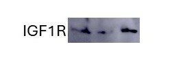
Western Blot
0.1 µg/mL
Sample: Recombinant Human IGF‑I R/IGF1R (Catalog # 391-GR) and Recombinant Mouse IGF‑I R/IGF1R (Catalog # 6630-GR).
Sample: Recombinant Human IGF‑I R/IGF1R (Catalog # 391-GR) and Recombinant Mouse IGF‑I R/IGF1R (Catalog # 6630-GR).
Neutralization
Measured by its ability to neutralize IGF-I-induced proliferation in the MCF-7 human breast cancer cell line. Karey, K.P. et al. (1988) Cancer Research 48:4083. The Neutralization Dose (ND50) is typically 0.5-1.5 µg/mL in the presence of 6 ng/mL
Recombinant
Human IGF-I/IGF-1 (Catalog # 291-G1).
Reviewed Applications
Read 3 reviews rated 4 using AF-305-NA in the following applications:
Formulation, Preparation, and Storage
Purification
Antigen Affinity-purified
Reconstitution
Reconstitute at 0.2 mg/mL in sterile PBS. For liquid material, refer to CoA for concentration.
Loading...
Formulation
Lyophilized from a 0.2 μm filtered solution in PBS with Trehalose. *Small pack size (SP) is supplied either lyophilized or as a 0.2 µm filtered solution in PBS.
Shipping
Lyophilized product is shipped at ambient temperature. Liquid small pack size (-SP) is shipped with polar packs. Upon receipt, store immediately at the temperature recommended below.
Stability & Storage
Use a manual defrost freezer and avoid repeated freeze-thaw cycles.
- 12 months from date of receipt, -20 to -70 °C as supplied.
- 1 month, 2 to 8 °C under sterile conditions after reconstitution.
- 6 months, -20 to -70 °C under sterile conditions after reconstitution.
Calculators
Background: IGF-I R/IGF1R
References
- Rechler, M.M. and S.P. Nissley (1990) in Insulin-Like Growth Factors. Sporn, M.B. and A.B. Roberts (eds): Peptide Growth Factors and Their Receptors I, New York: Springer-Verlag, p. 263.
Long Name
Insulin-like Growth Factor I Receptor
Alternate Names
CD221, IGF-1R, IGF-IR, IGF1R, IGFIR
Entrez Gene IDs
3480 (Human)
Gene Symbol
IGF1R
UniProt
Additional IGF-I R/IGF1R Products
Product Documents for Human/Mouse IGF-I R/IGF1R Antibody
Certificate of Analysis
To download a Certificate of Analysis, please enter a lot or batch number in the search box below.
Note: Certificate of Analysis not available for kit components.
Product Specific Notices for Human/Mouse IGF-I R/IGF1R Antibody
For research use only
Citations for Human/Mouse IGF-I R/IGF1R Antibody
 Powered by Bioz
Customer Reviews for Human/Mouse IGF-I R/IGF1R Antibody (3)
4 out of 5
3 Customer Ratings
Have you used Human/Mouse IGF-I R/IGF1R Antibody?
Submit a review and receive an Amazon gift card!
$25/€18/£15/$25CAN/¥2500 Yen for a review with an image
$10/€7/£6/$10CAN/¥1110 Yen for a review without an image
Submit a review
Customer Images
Showing
1
-
3 of
3 reviews
Showing All
Filter By:
-
Application: Western BlotVerified Customer | Posted 12/18/2023

-
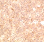
Application: ImmunohistochemistrySample Tested: Cancer TissueSpecies: HumanVerified Customer | Posted 06/18/2021
-
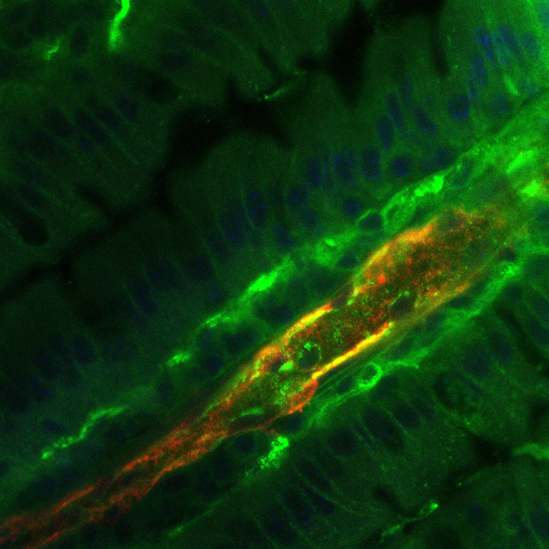
Application: Immunocytochemistry/ImmunofluorescenceSample Tested: Intestinal villiSpecies: MouseVerified Customer | Posted 07/30/2019IGF1R in green
There are no reviews that match your criteria.
Protocols
Find general support by application which include: protocols, troubleshooting, illustrated assays, videos and webinars.
- Antigen Retrieval Protocol (PIER)
- Antigen Retrieval for Frozen Sections Protocol
- Appropriate Fixation of IHC/ICC Samples
- Cellular Response to Hypoxia Protocols
- Chromogenic IHC Staining of Formalin-Fixed Paraffin-Embedded (FFPE) Tissue Protocol
- Chromogenic Immunohistochemistry Staining of Frozen Tissue
- ClariTSA™ Fluorophore Kits
- Detection & Visualization of Antibody Binding
- Fluorescent IHC Staining of Frozen Tissue Protocol
- Graphic Protocol for Heat-induced Epitope Retrieval
- Graphic Protocol for the Preparation and Fluorescent IHC Staining of Frozen Tissue Sections
- Graphic Protocol for the Preparation and Fluorescent IHC Staining of Paraffin-embedded Tissue Sections
- Graphic Protocol for the Preparation of Gelatin-coated Slides for Histological Tissue Sections
- ICC Cell Smear Protocol for Suspension Cells
- ICC Immunocytochemistry Protocol Videos
- ICC for Adherent Cells
- IHC Sample Preparation (Frozen sections vs Paraffin)
- Immunocytochemistry (ICC) Protocol
- Immunocytochemistry Troubleshooting
- Immunofluorescence of Organoids Embedded in Cultrex Basement Membrane Extract
- Immunofluorescent IHC Staining of Formalin-Fixed Paraffin-Embedded (FFPE) Tissue Protocol
- Immunohistochemistry (IHC) and Immunocytochemistry (ICC) Protocols
- Immunohistochemistry Frozen Troubleshooting
- Immunohistochemistry Paraffin Troubleshooting
- Preparing Samples for IHC/ICC Experiments
- Preventing Non-Specific Staining (Non-Specific Binding)
- Primary Antibody Selection & Optimization
- Protocol for Heat-Induced Epitope Retrieval (HIER)
- Protocol for Making a 4% Formaldehyde Solution in PBS
- Protocol for VisUCyte™ HRP Polymer Detection Reagent
- Protocol for the Fluorescent ICC Staining of Cell Smears - Graphic
- Protocol for the Fluorescent ICC Staining of Cultured Cells on Coverslips - Graphic
- Protocol for the Preparation & Fixation of Cells on Coverslips
- Protocol for the Preparation and Chromogenic IHC Staining of Frozen Tissue Sections
- Protocol for the Preparation and Chromogenic IHC Staining of Frozen Tissue Sections - Graphic
- Protocol for the Preparation and Chromogenic IHC Staining of Paraffin-embedded Tissue Sections
- Protocol for the Preparation and Chromogenic IHC Staining of Paraffin-embedded Tissue Sections - Graphic
- Protocol for the Preparation and Fluorescent ICC Staining of Cells on Coverslips
- Protocol for the Preparation and Fluorescent ICC Staining of Non-adherent Cells
- Protocol for the Preparation and Fluorescent ICC Staining of Stem Cells on Coverslips
- Protocol for the Preparation and Fluorescent IHC Staining of Frozen Tissue Sections
- Protocol for the Preparation and Fluorescent IHC Staining of Paraffin-embedded Tissue Sections
- Protocol for the Preparation of Gelatin-coated Slides for Histological Tissue Sections
- Protocol for the Preparation of a Cell Smear for Non-adherent Cell ICC - Graphic
- R&D Systems Quality Control Western Blot Protocol
- TUNEL and Active Caspase-3 Detection by IHC/ICC Protocol
- The Importance of IHC/ICC Controls
- Troubleshooting Guide: Immunohistochemistry
- Troubleshooting Guide: Western Blot Figures
- Western Blot Conditions
- Western Blot Protocol
- Western Blot Protocol for Cell Lysates
- Western Blot Troubleshooting
- Western Blot Troubleshooting Guide
- View all Protocols, Troubleshooting, Illustrated assays and Webinars
Loading...
Associated Pathways


