Insulin-like growth factor I receptor (IGF-I R) is a disulfide-linked heterotetrameric transmembrane protein consisting of two alpha and two beta subunits. Both the alpha and beta subunits are encoded within a single receptor precursor cDNA. The proreceptor polypeptide is proteolytically cleaved and disulfide-linked to yield the mature heterotetrameric receptor. The alpha subunit of IGF-I R is extracellular while the beta subunit has an extracellular domain, a transmembrane domain and a cytoplasmic tyrosine kinase domain. IGF-I R is highly expressed in all cell types and tissues. Essentially all of the biological activities of IGF-I and -II have been shown to be mediated via IGF-I R.

Key Product Details
Species Reactivity
Validated:
Human, Mouse
Cited:
Human, Mouse, Bovine
Applications
Validated:
Immunohistochemistry, Western Blot, ELISA Capture (Matched Antibody Pair), Neutralization, Flow Cytometry, Immunocytochemistry
Cited:
Immunohistochemistry, Immunohistochemistry-Paraffin, Western Blot, Neutralization, Flow Cytometry, Immunocytochemistry, ELISA Capture, ELISA Development, In vivo assay, Functional Assay
Label
Unconjugated
Antibody Source
Monoclonal Mouse IgG1 Clone # 33255
Loading...
Product Specifications
Immunogen
S. frugiperda insect ovarian cell line Sf 21-derived recombinant human IGF-I R/IGF1R
Glu31-Asn932
Accession # P08069
Glu31-Asn932
Accession # P08069
Specificity
Detects human IGF-I R/IGF1R in sandwich ELISAs and Western blots. Detects mouse IGF-I R/IGF1R in Immunohistochemistry. In sandwich immunoassays, less than 0.15% cross-reactivity or interference was observed with recombinant human (rh) IGF-I, rhIGF‑II, rhIL‑3 R alpha, rhIL‑9 R, and rhTGF‑ beta RII.
Clonality
Monoclonal
Host
Mouse
Isotype
IgG1
Endotoxin Level
<0.10 EU per 1 μg of the antibody by the LAL method.
Scientific Data Images for IGF-I R/IGF1R Antibody (33255)
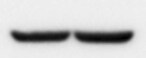
Detection of Human IGF-I R/IGF1R by Western Blot.
Western blot shows lysates of NTera-2 human testicular embryonic carcinoma cell line, SK-Mel-28 human malignant melanoma cell line, and G361 human melanoma cell line. PVDF membrane was probed with 1 µg/mL of Mouse Anti-Human/Mouse IGF-I R/IGF1R Monoclonal Antibody (Catalog # MAB391) followed by HRP-conjugated Anti-Mouse IgG Secondary Antibody (Catalog # HAF007). A specific band was detected for IGF-I R/IGF1R at approximately 275 kDa (as indicated). This experiment was conducted under non-reducing conditions and using Immunoblot Buffer Group 2.
Detection of IGF-I R/IGF1R in MCF‑7 Human Cell Line by Flow Cytometry.
MCF-7 human breast cancer cell line was stained with Mouse Anti-Human IGF-I R/IGF1R Monoclonal Antibody (Catalog # MAB391, filled histogram) or isotype control antibody (Catalog # MAB002, open histogram) followed by anti-mouse IgG PE-conjugated secondary antibody (Catalog # F0102B). View our protocol for Staining Membrane-associated Proteins.
IGF-I R/IGF1R in MCF‑7 and HDLM Human Cell Lines.
IGF-I R/IGF1R was detected in immersion fixed MCF-7 human breast cancer cell line (positive staining; left panel) and HDLM human Hodgkin's lymphoma cell line (negative staining; right panel) using Mouse Anti-Human/Mouse IGF-I R/IGF1R Monoclonal Antibody (Catalog # MAB391) at 3 µg/mL for 3 hours at room temperature. Cells were stained using the NorthernLights™ 557-conjugated Anti-Mouse IgG Secondary Antibody (red; Catalog # NL007) and counterstained with DAPI (blue). Specific staining was localized to cell membrane in MCF-7 cell line. View our protocol for Fluorescent ICC Staining of Cells on Coverslips.
IGF-I R/IGF1R in Mouse Heart.
IGF-I R/IGF1R was detected in perfusion fixed paraffin-embedded sections of mouse heart using Mouse Anti-Human/Mouse IGF-I R/IGF1R Monoclonal Antibody (Catalog # MAB391) at 15 µg/mL for 1 hour at room temperature followed by incubation with the Anti-Mouse IgG VisUCyte™ HRP Polymer Antibody (Catalog # VC001). Tissue was stained using DAB (brown) and counterstained with hematoxylin (blue). Specific staining was localized to plasma membrane and cytoplasm. View our protocol for IHC Staining with VisUCyte HRP Polymer Detection Reagents.
Cell Proliferation Induced by IGF‑I and Neutralization by Human IGF-I R/IGF1R Antibody.
Recombinant Human (rh) IGF-I/IGF-1 (Catalog # 291-G1) stimulates proliferation in the MCF-7 human breast cancer cell line in a dose-dependent manner (orange line). Proliferation elicited by rhIGF-I/IGF-1 (6 ng/mL) is neutralized (green line) by increasing concentrations of Mouse Anti-Human/Mouse IGF-I R/IGF1R Monoclonal Antibody (Catalog # MAB391). At 11 µg/mL, this antibody will neutralize 50-75% rhIGF-1 induced activity.
Detection of IGF-I R/IGF1R by Immunocytochemistry/ Immunofluorescence
IGF1-R inhibition decreases autophagosome precursor formation by reducing clathrin-dependent endocytosis. (D–F) HeLa cells were transfected with 0.5 μg of the GFP-Atg16L1 construct. After 24 h, the cells were treated for 24 h with MAB391, fixed&analysed under fluorescence microscope. The P-values for assessing the %age of cells showing ≥20 GFP-Atg16-positive vesicles as well as the number of GFP-Atg16 vesicles/cell determined by Student's t-test (E: n = 3; CTRL versus 24 h MAB391, P = 0.00272; F: n = 3; CTRL versus 24 h MAB391, P = 0.00496). Image collected & cropped by CiteAb from the following open publication (https://pubmed.ncbi.nlm.nih.gov/23804751), licensed under a CC-BY license. Not internally tested by R&D Systems.
Human IGF-I R ELISA Standard Curve
Recombinant Human IGF-I R/IGF1R (Catalog # 391-GR) was serially diluted and captured by Mouse Anti-Human/Mouse IGF-I R/IGF1R Monoclonal Antibody (Catalog # MAB391) coated on a Clear Polystyrene Microplate (Catalog # DY990). Goat Anti-Human/Mouse IGF-I R/IGF1R Antigen Affinity-purified Polyclonal Antibody (Catalog # AF-305-NA) was biotinylated and incubated with the protein captured on the plate. Detection of the standard curve was achieved by incubating Streptavidin-HRP (Catalog # DY998)Applications for IGF-I R/IGF1R Antibody (33255)
Application
Recommended Usage
Flow Cytometry
0.25 µg/106 cells
Sample: MCF-7 human breast cancer cell line
Sample: MCF-7 human breast cancer cell line
Immunocytochemistry
3-25 µg/mL
Sample: Immersion fixed MCF‑7 human breast cancer cell line
Sample: Immersion fixed MCF‑7 human breast cancer cell line
Immunohistochemistry
5-25 µg/mL
Sample: Perfusion fixed paraffin-embedded sections of mouse heart
Sample: Perfusion fixed paraffin-embedded sections of mouse heart
Western Blot
1 µg/mL
Sample: NTera-2 human testicular embryonic carcinoma cell line, SK-Mel-28 human malignant melanoma cell line, and G361 human melanoma cell line under non-reducing conditions only
Sample: NTera-2 human testicular embryonic carcinoma cell line, SK-Mel-28 human malignant melanoma cell line, and G361 human melanoma cell line under non-reducing conditions only
Neutralization
Measured by its ability to neutralize IGF‑I-induced proliferation in the MCF‑7 human breast cancer cell line. Karey, K.P. et al. (1988) Cancer Research 48:4083. At 11 µg/mL, this antibody will neutralize approximately 50-75% of the bioactivity due to 6 ng/mL Recombinant Human IGF‑I.
Human IGF-I R/IGF1R Sandwich Immunoassay
Please Note: Optimal dilutions of this antibody should be experimentally determined.
Reviewed Applications
Read 1 review rated 5 using MAB391 in the following applications:
Flow Cytometry Panel Builder
Bio-Techne Knows Flow Cytometry
Save time and reduce costly mistakes by quickly finding compatible reagents using the Panel Builder Tool.
Advanced Features
- Spectra Viewer - Custom analysis of spectra from multiple fluorochromes
- Spillover Popups - Visualize the spectra of individual fluorochromes
- Antigen Density Selector - Match fluorochrome brightness with antigen density

Formulation, Preparation, and Storage
Purification
Protein A or G purified from hybridoma culture supernatant
Reconstitution
Reconstitute at 0.5 mg/mL in sterile PBS. For liquid material, refer to CoA for concentration.
Loading...
Formulation
Lyophilized from a 0.2 μm filtered solution in PBS with Trehalose. *Small pack size (SP) is supplied either lyophilized or as a 0.2 µm filtered solution in PBS.
Shipping
Lyophilized product is shipped at ambient temperature. Liquid small pack size (-SP) is shipped with polar packs. Upon receipt, store immediately at the temperature recommended below.
Stability & Storage
Use a manual defrost freezer and avoid repeated freeze-thaw cycles.
- 12 months from date of receipt, -20 to -70 °C as supplied.
- 1 month, 2 to 8 °C under sterile conditions after reconstitution.
- 6 months, -20 to -70 °C under sterile conditions after reconstitution.
Calculators
Background: IGF-I R/IGF1R
Long Name
Insulin-like Growth Factor I Receptor
Alternate Names
CD221, IGF-1R, IGF-IR, IGF1R, IGFIR
Entrez Gene IDs
3480 (Human)
Gene Symbol
IGF1R
UniProt
Additional IGF-I R/IGF1R Products
Product Documents for IGF-I R/IGF1R Antibody (33255)
Certificate of Analysis
To download a Certificate of Analysis, please enter a lot or batch number in the search box below.
Note: Certificate of Analysis not available for kit components.
Product Specific Notices for IGF-I R/IGF1R Antibody (33255)
For research use only
Citations for IGF-I R/IGF1R Antibody (33255)
 Powered by Bioz
Customer Reviews for IGF-I R/IGF1R Antibody (33255) (1)
5 out of 5
1 Customer Rating
Have you used IGF-I R/IGF1R Antibody (33255)?
Submit a review and receive an Amazon gift card!
$25/€18/£15/$25CAN/¥2500 Yen for a review with an image
$10/€7/£6/$10CAN/¥1110 Yen for a review without an image
Submit a review
Customer Images
Showing
1
-
1 of
1 review
Showing All
Filter By:
-
Application: Western BlotSample Tested: Heart tissueSpecies: MouseVerified Customer | Posted 05/20/2022

There are no reviews that match your criteria.
Protocols
Find general support by application which include: protocols, troubleshooting, illustrated assays, videos and webinars.
- 7-Amino Actinomycin D (7-AAD) Cell Viability Flow Cytometry Protocol
- Antigen Retrieval Protocol (PIER)
- Antigen Retrieval for Frozen Sections Protocol
- Appropriate Fixation of IHC/ICC Samples
- Cellular Response to Hypoxia Protocols
- Chromogenic IHC Staining of Formalin-Fixed Paraffin-Embedded (FFPE) Tissue Protocol
- Chromogenic Immunohistochemistry Staining of Frozen Tissue
- ClariTSA™ Fluorophore Kits
- Detection & Visualization of Antibody Binding
- Extracellular Membrane Flow Cytometry Protocol
- Flow Cytometry Protocol for Cell Surface Markers
- Flow Cytometry Protocol for Staining Membrane Associated Proteins
- Flow Cytometry Staining Protocols
- Flow Cytometry Troubleshooting Guide
- Fluorescent IHC Staining of Frozen Tissue Protocol
- Graphic Protocol for Heat-induced Epitope Retrieval
- Graphic Protocol for the Preparation and Fluorescent IHC Staining of Frozen Tissue Sections
- Graphic Protocol for the Preparation and Fluorescent IHC Staining of Paraffin-embedded Tissue Sections
- Graphic Protocol for the Preparation of Gelatin-coated Slides for Histological Tissue Sections
- ICC Cell Smear Protocol for Suspension Cells
- ICC Immunocytochemistry Protocol Videos
- ICC for Adherent Cells
- IHC Sample Preparation (Frozen sections vs Paraffin)
- Immunocytochemistry (ICC) Protocol
- Immunocytochemistry Troubleshooting
- Immunofluorescence of Organoids Embedded in Cultrex Basement Membrane Extract
- Immunofluorescent IHC Staining of Formalin-Fixed Paraffin-Embedded (FFPE) Tissue Protocol
- Immunohistochemistry (IHC) and Immunocytochemistry (ICC) Protocols
- Immunohistochemistry Frozen Troubleshooting
- Immunohistochemistry Paraffin Troubleshooting
- Intracellular Flow Cytometry Protocol Using Alcohol (Methanol)
- Intracellular Flow Cytometry Protocol Using Detergents
- Intracellular Nuclear Staining Flow Cytometry Protocol Using Detergents
- Intracellular Staining Flow Cytometry Protocol Using Alcohol Permeabilization
- Intracellular Staining Flow Cytometry Protocol Using Detergents to Permeabilize Cells
- Preparing Samples for IHC/ICC Experiments
- Preventing Non-Specific Staining (Non-Specific Binding)
- Primary Antibody Selection & Optimization
- Propidium Iodide Cell Viability Flow Cytometry Protocol
- Protocol for Heat-Induced Epitope Retrieval (HIER)
- Protocol for Liperfluo
- Protocol for Making a 4% Formaldehyde Solution in PBS
- Protocol for VisUCyte™ HRP Polymer Detection Reagent
- Protocol for the Characterization of Human Th22 Cells
- Protocol for the Characterization of Human Th9 Cells
- Protocol for the Fluorescent ICC Staining of Cell Smears - Graphic
- Protocol for the Fluorescent ICC Staining of Cultured Cells on Coverslips - Graphic
- Protocol for the Preparation & Fixation of Cells on Coverslips
- Protocol for the Preparation and Chromogenic IHC Staining of Frozen Tissue Sections
- Protocol for the Preparation and Chromogenic IHC Staining of Frozen Tissue Sections - Graphic
- Protocol for the Preparation and Chromogenic IHC Staining of Paraffin-embedded Tissue Sections
- Protocol for the Preparation and Chromogenic IHC Staining of Paraffin-embedded Tissue Sections - Graphic
- Protocol for the Preparation and Fluorescent ICC Staining of Cells on Coverslips
- Protocol for the Preparation and Fluorescent ICC Staining of Non-adherent Cells
- Protocol for the Preparation and Fluorescent ICC Staining of Stem Cells on Coverslips
- Protocol for the Preparation and Fluorescent IHC Staining of Frozen Tissue Sections
- Protocol for the Preparation and Fluorescent IHC Staining of Paraffin-embedded Tissue Sections
- Protocol for the Preparation of Gelatin-coated Slides for Histological Tissue Sections
- Protocol for the Preparation of a Cell Smear for Non-adherent Cell ICC - Graphic
- Protocol: Annexin V and PI Staining by Flow Cytometry
- Protocol: Annexin V and PI Staining for Apoptosis by Flow Cytometry
- R&D Systems Quality Control Western Blot Protocol
- TUNEL and Active Caspase-3 Detection by IHC/ICC Protocol
- The Importance of IHC/ICC Controls
- Troubleshooting Guide: Fluorokine Flow Cytometry Kits
- Troubleshooting Guide: Immunohistochemistry
- Troubleshooting Guide: Western Blot Figures
- Western Blot Conditions
- Western Blot Protocol
- Western Blot Protocol for Cell Lysates
- Western Blot Troubleshooting
- Western Blot Troubleshooting Guide
- View all Protocols, Troubleshooting, Illustrated assays and Webinars
Loading...
Associated Pathways


