40S Ribosomal Protein S6 (RPS6) is the major substrate of protein kinases, particularly p70 S6 kinase, in eukaryotic ribosomes. RPS6 phosphorylation at S235, S236, S240, and S244 upregulates the translation of mRNAs containing an oligopyrimidine tract at their transcriptional start sites. This phosphorylation is stimulated by growth factors, tumor promoting agents, and other mitogens. RPS6 is dephosphorylated during growth arrest.
Ribosomal Protein S6/RPS6 Antibody (522731)
R&D Systems | Catalog # MAB5436

Key Product Details
Species Reactivity
Validated:
Human, Mouse, Rat
Cited:
Human
Applications
Validated:
Western Blot, Immunocytochemistry
Cited:
Flow Cytometry, Immunocytochemistry
Label
Unconjugated
Antibody Source
Monoclonal Mouse IgG2B Clone # 522731
Loading...
Product Specifications
Immunogen
E. coli-derived recombinant human RPS6
Met1-Lys249
Accession # P62753
Met1-Lys249
Accession # P62753
Specificity
The antibody detects endogenous human, mouse and rat RPS6 in Western blots.
Clonality
Monoclonal
Host
Mouse
Isotype
IgG2B
Scientific Data Images for Ribosomal Protein S6/RPS6 Antibody (522731)
Detection of Human and Rat Ribosomal Protein S6 by Western Blot.
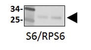
Western blot shows lysates of HeLa human cervical epithelial carcinoma cell line, HepG2 human hepatocellular carcinoma cell line, and NRK rat normal kidney cell line. PVDF membrane was probed with 0.25 µg/mL of Mouse Anti-Human/Mouse/Rat Ribosomal Protein S6 Monoclonal Antibody (Catalog # MAB5436) followed by HRP-conjugated Anti-Mouse IgG Secondary Antibody (Catalog # HAF007). A specific band was detected for Ribosomal Protein S6 at approximately 32 kDa (as indicated). This experiment was conducted under reducing conditions and using Immunoblot Buffer Group 3.
Ribosomal Protein S6/RPS6 in MM55K Mouse Cell Line.
Ribosomal Protein S6/RPS6 was detected in immersion fixed MM55K mouse kidney cell line using Mouse Anti-Human/Mouse/Rat Ribosomal Protein S6/RPS6 Monoclonal Antibody (Catalog # MAB5436) at 25 µg/mL for 3 hours at room temperature. Cells were stained using the NorthernLights™ 557-conjugated Anti-Mouse IgG Secondary Antibody (red; Catalog # NL007) and counterstained with DAPI (blue). Specific staining was localized to cytoplasm. View our protocol for Fluorescent ICC Staining of Cells on Coverslips.Applications for Ribosomal Protein S6/RPS6 Antibody (522731)
Application
Recommended Usage
Immunocytochemistry
8-25 µg/mL
Sample: Immersion fixed MM55K mouse kidney cell line
Sample: Immersion fixed MM55K mouse kidney cell line
Western Blot
0.25 µg/mL
Sample: HeLa human cervical epithelial carcinoma cell line, HepG2 human hepatocellular carcinoma cell line, and NRK rat normal kidney cells
Sample: HeLa human cervical epithelial carcinoma cell line, HepG2 human hepatocellular carcinoma cell line, and NRK rat normal kidney cells
Reviewed Applications
Read 2 reviews rated 4 using MAB5436 in the following applications:
Formulation, Preparation, and Storage
Purification
Protein A or G purified from hybridoma culture supernatant
Reconstitution
Reconstitute at 0.5 mg/mL in sterile PBS. For liquid material, refer to CoA for concentration.
Loading...
Formulation
Lyophilized from a 0.2 μm filtered solution in PBS with Trehalose. *Small pack size (SP) is supplied either lyophilized or as a 0.2 µm filtered solution in PBS.
Shipping
Lyophilized product is shipped at ambient temperature. Liquid small pack size (-SP) is shipped with polar packs. Upon receipt, store immediately at the temperature recommended below.
Stability & Storage
Use a manual defrost freezer and avoid repeated freeze-thaw cycles.
- 12 months from date of receipt, -20 to -70 °C as supplied.
- 1 month, 2 to 8 °C under sterile conditions after reconstitution.
- 6 months, -20 to -70 °C under sterile conditions after reconstitution.
Calculators
Background: Ribosomal Protein S6/RPS6
Alternate Names
RPS6
Gene Symbol
RPS6
UniProt
Additional Ribosomal Protein S6/RPS6 Products
Product Documents for Ribosomal Protein S6/RPS6 Antibody (522731)
Certificate of Analysis
To download a Certificate of Analysis, please enter a lot or batch number in the search box below.
Note: Certificate of Analysis not available for kit components.
Product Specific Notices for Ribosomal Protein S6/RPS6 Antibody (522731)
For research use only
Related Research Areas
Citations for Ribosomal Protein S6/RPS6 Antibody (522731)
 Powered by Bioz
Customer Reviews for Ribosomal Protein S6/RPS6 Antibody (522731) (2)
4 out of 5
2 Customer Ratings
Have you used Ribosomal Protein S6/RPS6 Antibody (522731)?
Submit a review and receive an Amazon gift card!
$25/€18/£15/$25CAN/¥2500 Yen for a review with an image
$10/€7/£6/$10CAN/¥1110 Yen for a review without an image
Submit a review
Customer Images
Showing
1
-
2 of
2 reviews
Showing All
Filter By:
-
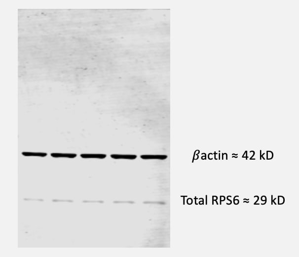
Application: Western BlotSample Tested: Bone marrow cellsSpecies: MouseVerified Customer | Posted 11/30/2022Mouse bone marrow mesenchymal stromal cells lysed with NP40 buffer for protein extraction. 10 ug of proteins were loaded in a 10% Acrylamide gel.

-
Application: Western BlotSample Tested: Skeletal muscle tissueSpecies: MouseVerified Customer | Posted 11/20/2017Western Blot: S6/RPS6 [MAB5436] - Total protein from mouse skeletal muscle tissue, separated on a 4-12% gel by SDS-PAGE, transferred to nitrocellulose membrane and blocked in 5% non-fat milk for 1h at room temperature. The membrane was probed with anti-S6/RPS6 1:2000 in non-fat milk.
There are no reviews that match your criteria.
Protocols
Find general support by application which include: protocols, troubleshooting, illustrated assays, videos and webinars.
- Appropriate Fixation of IHC/ICC Samples
- Cellular Response to Hypoxia Protocols
- ClariTSA™ Fluorophore Kits
- Detection & Visualization of Antibody Binding
- ICC Cell Smear Protocol for Suspension Cells
- ICC Immunocytochemistry Protocol Videos
- ICC for Adherent Cells
- Immunocytochemistry (ICC) Protocol
- Immunocytochemistry Troubleshooting
- Immunofluorescence of Organoids Embedded in Cultrex Basement Membrane Extract
- Immunohistochemistry (IHC) and Immunocytochemistry (ICC) Protocols
- Preparing Samples for IHC/ICC Experiments
- Preventing Non-Specific Staining (Non-Specific Binding)
- Primary Antibody Selection & Optimization
- Protocol for VisUCyte™ HRP Polymer Detection Reagent
- Protocol for the Fluorescent ICC Staining of Cell Smears - Graphic
- Protocol for the Fluorescent ICC Staining of Cultured Cells on Coverslips - Graphic
- Protocol for the Preparation and Fluorescent ICC Staining of Cells on Coverslips
- Protocol for the Preparation and Fluorescent ICC Staining of Non-adherent Cells
- Protocol for the Preparation and Fluorescent ICC Staining of Stem Cells on Coverslips
- Protocol for the Preparation of a Cell Smear for Non-adherent Cell ICC - Graphic
- R&D Systems Quality Control Western Blot Protocol
- TUNEL and Active Caspase-3 Detection by IHC/ICC Protocol
- The Importance of IHC/ICC Controls
- Troubleshooting Guide: Western Blot Figures
- Western Blot Conditions
- Western Blot Protocol
- Western Blot Protocol for Cell Lysates
- Western Blot Troubleshooting
- Western Blot Troubleshooting Guide
- View all Protocols, Troubleshooting, Illustrated assays and Webinars
Loading...
Associated Pathways

