Signal transduction and activator of transcription 5 (STAT5) is a member of the Jak/STAT signal transduction pathway and is activated by a variety of cytokines (IL22, IL6, IFN-α). STAT5 has two isoforms (A and B) that share 93% amino acid identity and bind the DNA consensus site TTCN3GAA. STAT5 mediates cytokine signaling by acting as a signal transducer in the cytoplasm and, upon phosphorylation, translocates to the nucleus and activates transcription of specific genes. STAT5 is involved in a wide array of biological processes ranging from regulating apoptosis to adult mammary gland proliferation, differentiation and survival.

Loading...
Key Product Details
Validated by
Knockout/Knockdown, Biological Validation
Species Reactivity
Validated:
Human, Mouse
Cited:
Human, Mouse
Applications
Validated:
Knockout Validated, Western Blot, Intracellular Staining by Flow Cytometry, Immunocytochemistry, Simple Western, Immunoprecipitation, CyTOF-ready
Cited:
Immunohistochemistry, Western Blot, Immunocytochemistry, Immunoprecipitation, Chromatin Immunoprecipitation (ChIP)
Label
Unconjugated
Antibody Source
Polyclonal Rabbit IgG
Loading...
Product Specifications
Immunogen
Human STAT5b synthetic peptide
aa 777-787
aa 777-787
Specificity
Detects human and mouse STAT5b.
Clonality
Polyclonal
Host
Rabbit
Isotype
IgG
Scientific Data Images for STAT5b Antibody
Detection of Human and Mouse STAT5b by Western Blot.
Western Blot shows lysates of HeLa human cervical epithelial carcinoma cell line, Daudi human Burkitt's lymphoma cell line, NIH‑3T3 mouse embryonic fibroblast cell line and M1 mouse myeloid leukemia cell line. PVDF membrane was probed with 0.2 µg/ml of Rabbit Anti-Human/Mouse STAT5b Antigen Affinity-purified Polyclonal Antibody (Catalog # AF1584) followed by HRP-conjugated Anti-Rabbit IgG Secondary Antibody (Catalog # HAF008). A specific band was detected for STAT5b at approximately 90 kDa (as indicated). This experiment was conducted under reducing conditions and using Western Blot Buffer Group 1.
STAT5b in HepG2 Human Cell Line.
STAT5b was detected in immersion fixed HepG2 human hepatocellular carcinoma cell line using Rabbit Anti-Human/Mouse STAT5b Antigen Affinity-purified Polyclonal Antibody (Catalog # AF1584) at 5 µg/mL for 3 hours at room temperature. Cells were stained using the NorthernLights™ 557-conjugated Anti-Rabbit IgG Secondary Antibody (red; NL004) and counterstained with DAPI(blue). Specific staining was localized to cytoplasm and nucleus. View our protocol for Fluorescent ICC Staining of Cells on Coverslips.
Immunoprecipitation of Human and Mouse STAT5b.
HeLa human cervical epithelial carcinoma and M1 mouse myeloid leukemia cell line were untreated (-) or activated (+) with 100 ng/mL Recombinant Human IFN-gamma (285-IF) for 15 minutes. STAT5b was immunoprecipitated from lysates of 5 x 10⁶cells following incubation with 2 µg Rabbit Anti-Human/Mouse STAT5b Antigen Affinity-purified Polyclonal Antibody (Catalog # AF1584) for 1 hour at room temperature. STAT5b-antibody complexes were absorbed using Protein A sepharose (Invitrogen, Catalog # 10-1041). Immunoprecipitated STAT5b was detected by Western blot using 1 µg/mL Human Phospho-STAT5a/b (Y699) Antigen Affinity-purified Polyclonal Antibody (AF4190). View our recommended buffer recipes for immunoprecipitation.
Detection of STAT5b in Jurkat Human Cell Line by Flow Cytometry.
Jurkat human acute T cell leukemia cell line was stained with Rabbit Anti-Human/Mouse STAT5b Antigen Affinity-purified Polyclonal Antibody (Catalog # AF1584, filled histogram) or control antibody (AB-105-C, open histogram), followed by Allophycocyanin-conjugated Anti-Rabbit IgG Secondary Antibody (Catalog # F0111). To facilitate intracellular staining, cells were fixed with paraformaldehyde and permeabilized with methanol.
Detection of Human and Mouse STAT5b by Simple WesternTM.
Simple Western lane view shows lysates of HeLa human cervical epithelial carcinoma cell line and NIH‑3T3 mouse embryonic fibroblast cell line, loaded at 0.2 mg/mL. A specific band was detected for STAT5b at approximately 92 kDa (as indicated) using 10 µg/mL of Rabbit Anti-Human/Mouse STAT5b Antigen Affinity-purified Polyclonal Antibody (Catalog # AF1584). This experiment was conducted under reducing conditions and using the 12-230 kDa separation system.Non-specific interaction with the 230 kDa Simple Western standard may be seen with this antibody.
Western Blot Shows Human STAT5b Specificity by Using Knockout Cell Line.
Western blot shows lysates of HeLa human cervical epithelial carcinoma parental cell line and STAT5b knockout HeLa cell line (KO). PVDF membrane was probed with 0.2 µg/mL of Rabbit Anti-Human/Mouse STAT5b Antigen Affinity-purified Polyclonal Antibody (Catalog # AF1584) followed by HRP-conjugated Anti-Rabbit IgG Secondary Antibody (HAF008). A specific band was detected for STAT5b at approximately 90 kDa (as indicated) in the parental HeLa cell line, but is not detectable in knockout HeLa cell line. GAPDH (AF5718) is shown as a loading control. This experiment was conducted under reducing conditions and using Immunoblot Buffer Group 1.
Western Blot Shows Human STAT5b Specificity Using Knockout Cell Line.
Western blot shows lysates of HAP1 human near-haploid cell line and STAT5b knockout HAP1 cell line (KO). Nitrocellulose membrane was probed with 0.2 µg/mL of Rabbit Anti-Human/Mouse STAT5b Antigen Affinity-purified Polyclonal Antibody (Catalog # AF1584) followed by HRP-conjugated Anti-Rabbit IgG Secondary Antibody. A specific band was detected for STAT5b at approximately 85 kDa (as indicated) in the parental HAP1 cell line, but is not detectable in knockout HAP1 cell line. The Ponceau stained transfer of the blot is shown. This experiment was conducted under reducing conditions. Image, protocol, and testing courtesy of YCharOS Inc. See ycharos.com for additional details.
Detection of Stat5b by Immunoprecipitation.
Immunoprecipitation was performed on cell lysate of HAP1 human near-haploid cell line using 2.0 μg of Rabbit Anti-Human Stat5b Polyclonal Antibody (Catalog # AF1584) pre-coupled to protein G or protein A beads. Immunoprecipitated Stat5b was detected with a Mouse Anti-Stat5b antibody. The Ponceau stained transfers of each blot are shown. SM=10% starting material; UB=10% unbound fraction; IP=immunoprecipitated. Image, protocol, and testing courtesy of YCharOS Inc. (ycharos.com).Applications for STAT5b Antibody
Application
Recommended Usage
CyTOF-ready
Ready to be labeled using established conjugation methods. No BSA or other carrier proteins that could interfere with conjugation.
Immunocytochemistry
5-15 µg/mL
Sample: Immersion fixed HepG2 human hepatocellular carcinoma cell line
Sample: Immersion fixed HepG2 human hepatocellular carcinoma cell line
Immunoprecipitation
1-5 µg/500 µg cell lysate
Sample: HeLa human cervical epithelial carcinoma cell line and M1 mouse myeloid leukemia cell line activated with Recombinant Human IFN‑ gamma (Catalog # 285-IF), see our available Western blot detection antibodies. Cell lysate of HAP1 human near-haploid cell line
Sample: HeLa human cervical epithelial carcinoma cell line and M1 mouse myeloid leukemia cell line activated with Recombinant Human IFN‑ gamma (Catalog # 285-IF), see our available Western blot detection antibodies. Cell lysate of HAP1 human near-haploid cell line
Intracellular Staining by Flow Cytometry
2.5 µg/106 cells
Sample: Jurkat human acute T cell leukemia cell line fixed with paraformaldehyde and permeabilized with methanol
Sample: Jurkat human acute T cell leukemia cell line fixed with paraformaldehyde and permeabilized with methanol
Knockout Validated
A) STAT5b
is specifically detected in HeLa human cervical epithelial carcinoma parental cell line but is not detectable in
STAT5b knockout HeLa cell line. B) STAT5b is specifically detected in the parental HAP1 cell line, but is not detectable in knockout HAP1 cell line.
Simple Western
10 µg/mL
Sample: HeLa human cervical epithelial carcinoma cell line and NIH‑3T3 mouse embryonic fibroblast cell line
Sample: HeLa human cervical epithelial carcinoma cell line and NIH‑3T3 mouse embryonic fibroblast cell line
Western Blot
0.2 µg/mL
Sample: HeLa human cervical epithelial carcinoma cell line, Daudi human Burkitt's lymphoma cell line, NIH‑3T3 mouse embryonic fibroblast cell line, and M1 mouse myeloid leukemia cell line
Sample: HeLa human cervical epithelial carcinoma cell line, Daudi human Burkitt's lymphoma cell line, NIH‑3T3 mouse embryonic fibroblast cell line, and M1 mouse myeloid leukemia cell line
Reviewed Applications
Read 3 reviews rated 4.7 using AF1584 in the following applications:
Flow Cytometry Panel Builder
Bio-Techne Knows Flow Cytometry
Save time and reduce costly mistakes by quickly finding compatible reagents using the Panel Builder Tool.
Advanced Features
- Spectra Viewer - Custom analysis of spectra from multiple fluorochromes
- Spillover Popups - Visualize the spectra of individual fluorochromes
- Antigen Density Selector - Match fluorochrome brightness with antigen density

Formulation, Preparation, and Storage
Purification
Antigen Affinity-purified
Reconstitution
Reconstitute at 0.2 mg/mL in sterile PBS. For liquid material, refer to CoA for concentration.
Loading...
Formulation
Lyophilized from a 0.2 μm filtered solution in PBS with Trehalose. See Certificate of Analysis for details.
*Small pack size (-SP) is supplied either lyophilized or as a 0.2 µm filtered solution in PBS.
*Small pack size (-SP) is supplied either lyophilized or as a 0.2 µm filtered solution in PBS.
Shipping
Lyophilized product is shipped at ambient temperature. Liquid small pack size (-SP) is shipped with polar packs. Upon receipt, store immediately at the temperature recommended below.
Stability & Storage
Use a manual defrost freezer and avoid repeated freeze-thaw cycles.
- 12 months from date of receipt, -20 to -70 °C as supplied.
- 1 month, 2 to 8 °C under sterile conditions after reconstitution.
- 6 months, -20 to -70 °C under sterile conditions after reconstitution.
Calculators
Background: STAT5b
Long Name
Signal Transducer and Activator of Transcription 5b
Alternate Names
signal transducer and activator of transcription 5B, STAT5, transcription factor STAT5B
Gene Symbol
STAT5B
Additional STAT5b Products
Product Documents for STAT5b Antibody
Certificate of Analysis
To download a Certificate of Analysis, please enter a lot or batch number in the search box below.
Note: Certificate of Analysis not available for kit components.
Product Specific Notices for STAT5b Antibody
For research use only
Citations for STAT5b Antibody
 Powered by Bioz
Customer Reviews for STAT5b Antibody (3)
4.7 out of 5
3 Customer Ratings
Have you used STAT5b Antibody?
Submit a review and receive an Amazon gift card!
$25/€18/£15/$25CAN/¥2500 Yen for a review with an image
$10/€7/£6/$10CAN/¥1110 Yen for a review without an image
Submit a review
Customer Images
Showing
1
-
3 of
3 reviews
Showing All
Filter By:
-
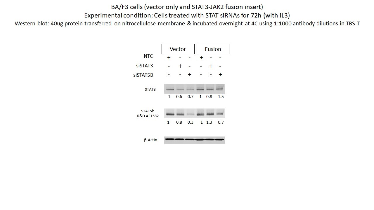
Application: Western BlotSample Tested: BaF3 mouse pro-B cell lineSpecies: MouseVerified Customer | Posted 09/13/2021BA/F3 cells (vector only and STAT3-JAK2 fusion insert) Experimental condition: Cells treated with STAT siRNAs for 72h (with iL3) Western blot: 40ug protein transferred on nitrocellulose membrane & incubated overnight at 4C using 1:1000 antibody dilutions in TBS-T

-
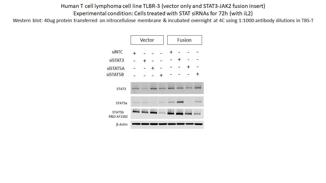
Application: Western BlotSample Tested: TLBR3 human T-cell lymphoma cellsSpecies: HumanVerified Customer | Posted 09/13/2021
-
Application: Western BlotSample Tested: See PMID 22729867Species: HumanVerified Customer | Posted 01/05/2015
There are no reviews that match your criteria.
Protocols
Find general support by application which include: protocols, troubleshooting, illustrated assays, videos and webinars.
- 7-Amino Actinomycin D (7-AAD) Cell Viability Flow Cytometry Protocol
- Appropriate Fixation of IHC/ICC Samples
- Cellular Response to Hypoxia Protocols
- ClariTSA™ Fluorophore Kits
- Detection & Visualization of Antibody Binding
- Extracellular Membrane Flow Cytometry Protocol
- Flow Cytometry Protocol for Cell Surface Markers
- Flow Cytometry Protocol for Staining Membrane Associated Proteins
- Flow Cytometry Staining Protocols
- Flow Cytometry Troubleshooting Guide
- ICC Cell Smear Protocol for Suspension Cells
- ICC Immunocytochemistry Protocol Videos
- ICC for Adherent Cells
- Immunocytochemistry (ICC) Protocol
- Immunocytochemistry Troubleshooting
- Immunofluorescence of Organoids Embedded in Cultrex Basement Membrane Extract
- Immunohistochemistry (IHC) and Immunocytochemistry (ICC) Protocols
- Immunoprecipitation Protocol
- Intracellular Flow Cytometry Protocol Using Alcohol (Methanol)
- Intracellular Flow Cytometry Protocol Using Detergents
- Intracellular Nuclear Staining Flow Cytometry Protocol Using Detergents
- Intracellular Staining Flow Cytometry Protocol Using Alcohol Permeabilization
- Intracellular Staining Flow Cytometry Protocol Using Detergents to Permeabilize Cells
- Preparing Samples for IHC/ICC Experiments
- Preventing Non-Specific Staining (Non-Specific Binding)
- Primary Antibody Selection & Optimization
- Propidium Iodide Cell Viability Flow Cytometry Protocol
- Protocol for Liperfluo
- Protocol for VisUCyte™ HRP Polymer Detection Reagent
- Protocol for the Characterization of Human Th22 Cells
- Protocol for the Characterization of Human Th9 Cells
- Protocol for the Fluorescent ICC Staining of Cell Smears - Graphic
- Protocol for the Fluorescent ICC Staining of Cultured Cells on Coverslips - Graphic
- Protocol for the Preparation and Fluorescent ICC Staining of Cells on Coverslips
- Protocol for the Preparation and Fluorescent ICC Staining of Non-adherent Cells
- Protocol for the Preparation and Fluorescent ICC Staining of Stem Cells on Coverslips
- Protocol for the Preparation of a Cell Smear for Non-adherent Cell ICC - Graphic
- Protocol: Annexin V and PI Staining by Flow Cytometry
- Protocol: Annexin V and PI Staining for Apoptosis by Flow Cytometry
- R&D Systems Quality Control Western Blot Protocol
- TUNEL and Active Caspase-3 Detection by IHC/ICC Protocol
- The Importance of IHC/ICC Controls
- Troubleshooting Guide: Fluorokine Flow Cytometry Kits
- Troubleshooting Guide: Western Blot Figures
- Western Blot Conditions
- Western Blot Protocol
- Western Blot Protocol for Cell Lysates
- Western Blot Troubleshooting
- Western Blot Troubleshooting Guide
- View all Protocols, Troubleshooting, Illustrated assays and Webinars
Loading...
Associated Pathways


IL-15 Signaling Pathways
 IL-21 Signaling Pathways
 Jak/STAT Signaling Pathway
 Th1 Differentiation Pathway
 Th17 Differentiation Pathway
