OX40 Ligand (OX40L), also known as gp34, is a type II transmembrane glycoprotein belonging to the TNF superfamily. Human OX40L cDNA encodes a 183 amino acids (aa) polypeptide with an amino-terminal cytoplasmic domain (aa 1¬23) and a carboxy¬terminal extracellular domain (aa 51-183). It shares 46% aa sequence identity with the mouse counterpart. OX40L is expressed on the surface of activated B cells, T cells, dendritic cells and endothelial cells. Similarly to other TNF superfamily members, membrane-bound OX40 Ligand exists as a homotrimer. OX40L binds to OX40 (CD134), a member of the TNF receptor superfamily that is expressed predominantly on activated CD4+ T cells. OX40 Ligand is one of the co¬stimulatory molecules in the immune system that includes B7, CD40 Ligand, CD30 Ligand, CD27 Ligand and 4¬1BB Ligand. Because OX40 appears as a late activation-induced T cell surface antigen, it has been speculated that the major function of OX40-OX40L interaction is to transmit a late co-stimulatory signal to promote the survival and proliferation of activated CD4+ T cells and prolong the immune response. Engagement of OX40 on activated T cells in situ in tumors has been shown to augment immune responses and subsequent tumor regression.

Key Product Details
Species Reactivity
Human
Applications
Immunohistochemistry
Label
Unconjugated
Antibody Source
Monoclonal Mouse IgG2B Clone # 1034083
Loading...
Product Specifications
Immunogen
Mouse myeloma cell line NS0-derived human OX40 Ligand/TNFRSF4
Gln51-Leu183
Accession # Q6FGS4
Gln51-Leu183
Accession # Q6FGS4
Specificity
Detects human OX40 Ligand/TNFRSF4 in direct ELISAs.
Clonality
Monoclonal
Host
Mouse
Isotype
IgG2B
Scientific Data Images for Human OX40 Ligand/TNFSF4 Antibody (1034083)
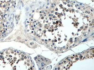
OX40 Ligand/TNFRSF4 in Human Testis.
OX40 Ligand/TNFRSF4 was detected in immersion fixed paraffin-embedded sections of human testis using Mouse Anti-Human OX40 Ligand/TNFRSF4 Monoclonal Antibody (Catalog # MAB10656) at 5 µg/mL for 1 hour at room temperature followed by incubation with the Anti-Mouse IgG VisUCyte™ HRP Polymer Antibody (VC001). Before incubation with the primary antibody, tissue was subjected to heat-induced epitope retrieval using Antigen Retrieval Reagent-Basic (CTS013). Tissue was stained using DAB (brown) and counterstained with hematoxylin (blue). Specific staining was localized to cell surface in sperm cells. Staining was performed using our protocol for IHC Staining with VisUCyte HRP Polymer Detection Reagents.Applications for Human OX40 Ligand/TNFSF4 Antibody (1034083)
Application
Recommended Usage
Immunohistochemistry
5-25 µg/mL
Sample: Immersion fixed paraffin-embedded sections of human testis
Sample: Immersion fixed paraffin-embedded sections of human testis
Reviewed Applications
Read 1 review rated 5 using MAB10656 in the following applications:
Formulation, Preparation, and Storage
Purification
Protein A or G purified from cell culture supernatant
Reconstitution
Reconstitute at 0.5 mg/mL in sterile PBS. For liquid material, refer to CoA for concentration.
Loading...
Formulation
Lyophilized from a 0.2 μm filtered solution in PBS with Trehalose. *Small pack size (SP) is supplied either lyophilized or as a 0.2 µm filtered solution in PBS.
Shipping
Lyophilized product is shipped at ambient temperature. Liquid small pack size (-SP) is shipped with polar packs. Upon receipt, store immediately at the temperature recommended below.
Stability & Storage
Use a manual defrost freezer and avoid repeated freeze-thaw cycles.
- 12 months from date of receipt, -20 to -70 °C as supplied.
- 1 month, 2 to 8 °C under sterile conditions after reconstitution.
- 6 months, -20 to -70 °C under sterile conditions after reconstitution.
Calculators
Background: OX40 Ligand/TNFSF4
References
- Godfrey, W.R. et al. (1994) J. Exp. Med. 180:757
- Baum, P.R. et al. (1994) EMBO J. 13:3992.
- AlShamkhani, A. et al. (1997) J. Biol. Chem. 272:5275.
- Kjaergaard, J. et al. (2000) Cancer Res. 60:5514.
Alternate Names
CD252, gp34, TNFSF4
Gene Symbol
TNFSF4
UniProt
Additional OX40 Ligand/TNFSF4 Products
Product Documents for Human OX40 Ligand/TNFSF4 Antibody (1034083)
Certificate of Analysis
To download a Certificate of Analysis, please enter a lot or batch number in the search box below.
Note: Certificate of Analysis not available for kit components.
Product Specific Notices for Human OX40 Ligand/TNFSF4 Antibody (1034083)
For research use only
Customer Reviews for Human OX40 Ligand/TNFSF4 Antibody (1034083) (1)
5 out of 5
1 Customer Rating
Have you used Human OX40 Ligand/TNFSF4 Antibody (1034083)?
Submit a review and receive an Amazon gift card!
$25/€18/£15/$25CAN/¥2500 Yen for a review with an image
$10/€7/£6/$10CAN/¥1110 Yen for a review without an image
Submit a review
Customer Images
Showing
1
-
1 of
1 review
Showing All
Filter By:
-
Application: ImmunohistochemistrySample Tested: Testis tissueSpecies: HumanVerified Customer | Posted 03/22/2022

There are no reviews that match your criteria.
Protocols
Find general support by application which include: protocols, troubleshooting, illustrated assays, videos and webinars.
- Antigen Retrieval Protocol (PIER)
- Antigen Retrieval for Frozen Sections Protocol
- Appropriate Fixation of IHC/ICC Samples
- Cellular Response to Hypoxia Protocols
- Chromogenic IHC Staining of Formalin-Fixed Paraffin-Embedded (FFPE) Tissue Protocol
- Chromogenic Immunohistochemistry Staining of Frozen Tissue
- ClariTSA™ Fluorophore Kits
- Detection & Visualization of Antibody Binding
- Fluorescent IHC Staining of Frozen Tissue Protocol
- Graphic Protocol for Heat-induced Epitope Retrieval
- Graphic Protocol for the Preparation and Fluorescent IHC Staining of Frozen Tissue Sections
- Graphic Protocol for the Preparation and Fluorescent IHC Staining of Paraffin-embedded Tissue Sections
- Graphic Protocol for the Preparation of Gelatin-coated Slides for Histological Tissue Sections
- IHC Sample Preparation (Frozen sections vs Paraffin)
- Immunofluorescent IHC Staining of Formalin-Fixed Paraffin-Embedded (FFPE) Tissue Protocol
- Immunohistochemistry (IHC) and Immunocytochemistry (ICC) Protocols
- Immunohistochemistry Frozen Troubleshooting
- Immunohistochemistry Paraffin Troubleshooting
- Preparing Samples for IHC/ICC Experiments
- Preventing Non-Specific Staining (Non-Specific Binding)
- Primary Antibody Selection & Optimization
- Protocol for Heat-Induced Epitope Retrieval (HIER)
- Protocol for Making a 4% Formaldehyde Solution in PBS
- Protocol for VisUCyte™ HRP Polymer Detection Reagent
- Protocol for the Preparation & Fixation of Cells on Coverslips
- Protocol for the Preparation and Chromogenic IHC Staining of Frozen Tissue Sections
- Protocol for the Preparation and Chromogenic IHC Staining of Frozen Tissue Sections - Graphic
- Protocol for the Preparation and Chromogenic IHC Staining of Paraffin-embedded Tissue Sections
- Protocol for the Preparation and Chromogenic IHC Staining of Paraffin-embedded Tissue Sections - Graphic
- Protocol for the Preparation and Fluorescent IHC Staining of Frozen Tissue Sections
- Protocol for the Preparation and Fluorescent IHC Staining of Paraffin-embedded Tissue Sections
- Protocol for the Preparation of Gelatin-coated Slides for Histological Tissue Sections
- TUNEL and Active Caspase-3 Detection by IHC/ICC Protocol
- The Importance of IHC/ICC Controls
- Troubleshooting Guide: Immunohistochemistry
- View all Protocols, Troubleshooting, Illustrated assays and Webinars
Loading...
Associated Pathways

