
Class IA phosphoinositide 3-kinases (PI 3-kinases) are a family of p85/p110 heterodimeric lipid kinases that function as signal transducers downstream of growth factor receptors, mainly receptor tyrosine kinases. Mammals express three class IA catalytic subunits: p110 alpha, p110 beta, and p110 delta. The p110 delta subunit is expressed predominantly in leukocytes, indicating that it may play a role in immune signaling.
Human PI 3-Kinase p110 delta Antibody (275144)
R&D Systems | Catalog # MAB2687


Scientific images may be resized, cropped, or adjusted for brightness or contrast for presentation purposes. These updates are limited to presentation and do not affect the underlying scientific interpretation of the data. Where available, additional source data may be provided upon request.
Key Product Details
Validated by
Biological Validation
Species Reactivity
Validated:
Human
Cited:
Human, Mouse
Applications
Validated:
Western Blot
Cited:
Western Blot
Label
Unconjugated
Antibody Source
Monoclonal Mouse IgG2B Clone # 275144
Loading...
Product Specifications
Immunogen
E. coli-derived recombinant human PI 3-Kinase p110 delta
Cys391-Phe542
Accession # O00329
Cys391-Phe542
Accession # O00329
Specificity
Detects endogenous human PI 3-kinase p110 delta in Western blots.
Clonality
Monoclonal
Host
Mouse
Isotype
IgG2B
Scientific Data Images for Human PI 3-Kinase p110 delta Antibody (275144)
Detection of Human PI 3-Kinase p110 delta by Western Blot.
Western blot shows lysates of Raji human Burkitt's lymphoma cell line, Jurkat human acute T cell leukemia cell line, and K562 human chronic myelogenous leukemia cell line. PVDF membrane was probed with 1 µg/mL of Human PI 3-Kinase p110d Monoclonal Antibody (Catalog # MAB2687) followed by HRP-conjugated Anti-Mouse IgG Secondary Antibody (Catalog # HAF007). A specific band was detected for PI 3-Kinase p110 delta at approximately 110 kDa (as indicated). This experiment was conducted using Immunoblot Buffer Group 1.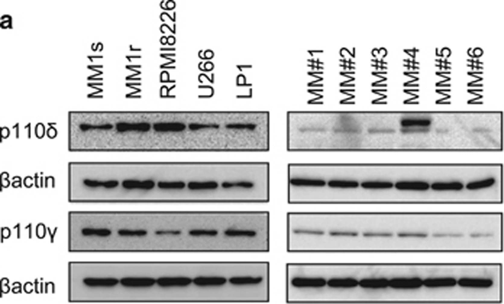
Detection of Human PI 3-Kinase p110 delta by Western Blot
Inhibition of PI3K delta and PI3K gamma subunits in MM proliferation. (a) Western blot analysis of PI3K delta and PI3K gamma subunits in MM cell lines and MM primary cells. Blots were probed with beta -actin to show sample loading. (b) MM primary cells were treated with idelalisib (1 μm), CZC24832 (1 μm) and duvelisib (1 μm) for 72 h and then assessed for cell viability using CellTiter Glo. (c) MM primary cells were treated with idelalisib (1 μm), CZC24832 (1 μm) and duvelisib (1 μm) for 72 h and then assessed for apoptosis by PI/Annexin V staining (n=4). Image collected and cropped by CiteAb from the following publication (https://www.nature.com/articles/bcj201716), licensed under a CC-BY license. Not internally tested by R&D Systems.Applications for Human PI 3-Kinase p110 delta Antibody (275144)
Application
Recommended Usage
Western Blot
1 µg/mL
Sample: Raji human Burkitt's lymphoma cell line, Jurkat human acute T cell leukemia cell line, and K562 human chronic myelogenous leukemia cell line
Sample: Raji human Burkitt's lymphoma cell line, Jurkat human acute T cell leukemia cell line, and K562 human chronic myelogenous leukemia cell line
Reviewed Applications
Read 4 reviews rated 4.3 using MAB2687 in the following applications:
Formulation, Preparation, and Storage
Purification
Protein A or G purified from hybridoma culture supernatant
Reconstitution
Reconstitute at 0.5 mg/mL in sterile PBS. For liquid material, refer to CoA for concentration.
Loading...
Formulation
Lyophilized from a 0.2 μm filtered solution in PBS with Trehalose. *Small pack size (SP) is supplied either lyophilized or as a 0.2 µm filtered solution in PBS.
Shipping
Lyophilized product is shipped at ambient temperature. Liquid small pack size (-SP) is shipped with polar packs. Upon receipt, store immediately at the temperature recommended below.
Stability & Storage
Use a manual defrost freezer and avoid repeated freeze-thaw cycles.
- 12 months from date of receipt, -20 to -70 °C as supplied.
- 1 month, 2 to 8 °C under sterile conditions after reconstitution.
- 6 months, -20 to -70 °C under sterile conditions after reconstitution.
Calculators
Background: PI 3-Kinase p110 delta
Long Name
Protein Tyrosine Phosphatase 1D
Alternate Names
PI 3Kinase p110 delta, PIK3CD
Entrez Gene IDs
5293 (Human)
Gene Symbol
PIK3CD
UniProt
Additional PI 3-Kinase p110 delta Products
Product Documents for Human PI 3-Kinase p110 delta Antibody (275144)
Certificate of Analysis
To download a Certificate of Analysis, please enter a lot or batch number in the search box below.
Note: Certificate of Analysis not available for kit components.
Product Specific Notices for Human PI 3-Kinase p110 delta Antibody (275144)
For research use only
Citations for Human PI 3-Kinase p110 delta Antibody (275144)
 Powered by Bioz
Customer Reviews for Human PI 3-Kinase p110 delta Antibody (275144) (4)
4.3 out of 5
4 Customer Ratings
Have you used Human PI 3-Kinase p110 delta Antibody (275144)?
Submit a review and receive an Amazon gift card!
$25/€18/£15/$25CAN/¥2500 for a review with an image
$10/€7/£6/$10CAN/¥1110 for a review without an image
Submit a review
Customer Images

Showing
1
-
4 of
4 reviews
Showing All
Filter By:
-
Application: MicroarraysSample Tested: EDTA PlasmaSpecies: HumanVerified Customer | Posted 01/14/2021

-
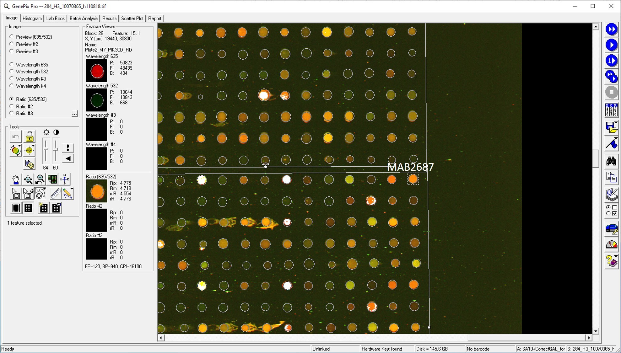
Application: MicroarraySample Tested: EDTA PlasmaSpecies: HumanVerified Customer | Posted 02/07/2020Antibody was printed on custom arrays and incubated with fluorescently labeled human EDTA plasma
-
Application: MicroarraysSample Tested: EDTA PlasmaSpecies: HumanVerified Customer | Posted 11/14/2018
-
Application: ELISASample Tested: Serum and PlasmaSpecies: Human and MouseVerified Customer | Posted 11/07/2018
There are no reviews that match your criteria.
Protocols
Find general support by application which include: protocols, troubleshooting, illustrated assays, videos and webinars.
- Cellular Response to Hypoxia Protocols
- R&D Systems Quality Control Western Blot Protocol
- Troubleshooting Guide: Western Blot Figures
- Western Blot Conditions
- Western Blot Protocol
- Western Blot Protocol for Cell Lysates
- Western Blot Troubleshooting
- Western Blot Troubleshooting Guide
- View all Protocols, Troubleshooting, Illustrated assays and Webinars
Loading...
Associated Pathways


Common Cytokine Receptor Gamma-Chain Family Signaling Pathways
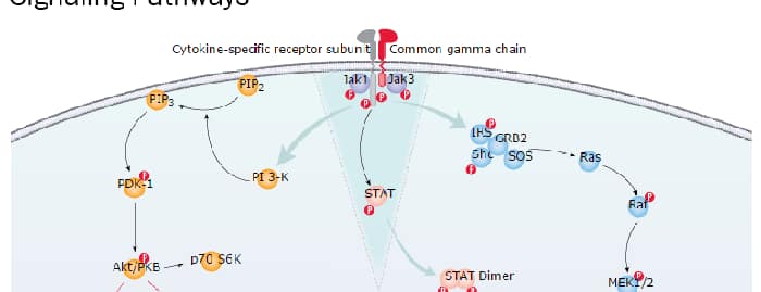 IL-2 Signaling Pathways
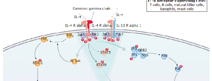
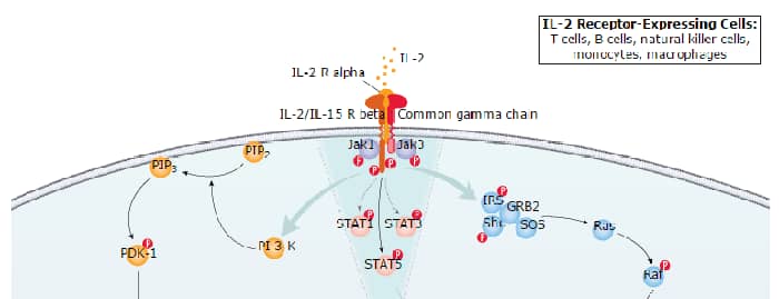 IL-4 Signaling Pathways
 IL-7 Signaling Pathways
 IL-9 Signaling Pathways
 IL-15 Signaling Pathways
 IL-21 Signaling Pathways
 mTOR Signaling Pathway
 Notch Signaling Pathways
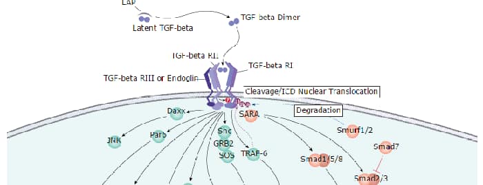
 TGF-beta Signaling Pathways
 VEGF - VEGF R2 Signaling Pathways
