Human prostate-specific membrane antigen (PSMA), a tumor marker in prostate cancer encoded by the FOLH1 gene, is a type II transmembrane zinc metallopeptidase that is most highly expressed in the nervous system, prostate, kidney, and small intestine (1, 2). The enzyme is also known as glutamate carboxypeptidase II (GCPII), folate hydrolase 1, folypoly-gamma-glutamate carboxypeptidase (FGCP), and N-acetylated-alpha-linked acidic dipeptidase I (NAALADase I). In the brain, PSMA hydrolyzes the neurotransmitter N-acetyl-Asp-Glu to produce glutamate, another neurotransmitter. Inhibition of brain PSMA activity is considered to be a promising approach for the treatment of neurological disorders associated with glutamate excitotoxicity, such as stroke, chronic pain, and amyotrophic lateral sclerosis (3). Intestinal PSMA hydrolyzes folylpoly-gamma -glutamates, facilitating the uptake of folate (4).
Human PSMA/FOLH1/NAALADase I Antibody (460420)
R&D Systems | Catalog # MAB4234

Key Product Details
Species Reactivity
Validated:
Human
Cited:
Human, Mouse, Rat, Primate - Macaca mulatta (Rhesus Macaque)
Applications
Validated:
Immunohistochemistry, Western Blot, Flow Cytometry, Immunocytochemistry, CyTOF-ready
Cited:
Immunohistochemistry, Western Blot, Immunocytochemistry, Simple Western
Label
Unconjugated
Antibody Source
Monoclonal Mouse IgG2A Clone # 460420
Loading...
Product Specifications
Immunogen
Chinese hamster ovary cell line CHO-derived recombinant human PSMA/FOLH1/NAALADase I
Lys44-Ala750
Accession # Q04609
Lys44-Ala750
Accession # Q04609
Specificity
Detects human PSMA/FOLH1/NAALADase I in direct ELISA and Western blot. In direct ELISAs, less than 10% cross-reactivity with recombinant human (rh) NAALADase-like 2 and no cross-reactivity with rhNAALADase-like 1, rhNAALADase-like 3, recombinant mouse (rm) NAALADase I, or rmNAALAase-like 2 is observed.
Clonality
Monoclonal
Host
Mouse
Isotype
IgG2A
Scientific Data Images for Human PSMA/FOLH1/NAALADase I Antibody (460420)
Detection of Human NAALADase I by Western Blot.
Western blot shows lysates of human prostate tissue. PVDF membrane was probed with 2 µg/mL of Mouse Anti-Human PSMA/FOLH1/NAALADase I Monoclonal Antibody (Catalog # MAB4234) followed by HRP-conjugated Anti-Mouse IgG Secondary Antibody (Catalog # HAF007). A specific band was detected for NAALADase I at approximately 120 kDa (as indicated). This experiment was conducted under reducing conditions and using Immunoblot Buffer Group 1.
Detection of NAALADase I in LNCaP Human Cell Line by Flow Cytometry.
LNCaP human prostate cancer cell line was stained with Mouse Anti-Human PSMA/FOLH1/NAALADase I Monoclonal Antibody (Catalog # MAB4234, filled histogram) or isotype control antibody (Catalog # MAB003, open histogram), followed by Phycoerythrin-conjugated Anti-Mouse IgG Secondary Antibody (Catalog # F0102B).
NAALADase I in PC‑3 Human Cell Line.
NAALADase I was detected in immersion fixed PC-3 human prostate cancer cell line using Mouse Anti-Human PSMA/FOLH1/ NAALADase I Monoclonal Antibody (Catalog # MAB4234) at 10 µg/mL for 3 hours at room temperature. Cells were stained using the NorthernLights™ 557-conjugated Anti-Mouse IgG Secondary Antibody (red; Catalog # NL007) and counterstained with DAPI (blue). Specific staining was localized to cell surfaces and cytoplasm. View our protocol for Fluorescent ICC Staining of Cells on Coverslips.
NAALADase I in Human Prostate.
NAALADase I was detected in formalin fixed paraffin-embedded sections of human prostate using Mouse Anti-Human PSMA/FOLH1/NAALADase I Monoclonal Antibody (Catalog # MAB4234) at 1 µg/mL overnight at 4 °C. Tissue was stained using the Anti-Mouse HRP-DAB Cell & Tissue Staining Kit (brown; Catalog # CTS002) and counterstained with hematoxylin (blue). Specific staining was localized to glandular epithelial cells. View our protocol for Chromogenic IHC Staining of Paraffin-embedded Tissue Sections.Applications for Human PSMA/FOLH1/NAALADase I Antibody (460420)
Application
Recommended Usage
CyTOF-ready
Ready to be labeled using established conjugation methods. No BSA or other carrier proteins that could interfere with conjugation.
Flow Cytometry
2.5 µg/106 cells
Sample: LNCaP human prostate cancer cell line
Sample: LNCaP human prostate cancer cell line
Immunocytochemistry
8-25 µg/mL
Sample: Immersion fixed PC‑3 human prostate cancer cell line
Sample: Immersion fixed PC‑3 human prostate cancer cell line
Immunohistochemistry
8-25 µg/mL
Sample: Immersion fixed paraffin-embedded sections of human prostate
Sample: Immersion fixed paraffin-embedded sections of human prostate
Western Blot
2 µg/mL
Sample: Human prostate tissue
Sample: Human prostate tissue
Reviewed Applications
Read 2 reviews rated 5 using MAB4234 in the following applications:
Flow Cytometry Panel Builder
Bio-Techne Knows Flow Cytometry
Save time and reduce costly mistakes by quickly finding compatible reagents using the Panel Builder Tool.
Advanced Features
- Spectra Viewer - Custom analysis of spectra from multiple fluorochromes
- Spillover Popups - Visualize the spectra of individual fluorochromes
- Antigen Density Selector - Match fluorochrome brightness with antigen density

Formulation, Preparation, and Storage
Purification
Protein A or G purified from hybridoma culture supernatant
Reconstitution
Reconstitute at 0.5 mg/mL in sterile PBS. For liquid material, refer to CoA for concentration.
Loading...
Formulation
Lyophilized from a 0.2 μm filtered solution in PBS with Trehalose. *Small pack size (SP) is supplied either lyophilized or as a 0.2 µm filtered solution in PBS.
Shipping
Lyophilized product is shipped at ambient temperature. Liquid small pack size (-SP) is shipped with polar packs. Upon receipt, store immediately at the temperature recommended below.
Stability & Storage
Use a manual defrost freezer and avoid repeated freeze-thaw cycles.
- 12 months from date of receipt, -20 to -70 °C as supplied.
- 1 month, 2 to 8 °C under sterile conditions after reconstitution.
- 6 months, -20 to -70 °C under sterile conditions after reconstitution.
Calculators
Background: PSMA/FOLH1/NAALADase I
References
- Silver, D.A. et al. (1997) Clin. Cancer Res. 3:81.
- Carter, R.E. et al. (1996) Pro. Natl. Acad. Sci. USA. 93:749.
- Jackson, P.F. and Slusher, B.S. (2001) Curr. Med. Chem. 8:949.
- Heston, W.D. (1997) Urology 49:104.
Long Name
Prostate-specific Membrane Antigen
Alternate Names
FGCP, FOLH1, GCP2, GCPII, mopsm, NAALAD1, NAALADase I
Gene Symbol
FOLH1
UniProt
Additional PSMA/FOLH1/NAALADase I Products
Product Documents for Human PSMA/FOLH1/NAALADase I Antibody (460420)
Certificate of Analysis
To download a Certificate of Analysis, please enter a lot or batch number in the search box below.
Note: Certificate of Analysis not available for kit components.
Product Specific Notices for Human PSMA/FOLH1/NAALADase I Antibody (460420)
For research use only
Citations for Human PSMA/FOLH1/NAALADase I Antibody (460420)
 Powered by Bioz
Customer Reviews for Human PSMA/FOLH1/NAALADase I Antibody (460420) (2)
5 out of 5
2 Customer Ratings
Have you used Human PSMA/FOLH1/NAALADase I Antibody (460420)?
Submit a review and receive an Amazon gift card!
$25/€18/£15/$25CAN/¥2500 Yen for a review with an image
$10/€7/£6/$10CAN/¥1110 Yen for a review without an image
Submit a review
Customer Images
Showing
1
-
2 of
2 reviews
Showing All
Filter By:
-
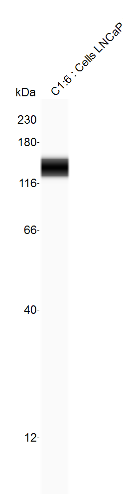
Application: Simple WesternSample Tested: LNCaP human prostate cancer cell lineSpecies: HumanVerified Customer | Posted 02/15/2019PSMA Expression in LNCaP cells by Simple Western. Run on 12-230 kda cartridge under reducing conditions.

-
Application: ELISASample Tested: Human recombinant antibodySpecies: HumanVerified Customer | Posted 10/31/2017
There are no reviews that match your criteria.
Protocols
Find general support by application which include: protocols, troubleshooting, illustrated assays, videos and webinars.
- 7-Amino Actinomycin D (7-AAD) Cell Viability Flow Cytometry Protocol
- Antigen Retrieval Protocol (PIER)
- Antigen Retrieval for Frozen Sections Protocol
- Appropriate Fixation of IHC/ICC Samples
- Cellular Response to Hypoxia Protocols
- Chromogenic IHC Staining of Formalin-Fixed Paraffin-Embedded (FFPE) Tissue Protocol
- Chromogenic Immunohistochemistry Staining of Frozen Tissue
- ClariTSA™ Fluorophore Kits
- Detection & Visualization of Antibody Binding
- Extracellular Membrane Flow Cytometry Protocol
- Flow Cytometry Protocol for Cell Surface Markers
- Flow Cytometry Protocol for Staining Membrane Associated Proteins
- Flow Cytometry Staining Protocols
- Flow Cytometry Troubleshooting Guide
- Fluorescent IHC Staining of Frozen Tissue Protocol
- Graphic Protocol for Heat-induced Epitope Retrieval
- Graphic Protocol for the Preparation and Fluorescent IHC Staining of Frozen Tissue Sections
- Graphic Protocol for the Preparation and Fluorescent IHC Staining of Paraffin-embedded Tissue Sections
- Graphic Protocol for the Preparation of Gelatin-coated Slides for Histological Tissue Sections
- ICC Cell Smear Protocol for Suspension Cells
- ICC Immunocytochemistry Protocol Videos
- ICC for Adherent Cells
- IHC Sample Preparation (Frozen sections vs Paraffin)
- Immunocytochemistry (ICC) Protocol
- Immunocytochemistry Troubleshooting
- Immunofluorescence of Organoids Embedded in Cultrex Basement Membrane Extract
- Immunofluorescent IHC Staining of Formalin-Fixed Paraffin-Embedded (FFPE) Tissue Protocol
- Immunohistochemistry (IHC) and Immunocytochemistry (ICC) Protocols
- Immunohistochemistry Frozen Troubleshooting
- Immunohistochemistry Paraffin Troubleshooting
- Intracellular Flow Cytometry Protocol Using Alcohol (Methanol)
- Intracellular Flow Cytometry Protocol Using Detergents
- Intracellular Nuclear Staining Flow Cytometry Protocol Using Detergents
- Intracellular Staining Flow Cytometry Protocol Using Alcohol Permeabilization
- Intracellular Staining Flow Cytometry Protocol Using Detergents to Permeabilize Cells
- Preparing Samples for IHC/ICC Experiments
- Preventing Non-Specific Staining (Non-Specific Binding)
- Primary Antibody Selection & Optimization
- Propidium Iodide Cell Viability Flow Cytometry Protocol
- Protocol for Heat-Induced Epitope Retrieval (HIER)
- Protocol for Liperfluo
- Protocol for Making a 4% Formaldehyde Solution in PBS
- Protocol for VisUCyte™ HRP Polymer Detection Reagent
- Protocol for the Characterization of Human Th22 Cells
- Protocol for the Characterization of Human Th9 Cells
- Protocol for the Fluorescent ICC Staining of Cell Smears - Graphic
- Protocol for the Fluorescent ICC Staining of Cultured Cells on Coverslips - Graphic
- Protocol for the Preparation & Fixation of Cells on Coverslips
- Protocol for the Preparation and Chromogenic IHC Staining of Frozen Tissue Sections
- Protocol for the Preparation and Chromogenic IHC Staining of Frozen Tissue Sections - Graphic
- Protocol for the Preparation and Chromogenic IHC Staining of Paraffin-embedded Tissue Sections
- Protocol for the Preparation and Chromogenic IHC Staining of Paraffin-embedded Tissue Sections - Graphic
- Protocol for the Preparation and Fluorescent ICC Staining of Cells on Coverslips
- Protocol for the Preparation and Fluorescent ICC Staining of Non-adherent Cells
- Protocol for the Preparation and Fluorescent ICC Staining of Stem Cells on Coverslips
- Protocol for the Preparation and Fluorescent IHC Staining of Frozen Tissue Sections
- Protocol for the Preparation and Fluorescent IHC Staining of Paraffin-embedded Tissue Sections
- Protocol for the Preparation of Gelatin-coated Slides for Histological Tissue Sections
- Protocol for the Preparation of a Cell Smear for Non-adherent Cell ICC - Graphic
- Protocol: Annexin V and PI Staining by Flow Cytometry
- Protocol: Annexin V and PI Staining for Apoptosis by Flow Cytometry
- R&D Systems Quality Control Western Blot Protocol
- TUNEL and Active Caspase-3 Detection by IHC/ICC Protocol
- The Importance of IHC/ICC Controls
- Troubleshooting Guide: Fluorokine Flow Cytometry Kits
- Troubleshooting Guide: Immunohistochemistry
- Troubleshooting Guide: Western Blot Figures
- Western Blot Conditions
- Western Blot Protocol
- Western Blot Protocol for Cell Lysates
- Western Blot Troubleshooting
- Western Blot Troubleshooting Guide
- View all Protocols, Troubleshooting, Illustrated assays and Webinars
Loading...
Associated Pathways
