SIRP alpha (Signal regulatory protein alpha; also SHPS1 and BIT) is a variably glycosylated 90-120 kDa member of the SIRP family of proteins. It is widely expressed, being found on neurons, microglia/macrophages, endothelium, and fibroblasts. SIRP alpha has a variety of functions, including presynsptic organization, inhibition of integrin action, and induction of myogenesis. It binds to CD47 and likely other ligands. Mature human SIRP alpha is a 477 amino acid (aa) type I transmembrane glycoprotein. It contains an extracellular region (aa 27-372) that shows one V-type Ig-like (aa 32-137) and two C2-type Ig-like domains (aa 147-347). Its cytoplasmic domain possesses two ITIMs which interact with protein tyrosine phosphatases. There is one alternative start site at Met102 plus a four aa insertion after Gln421. Over aa 27-370, human SIRP alpha shares 61% aa identity with mouse SIRP alpha.
Human SIRP alpha/CD172a Antibody (602411)
R&D Systems | Catalog # MAB4546

Key Product Details
Species Reactivity
Validated:
Human
Cited:
Human
Applications
Validated:
Western Blot, Flow Cytometry, Immunocytochemistry, CyTOF-ready
Cited:
Flow Cytometry
Label
Unconjugated
Antibody Source
Monoclonal Mouse IgG2B Clone # 602411
Loading...
Product Specifications
Immunogen
Chinese hamster ovary cell line CHO-derived recombinant human SIRP alpha /CD172a
Gly27-Asn370 (predicted)
Accession # P78324
Gly27-Asn370 (predicted)
Accession # P78324
Specificity
Detects human SIRP alpha /CD172a in direct ELISAs and Western blots. In direct ELISAs, 50-100% cross-reactivity with recombinant human (rh) SIRP beta 1 and no cross-reactivity with rhSIRP beta 2 is observed. In Western blots, approximately 10% cross-reactivity with rhSIRP beta 1 and no cross-reactivity with rhSIRP beta 2 is observed.
Clonality
Monoclonal
Host
Mouse
Isotype
IgG2B
Scientific Data Images for Human SIRP alpha/CD172a Antibody (602411)
Detection of Human SIRP alpha /CD172a
by Western Blot.
Western blot shows lysates of A549 human lung carcinoma cell line, U2OS human osteosarcoma cell line, U937 human histiocytic lymphoma cell line, and human granulocytes. PVDF membrane was probed with 2 µg/mL of Mouse Anti-Human SIRP alpha /CD172aMonoclonal Antibody (Catalog # MAB4546) followed by HRP-conjugated Anti-Mouse IgG Secondary Antibody (Catalog # HAF018). A specific band was detected for SIRP alpha /CD172a
at approximately 70-95 kDa (as indicated). This experiment was conducted under non-reducing conditions and using Western Blot Buffer Group 1.

Detection of SIRP alpha /CD172a in U937 Human Cell Line by Flow Cytometry.
U937 human histiocytic lymphoma cell line was stained with Mouse Anti-Human SIRPa/CD172a Monoclonal Antibody (Catalog # MAB4546, filled histogram) or isotype control antibody (Catalog # MAB0041, open histogram), followed by Allophycocyanin-conjugated Anti-Mouse IgG F(ab')2Secondary Antibody (Catalog # F0101B).
SIRP alpha /CD172a in THP‑1 Human Cell Line.
SIRPa/CD172a was detected in immersion fixed THP-1 human acute monocytic leukemia cell line using Mouse Anti-Human SIRPa/CD172a Monoclonal Antibody (Catalog # MAB4546) at 10 µg/mL for 3 hours at room temperature. Cells were stained using the NorthernLights™ 557-conjugated Anti-Mouse IgG Secondary Antibody (red; Catalog # NL007) and counterstained with DAPI. Specific staining was localized to cell membranes. View our protocol for Fluorescent ICC Staining of Non-adherent Cells.Applications for Human SIRP alpha/CD172a Antibody (602411)
Application
Recommended Usage
CyTOF-ready
Ready to be labeled using established conjugation methods. No BSA or other carrier proteins that could interfere with conjugation.
Flow Cytometry
2.5 µg/106 cells
Sample: U937 human histiocytic lymphoma cell line
Sample: U937 human histiocytic lymphoma cell line
Immunocytochemistry
8-25 µg/mL
Sample: Immersion fixed THP‑1 human acute monocytic leukemia cell line
Sample: Immersion fixed THP‑1 human acute monocytic leukemia cell line
Western Blot
2 µg/mL
Sample: A549 human lung carcinoma cell line, U2OS human osteosarcoma cell line, U937 human histiocytic lymphoma cell line, and human granulocytes
Sample: A549 human lung carcinoma cell line, U2OS human osteosarcoma cell line, U937 human histiocytic lymphoma cell line, and human granulocytes
Reviewed Applications
Read 3 reviews rated 4.3 using MAB4546 in the following applications:
Flow Cytometry Panel Builder
Bio-Techne Knows Flow Cytometry
Save time and reduce costly mistakes by quickly finding compatible reagents using the Panel Builder Tool.
Advanced Features
- Spectra Viewer - Custom analysis of spectra from multiple fluorochromes
- Spillover Popups - Visualize the spectra of individual fluorochromes
- Antigen Density Selector - Match fluorochrome brightness with antigen density

Formulation, Preparation, and Storage
Purification
Protein A or G purified from hybridoma culture supernatant
Reconstitution
Sterile PBS to a final concentration of 0.5 mg/mL. For liquid material, refer to CoA for concentration.
Loading...
Formulation
Lyophilized from a 0.2 μm filtered solution in PBS with Trehalose. *Small pack size (SP) is supplied either lyophilized or as a 0.2 µm filtered solution in PBS.
Shipping
Lyophilized product is shipped at ambient temperature. Liquid small pack size (-SP) is shipped with polar packs. Upon receipt, store immediately at the temperature recommended below.
Stability & Storage
Use a manual defrost freezer and avoid repeated freeze-thaw cycles.
- 12 months from date of receipt, -20 to -70 °C as supplied.
- 1 month, 2 to 8 °C under sterile conditions after reconstitution.
- 6 months, -20 to -70 °C under sterile conditions after reconstitution.
Calculators
Background: SIRP alpha/CD172a
Long Name
Signal-regulatory Protein alpha
Alternate Names
BIT, CD172a, MFR, MYD-1, SHPS1, SIRPA
Entrez Gene IDs
Gene Symbol
SIRPA
UniProt
Additional SIRP alpha/CD172a Products
Product Documents for Human SIRP alpha/CD172a Antibody (602411)
Certificate of Analysis
To download a Certificate of Analysis, please enter a lot or batch number in the search box below.
Note: Certificate of Analysis not available for kit components.
Product Specific Notices for Human SIRP alpha/CD172a Antibody (602411)
For research use only
Citations for Human SIRP alpha/CD172a Antibody (602411)
 Powered by Bioz
Customer Reviews for Human SIRP alpha/CD172a Antibody (602411) (3)
4.3 out of 5
3 Customer Ratings
Have you used Human SIRP alpha/CD172a Antibody (602411)?
Submit a review and receive an Amazon gift card!
$25/€18/£15/$25CAN/¥2500 Yen for a review with an image
$10/€7/£6/$10CAN/¥1110 Yen for a review without an image
Submit a review
Customer Images
Showing
1
-
3 of
3 reviews
Showing All
Filter By:
-
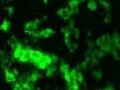
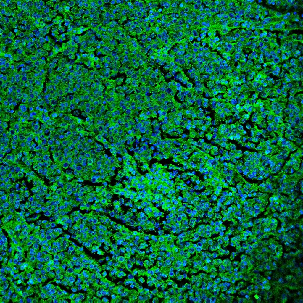
Application: Immunocytochemistry/ImmunofluorescenceSample Tested: fibroblastsSpecies: HumanVerified Customer | Posted 11/10/2021

-
Application: Immunocytochemistry/ImmunofluorescenceSample Tested: Melanoma tissueSpecies: HumanVerified Customer | Posted 11/23/2020
-
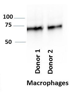
Application: Western BlotSample Tested: MACROPHAGESpecies: HumanVerified Customer | Posted 04/29/2020
There are no reviews that match your criteria.
Protocols
Find general support by application which include: protocols, troubleshooting, illustrated assays, videos and webinars.
- 7-Amino Actinomycin D (7-AAD) Cell Viability Flow Cytometry Protocol
- Appropriate Fixation of IHC/ICC Samples
- Cellular Response to Hypoxia Protocols
- ClariTSA™ Fluorophore Kits
- Detection & Visualization of Antibody Binding
- Extracellular Membrane Flow Cytometry Protocol
- Flow Cytometry Protocol for Cell Surface Markers
- Flow Cytometry Protocol for Staining Membrane Associated Proteins
- Flow Cytometry Staining Protocols
- Flow Cytometry Troubleshooting Guide
- ICC Cell Smear Protocol for Suspension Cells
- ICC Immunocytochemistry Protocol Videos
- ICC for Adherent Cells
- Immunocytochemistry (ICC) Protocol
- Immunocytochemistry Troubleshooting
- Immunofluorescence of Organoids Embedded in Cultrex Basement Membrane Extract
- Immunohistochemistry (IHC) and Immunocytochemistry (ICC) Protocols
- Intracellular Flow Cytometry Protocol Using Alcohol (Methanol)
- Intracellular Flow Cytometry Protocol Using Detergents
- Intracellular Nuclear Staining Flow Cytometry Protocol Using Detergents
- Intracellular Staining Flow Cytometry Protocol Using Alcohol Permeabilization
- Intracellular Staining Flow Cytometry Protocol Using Detergents to Permeabilize Cells
- Preparing Samples for IHC/ICC Experiments
- Preventing Non-Specific Staining (Non-Specific Binding)
- Primary Antibody Selection & Optimization
- Propidium Iodide Cell Viability Flow Cytometry Protocol
- Protocol for Liperfluo
- Protocol for VisUCyte™ HRP Polymer Detection Reagent
- Protocol for the Characterization of Human Th22 Cells
- Protocol for the Characterization of Human Th9 Cells
- Protocol for the Fluorescent ICC Staining of Cell Smears - Graphic
- Protocol for the Fluorescent ICC Staining of Cultured Cells on Coverslips - Graphic
- Protocol for the Preparation and Fluorescent ICC Staining of Cells on Coverslips
- Protocol for the Preparation and Fluorescent ICC Staining of Non-adherent Cells
- Protocol for the Preparation and Fluorescent ICC Staining of Stem Cells on Coverslips
- Protocol for the Preparation of a Cell Smear for Non-adherent Cell ICC - Graphic
- Protocol: Annexin V and PI Staining by Flow Cytometry
- Protocol: Annexin V and PI Staining for Apoptosis by Flow Cytometry
- R&D Systems Quality Control Western Blot Protocol
- TUNEL and Active Caspase-3 Detection by IHC/ICC Protocol
- The Importance of IHC/ICC Controls
- Troubleshooting Guide: Fluorokine Flow Cytometry Kits
- Troubleshooting Guide: Western Blot Figures
- Western Blot Conditions
- Western Blot Protocol
- Western Blot Protocol for Cell Lysates
- Western Blot Troubleshooting
- Western Blot Troubleshooting Guide
- View all Protocols, Troubleshooting, Illustrated assays and Webinars

