Human STING/TMEM173 Antibody (723505)
R&D Systems | Catalog # MAB7169

Key Product Details
Validated by
Knockout/Knockdown
Species Reactivity
Validated:
Human
Cited:
Human, Mouse, Primate
Applications
Validated:
Western Blot, Intracellular Staining by Flow Cytometry, Immunocytochemistry, Immunoprecipitation, CyTOF-ready
Cited:
Immunohistochemistry, Western Blot, Flow Cytometry, Immunocytochemistry, Chromatin Immunoprecipitation (ChIP)
Label
Unconjugated
Antibody Source
Monoclonal Mouse IgG2B Clone # 723505
Loading...
Product Specifications
Immunogen
E. coli-derived recombinant human STING/TMEM173
Ala215-Ser379
Accession # Q86WV6
Ala215-Ser379
Accession # Q86WV6
Specificity
Detects human STING/TMEM173 in direct ELISAs and Western blots.
Clonality
Monoclonal
Host
Mouse
Isotype
IgG2B
Scientific Data Images for Human STING/TMEM173 Antibody (723505)
Detection of Human STING/TMEM173 by Western Blot.
Western blot shows lysates of THP-1 human acute monocytic leukemia cell line and U937 human histiocytic lymphoma cell line. PVDF membrane was probed with 0.2 µg/mL of Mouse Anti-Human STING/TMEM173 Monoclonal Antibody (Catalog # MAB7169) followed by HRP-conjugated Anti-Mouse IgG Secondary Antibody (HAF007). A specific band was detected for STING/TMEM173 at approximately 40 kDa (as indicated). This experiment was conducted under reducing conditions and using Immunoblot Buffer Group 1.
Detection of STING/TMEM173 in Human PBMC Monocytes by Flow Cytometry.
Human peripheral blood mononuclear cell (PBMC) monocytes were stained with Mouse Anti-Human STING/TMEM173 Monoclonal Antibody (Catalog # MAB7169, filled histogram) or isotype control antibody (MAB0041, open histogram), followed by Allophycocyanin-conjugated Anti-Mouse IgG Secondary Antibody (F0101B). To facilitate intracellular staining, cells were fixed with paraformaldehyde and permeabilized with saponin.
Detection of STING/TMEM173 in THP‑1 Human Cell Line by Flow Cytometry.
THP-1 human acute monocytic leukemia cell line was stained with Mouse Anti-Human STING/TMEM173 Monoclonal Antibody (Catalog # MAB7169, filled histogram) or isotype control antibody (MAB0041, open histogram), followed by Allophycocyanin-conjugated Anti-Mouse IgG Secondary Antibody (F0101B). To facilitate intracellular staining, cells were fixed with paraformaldehyde and permeabilized with saponin.
Detection of STING/TMEM173 in U937 Human Cell Line by Flow Cytometry.
U937 human histiocytic lymphoma cell line was stained with Mouse Anti-Human STING/TMEM173 Monoclonal Antibody (Catalog # MAB7169, filled histogram) or isotype control antibody (MAB0041, open histogram), followed by Allophycocyanin-conjugated Anti-Mouse IgG Secondary Antibody (F0101B). To facilitate intracellular staining, cells were fixed with paraformaldehyde and permeabilized with saponin.
STING/TMEM173 in U937 Human Cell Line.
STING/TMEM173 was detected in immersion fixed U937 human histiocytic lymphoma cell line using Mouse Anti-Human STING/TMEM173 Monoclonal Antibody (Catalog # MAB7169) at 3 µg/mL for 3 hours at room temperature. Cells were stained using the NorthernLights™ 557-conjugated Anti-Mouse IgG Secondary Antibody (red; NL007) and counterstained with DAPI (blue). Specific staining was localized to cytoplasm. View our protocol for Fluorescent ICC Staining of Non-adherent Cells.
Detection of Mouse STING/TMEM173 by Western Blot
CYLD interacts with STING in a ubiquitination-dependent manner.(A) HEK293T cells were transfected with the indicated plasmids. Thirty-six hours after transfection, the cell lysates were immunoprecipitated with an anti-Myc antibody and then immunoblotted with the indicated antibodies. (B) HEK293T cells were transfected with the indicated plasmids. Thirty-six hours after transfection, cell lysates were immunoprecipitated with an anti-HA antibody and then immunoblotted with the indicated antibodies. (C and D) HEK293T cells were transfected with the indicated plasmids. Thirty-six hours after transfection, the cell lysates were immunoprecipitated with an anti-Myc antibody (C) or an anti-HA antibody (D) and then immunoblotted with the indicated antibodies. (E) HEK293T cells were transfected with the indicated plasmids. Thirty-six hours after transfection, the cell lysates were immunoprecipitated with an anti-Myc antibody and then immunoblotted with the indicated antibodies. (F) After stimulation with HSV-1 (MOI = 10) for the indicated time periods in the presence of MG132 (10 μM), lysates of MEFs were immunoprecipitated with an anti-STING antibody and then immunoblotted with the indicated antibodies. (G) HeLa cells were transfected with empty vector or Flag-tagged RNF5 for 24 h and then stained with the indicated antibodies before imaging by confocal microscopy. STING (Green) (R&D Systems); CYLD (Red) (Abcam); Nucleus (Blue). Scale bars represent 50 μm. Image collected and cropped by CiteAb from the following publication (https://pubmed.ncbi.nlm.nih.gov/30388174), licensed under a CC-BY license. Not internally tested by R&D Systems.
Detection of Human STING/TMEM173 by Western Blot
HCT116 cells surviving PARP1 depletion activate innate immune signaling.(A-B) RNA-Seq data from HCT116EV and HCT116PARP1-/- cells (clones C2 and C4) was analyzed using Gene Set Enrichment Analysis (GSEA). The top category of differentially expressed genes was “Interferon Alpha Response”. The enrichment plot is shown in (A) and the heat map for the 97 mRNAs in this category in shown in (B). Map was generated using Cufflinks software (version 2.2.1) and shows absolute expression values independently normalized and analyzed for each comparison pair (EV/C2 and EV/C4). (C) The same RNA-Seq dataset was analyzed using Ingenuity Pathway Analysis (IPA). A representative plot highlighting enrichment for Interferon-Stimulated Genes (ISGs) is shown. (D) The induction of multiple ISGs observed by RNA-Seq was confirmed by q-RT-PCR. Bars represent the average and standard deviation of quadruplicate samples in each experiment and data is representative of 2–4 independent experiments. (E-F) The induction of factors involved in the sensing/signaling of cytoplasmic nucleic acids observed by RNA-Seq was confirmed by Q-RT-PCR (E). Bars represent the average and standard deviation of quadruplicate samples in each experiment and data is representative of 2–3 independent experiments. Protein expression for the same factors was assessed by immunoblotting (F). Blots are representative of 2–3 independent experiments. (G) Fixed cells were stained with an antibody to IRF3 and counterstained with DAPI. Images are representative of 5 random fields per slide. The experiment was repeated twice with similar results. Image collected and cropped by CiteAb from the following publication (https://pubmed.ncbi.nlm.nih.gov/29590171), licensed under a CC-BY license. Not internally tested by R&D Systems.
Detection of Human STING/TMEM173 by Western Blot
ISG induction in cells surviving PARP1 depletion is dependent on RIG-I and MAVS.(A-E) To assess the efficiency of RNA silencing to STING (A), RIG-I (B), MDA-5 (C), TLR3 (D) or MAVS (E), cells transfected with the specific pooled siRNAs and control cells transfected with a scrambled siRNA or untreated were harvested 4 days after transfection and probed with the indicated antibodies. GAPDH (A, B, C, E) or alpha -tubulin (D) served as loading controls. In blots where multiple bands are observed (A, D, E), black arrows point to specific bands and asterisks to unspecific bands. (F) The expression of ISGs OAS1 and IFIT3 was quantified by qRT-PCR after knock down. Data is normalized to the expression in HCT116EV cells (= 1). Bars represent the average and standard deviation of quadruplicates. Data is representative of two independent experiments. Image collected and cropped by CiteAb from the following publication (https://pubmed.ncbi.nlm.nih.gov/29590171), licensed under a CC-BY license. Not internally tested by R&D Systems.
Detection of STING/TMEM173 by Immunoprecipitation.
PMA-treated THP-1 lysates were prepared, and immunoprecipitation was performed using 2.0 μg of Mouse Anti-Human STING/TMEM173 Monoclonal Antibody (Catalog # MAB7169) pre-coupled to Dynabeads protein G. Immunoprecipitated STING/TMEM173 was detected with a Rabbit Anti-STING/TMEM173 antibody. For western blot, the Rabbit Anti-STING/TMEM173 antibody was used at 1/1000. The Ponceau stained transfers of each blot are shown. SM=4% starting material; UB=4% unbound fraction; IP=immunoprecipitate; HC=antibody heavy chain. *=monoclonal antibody, **=recombinant antibody. Image, protocol and testing courtesy of YCharOS Inc. (ycharos.com).Applications for Human STING/TMEM173 Antibody (723505)
Application
Recommended Usage
CyTOF-ready
Ready to be labeled using established conjugation methods. No BSA or other carrier proteins that could interfere with conjugation.
Immunocytochemistry
8-25 µg/mL
Sample: Immersion fixed U937 human histiocytic lymphoma cell line
Sample: Immersion fixed U937 human histiocytic lymphoma cell line
Immunoprecipitation
2 µg/mL
Sample: Cell lysate of PMA-treated THP-1 human acute monocytic leukemia cell line
Sample: Cell lysate of PMA-treated THP-1 human acute monocytic leukemia cell line
Intracellular Staining by Flow Cytometry
0.25 µg/106 cells
Sample: Human peripheral blood mononuclear cell (PBMC) monocytes, THP‑1 human acute monocytic leukemia cell line, and U937 human histiocytic lymphoma cell line fixed with paraformaldehyde and permeabilized with saponin
Sample: Human peripheral blood mononuclear cell (PBMC) monocytes, THP‑1 human acute monocytic leukemia cell line, and U937 human histiocytic lymphoma cell line fixed with paraformaldehyde and permeabilized with saponin
Western Blot
0.2 µg/mL
Sample: THP‑1 human acute monocytic leukemia cell line and U937 human histiocytic lymphoma cell line
Sample: THP‑1 human acute monocytic leukemia cell line and U937 human histiocytic lymphoma cell line
Reviewed Applications
Read 1 review rated 3 using MAB7169 in the following applications:
Flow Cytometry Panel Builder
Bio-Techne Knows Flow Cytometry
Save time and reduce costly mistakes by quickly finding compatible reagents using the Panel Builder Tool.
Advanced Features
- Spectra Viewer - Custom analysis of spectra from multiple fluorochromes
- Spillover Popups - Visualize the spectra of individual fluorochromes
- Antigen Density Selector - Match fluorochrome brightness with antigen density

Formulation, Preparation, and Storage
Purification
Protein A or G purified from hybridoma culture supernatant
Reconstitution
Sterile PBS to a final concentration of 0.5 mg/mL. For liquid material, refer to CoA for concentration.
Loading...
Formulation
Lyophilized from a 0.2 μm filtered solution in PBS with Trehalose. See Certificate of Analysis for details.
*Small pack size (-SP) is supplied either lyophilized or as a 0.2 µm filtered solution in PBS.
*Small pack size (-SP) is supplied either lyophilized or as a 0.2 µm filtered solution in PBS.
Shipping
Lyophilized product is shipped at ambient temperature. Liquid small pack size (-SP) is shipped with polar packs. Upon receipt, store immediately at the temperature recommended below.
Stability & Storage
Use a manual defrost freezer and avoid repeated freeze-thaw cycles.
- 12 months from date of receipt, -20 to -70 °C as supplied.
- 1 month, 2 to 8 °C under sterile conditions after reconstitution.
- 6 months, -20 to -70 °C under sterile conditions after reconstitution.
Calculators
Background: STING/TMEM173
Long Name
Stimulator of Interferon Genes Protein/Transmembrane protein 173
Alternate Names
ERIS, MITA, MPYS, NET23, TMEM173
Gene Symbol
STING1
UniProt
Additional STING/TMEM173 Products
Product Documents for Human STING/TMEM173 Antibody (723505)
Certificate of Analysis
To download a Certificate of Analysis, please enter a lot or batch number in the search box below.
Note: Certificate of Analysis not available for kit components.
Product Specific Notices for Human STING/TMEM173 Antibody (723505)
For research use only
Related Research Areas
Citations for Human STING/TMEM173 Antibody (723505)
 Powered by Bioz
Customer Reviews for Human STING/TMEM173 Antibody (723505) (1)
3 out of 5
1 Customer Rating
Have you used Human STING/TMEM173 Antibody (723505)?
Submit a review and receive an Amazon gift card!
$25/€18/£15/$25CAN/¥2500 Yen for a review with an image
$10/€7/£6/$10CAN/¥1110 Yen for a review without an image
Submit a review
Customer Images
Showing
1
-
1 of
1 review
Showing All
Filter By:
-
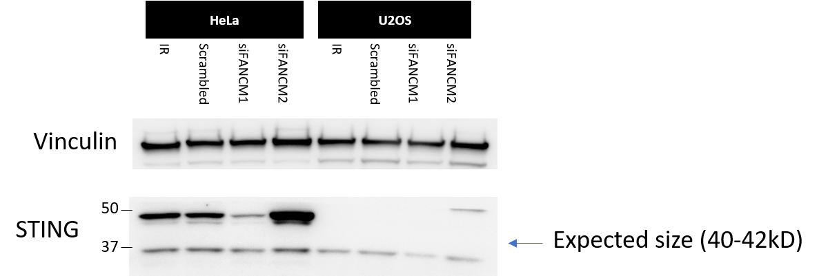
Application: Western BlotSample Tested: U2OS cells and HeLa cellsSpecies: HumanVerified Customer | Posted 10/11/2020ALT cells, U2OS shown here, are shown to lack STING. Based on this the apparent band of interest of 40-42kD seems to be migrating further up closer to the 50kD instead of the 37kD ladder.Antibody used at 0.105ug/mL or 1/2000. Blocked and incubated with 5% milk in TBST overnight at 4C. Blot detected using PICO Plus chemiluminescence reagent (Life Technologies).

There are no reviews that match your criteria.
Protocols
Find general support by application which include: protocols, troubleshooting, illustrated assays, videos and webinars.
- 7-Amino Actinomycin D (7-AAD) Cell Viability Flow Cytometry Protocol
- Appropriate Fixation of IHC/ICC Samples
- Cellular Response to Hypoxia Protocols
- ClariTSA™ Fluorophore Kits
- Detection & Visualization of Antibody Binding
- Extracellular Membrane Flow Cytometry Protocol
- Flow Cytometry Protocol for Cell Surface Markers
- Flow Cytometry Protocol for Staining Membrane Associated Proteins
- Flow Cytometry Staining Protocols
- Flow Cytometry Troubleshooting Guide
- ICC Cell Smear Protocol for Suspension Cells
- ICC Immunocytochemistry Protocol Videos
- ICC for Adherent Cells
- Immunocytochemistry (ICC) Protocol
- Immunocytochemistry Troubleshooting
- Immunofluorescence of Organoids Embedded in Cultrex Basement Membrane Extract
- Immunohistochemistry (IHC) and Immunocytochemistry (ICC) Protocols
- Immunoprecipitation Protocol
- Intracellular Flow Cytometry Protocol Using Alcohol (Methanol)
- Intracellular Flow Cytometry Protocol Using Detergents
- Intracellular Nuclear Staining Flow Cytometry Protocol Using Detergents
- Intracellular Staining Flow Cytometry Protocol Using Alcohol Permeabilization
- Intracellular Staining Flow Cytometry Protocol Using Detergents to Permeabilize Cells
- Preparing Samples for IHC/ICC Experiments
- Preventing Non-Specific Staining (Non-Specific Binding)
- Primary Antibody Selection & Optimization
- Propidium Iodide Cell Viability Flow Cytometry Protocol
- Protocol for Liperfluo
- Protocol for VisUCyte™ HRP Polymer Detection Reagent
- Protocol for the Characterization of Human Th22 Cells
- Protocol for the Characterization of Human Th9 Cells
- Protocol for the Fluorescent ICC Staining of Cell Smears - Graphic
- Protocol for the Fluorescent ICC Staining of Cultured Cells on Coverslips - Graphic
- Protocol for the Preparation and Fluorescent ICC Staining of Cells on Coverslips
- Protocol for the Preparation and Fluorescent ICC Staining of Non-adherent Cells
- Protocol for the Preparation and Fluorescent ICC Staining of Stem Cells on Coverslips
- Protocol for the Preparation of a Cell Smear for Non-adherent Cell ICC - Graphic
- Protocol: Annexin V and PI Staining by Flow Cytometry
- Protocol: Annexin V and PI Staining for Apoptosis by Flow Cytometry
- R&D Systems Quality Control Western Blot Protocol
- TUNEL and Active Caspase-3 Detection by IHC/ICC Protocol
- The Importance of IHC/ICC Controls
- Troubleshooting Guide: Fluorokine Flow Cytometry Kits
- Troubleshooting Guide: Western Blot Figures
- Western Blot Conditions
- Western Blot Protocol
- Western Blot Protocol for Cell Lysates
- Western Blot Troubleshooting
- Western Blot Troubleshooting Guide
- View all Protocols, Troubleshooting, Illustrated assays and Webinars
Loading...