Human TGF-beta RI/ALK-5 Antibody
R&D Systems | Catalog # AF3025

Key Product Details
Species Reactivity
Validated:
Cited:
Applications
Validated:
Cited:
Label
Antibody Source
Product Specifications
Immunogen
Leu34-Glu125
Accession # P36897
Specificity
Clonality
Host
Isotype
Scientific Data Images for Human TGF-beta RI/ALK-5 Antibody
Detection of Human TGF‑ beta RI/ALK‑5 by Western Blot.
Western blot shows lysates of HeLa human cervical epithelial carcinoma cell line, HepG2 human hepatocellular carcinoma cell line, and Raji human Burkitt's lymphoma cell line. PVDF membrane was probed with 0.2 µg/mL of Goat Anti-Human TGF-beta RI/ALK-5 Antigen Affinity-purified Polyclonal Antibody (Catalog # AF3025) followed by HRP-conjugated Anti-Goat IgG Secondary Antibody (Catalog # HAF019). Specific bands were detected for TGF-beta RI/ALK-5 at approximately 50 & 56 kDa (as indicated). This experiment was conducted under reducing conditions and using Immunoblot Buffer Group 1.
TGF-beta RI/ALK‑5 in Human PBMCs.
TGF-beta RI/ALK-5 was detected in immersion fixed human peripheral blood mononuclear cells (PBMCs) stimulated with LPS and monensin using Goat Anti-Human TGF-beta RI/ALK-5 Antigen Affinity-purified Polyclonal Antibody (Catalog # AF3025) at 10 µg/mL for 3 hours at room temperature. Cells were stained using the NorthernLights™ 557-conjugated Anti-Goat IgG Secondary Antibody (yellow; Catalog # NL001) and counter-stained with DAPI (blue). View our protocol for Fluorescent ICC Staining of Non-adherent Cells.
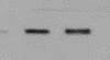
Detection of Porcine TGF-beta RI/ALK-5 by Western Blot
Myostatin signaling in longissimus dorsi muscle of low-birth-weight (LBWT) and normal-birth-weight (NBWT) neonatal pigs. (A): Representative western blots from four pairs of LBWT and NBWT pigs (N, NBWT; L, LBWT). (B–G): Protein abundance of myostain, ALK5 and ActRIIB, and protein abundance phosphorylation of smad2/3. Abundance was normalized to alpha -tubulin and phosphorylation normalized to the corresponding non-phospho-proteins. Results are means ± SE. n = 12. Values with different letters differ significantly (P ≤ 0.05). Image collected and cropped by CiteAb from the following publication (https://journal.frontiersin.org/article/10.3389/fphys.2017.00482/full), licensed under a CC-BY license. Not internally tested by R&D Systems.
Detection of TGF-beta RI/ALK-5 by Western Blot
WT1 regulates the STAT1/3 pathway in CAFs. A Protein levels of WT1, STAT1, STAT3, PD-L1 and the TGF beta receptor were determined in WT1 knockdown (A) and WT1 overexpressed (B) CAFs by Western blotting. Actin was used as a loading control. IDO release was determined by ELISA. RNA levels of PD-L1 was determined by qRT-PCR. TBP was used as a loading control. Representative experiment is shown of n = 3–5 biological replicates. Data represents mean ± SEM; * P < 0.05, ** P < 0.01, *** P < 0.001, and **** P < 0.0001 Image collected and cropped by CiteAb from the following open publication (https://pubmed.ncbi.nlm.nih.gov/40598415), licensed under a CC-BY license. Not internally tested by R&D Systems.Applications for Human TGF-beta RI/ALK-5 Antibody
ELISA
This antibody functions as an ELISA detection antibody when paired with Mouse Anti-Human TGF‑ beta RI/ALK‑5 Monoclonal Antibody (Catalog # MAB3025).
This product is intended for assay development on various assay platforms requiring antibody pairs.
Immunocytochemistry
Sample: Immersion fixed human peripheral blood mononuclear cells
Western Blot
Sample: HeLa human cervical epithelial carcinoma cell line, HepG2 human hepatocellular carcinoma cell line, and Raji human Burkitt's lymphoma cell line
Reviewed Applications
Read 2 reviews rated 5 using AF3025 in the following applications:
Formulation, Preparation, and Storage
Purification
Reconstitution
Reconstitute at 0.2 mg/mL in sterile PBS. For liquid material, refer to CoA for concentration.
Formulation
Shipping
Stability & Storage
- 12 months from date of receipt, -20 to -70 °C as supplied.
- 1 month, 2 to 8 °C under sterile conditions after reconstitution.
- 6 months, -20 to -70 °C under sterile conditions after reconstitution.
Calculators
Background: TGF-beta RI/ALK-5
Long Name
Alternate Names
Gene Symbol
UniProt
Additional TGF-beta RI/ALK-5 Products
Product Documents for Human TGF-beta RI/ALK-5 Antibody
Certificate of Analysis
To download a Certificate of Analysis, please enter a lot or batch number in the search box below.
Note: Certificate of Analysis not available for kit components.
Product Specific Notices for Human TGF-beta RI/ALK-5 Antibody
For research use only
Citations for Human TGF-beta RI/ALK-5 Antibody
 Powered by Bioz
Customer Reviews for Human TGF-beta RI/ALK-5 Antibody (2)
Have you used Human TGF-beta RI/ALK-5 Antibody?
Submit a review and receive an Amazon gift card!
$25/€18/£15/$25CAN/¥2500 Yen for a review with an image
$10/€7/£6/$10CAN/¥1110 Yen for a review without an image
Submit a review
Customer Images
-
Application: Affinity PurificationSample Tested: 3T3-L1 mouse embryonic fibroblast adipose-like cell lineSpecies: MouseVerified Customer | Posted 05/07/2019
-
Application: Western BlotSample Tested: Liver cellsSpecies: HumanVerified Customer | Posted 08/20/2018

There are no reviews that match your criteria.
Protocols
Find general support by application which include: protocols, troubleshooting, illustrated assays, videos and webinars.
- Appropriate Fixation of IHC/ICC Samples
- Cellular Response to Hypoxia Protocols
- ClariTSA™ Fluorophore Kits
- Detection & Visualization of Antibody Binding
- ELISA Sample Preparation & Collection Guide
- ELISA Troubleshooting Guide
- How to Run an R&D Systems DuoSet ELISA
- How to Run an R&D Systems Quantikine ELISA
- How to Run an R&D Systems Quantikine™ QuicKit™ ELISA
- ICC Cell Smear Protocol for Suspension Cells
- ICC Immunocytochemistry Protocol Videos
- ICC for Adherent Cells
- Immunocytochemistry (ICC) Protocol
- Immunocytochemistry Troubleshooting
- Immunofluorescence of Organoids Embedded in Cultrex Basement Membrane Extract
- Immunohistochemistry (IHC) and Immunocytochemistry (ICC) Protocols
- Preparing Samples for IHC/ICC Experiments
- Preventing Non-Specific Staining (Non-Specific Binding)
- Primary Antibody Selection & Optimization
- Protocol for VisUCyte™ HRP Polymer Detection Reagent
- Protocol for the Fluorescent ICC Staining of Cell Smears - Graphic
- Protocol for the Fluorescent ICC Staining of Cultured Cells on Coverslips - Graphic
- Protocol for the Preparation and Fluorescent ICC Staining of Cells on Coverslips
- Protocol for the Preparation and Fluorescent ICC Staining of Non-adherent Cells
- Protocol for the Preparation and Fluorescent ICC Staining of Stem Cells on Coverslips
- Protocol for the Preparation of a Cell Smear for Non-adherent Cell ICC - Graphic
- Quantikine HS ELISA Kit Assay Principle, Alkaline Phosphatase
- Quantikine HS ELISA Kit Principle, Streptavidin-HRP Polymer
- R&D Systems Quality Control Western Blot Protocol
- Sandwich ELISA (Colorimetric) – Biotin/Streptavidin Detection Protocol
- Sandwich ELISA (Colorimetric) – Direct Detection Protocol
- TUNEL and Active Caspase-3 Detection by IHC/ICC Protocol
- The Importance of IHC/ICC Controls
- Troubleshooting Guide: ELISA
- Troubleshooting Guide: Western Blot Figures
- Western Blot Conditions
- Western Blot Protocol
- Western Blot Protocol for Cell Lysates
- Western Blot Troubleshooting
- Western Blot Troubleshooting Guide
- View all Protocols, Troubleshooting, Illustrated assays and Webinars


