
Key Product Details
Species Reactivity
Validated:
Human, Mouse, Rat
Cited:
Human, Mouse, Rat
Applications
Validated:
Immunohistochemistry, Western Blot
Cited:
Immunohistochemistry, Immunohistochemistry-Paraffin, Western Blot, Flow Cytometry, Immunocytochemistry
Label
Unconjugated
Antibody Source
Monoclonal Mouse IgG2B Clone # 75133
Loading...
Product Specifications
Immunogen
Mouse myeloma cell line NS0-derived recombinant human TrkB
Cys32-His430
Accession # Q16620
Cys32-His430
Accession # Q16620
Specificity
Detects human TrkB in direct ELISAs. Detects human, mouse, and rat TrkB in Western blots. In ELISAs and Western blots, no cross-reactivity with recombinant human (rh) TrkA, rhTrkC, or recombinant rat TrkA is observed.
Clonality
Monoclonal
Host
Mouse
Isotype
IgG2B
Scientific Data Images for TrkB Antibody (75133)
Detection of Human, Mouse, and Rat TrkB by Western Blot.
Western blot shows lysates of human brain (motor cortex) tissue, mouse brain (cortex) tissue, and rat brain (hippocampus) tissue. PVDF membrane was probed with 2 µg/mL of Mouse Anti-Human/Mouse/Rat TrkB Monoclonal Antibody (Catalog # MAB397) followed by HRP-conjugated Anti-Mouse IgG Secondary Antibody (Catalog # HAF018). Specific bands were detected for TrkB at approximately 95 kDa and 145 kDa (as indicated). This experiment was conducted under reducing conditions and using Immunoblot Buffer Group 1.
TrkB in Human Brain.
TrkB was detected in immersion fixed paraffin-embedded sections of human brain (hippocampus) using Mouse Anti-Human/Mouse/Rat TrkB Monoclonal Antibody (Catalog # MAB397) at 25 µg/mL overnight at 4 °C. Before incubation with the primary antibody tissue was subjected to heat-induced epitope retrieval using Antigen Retrieval Reagent-Basic (Catalog # CTS013). Tissue was stained using the Anti-Mouse HRP-DAB Cell & Tissue Staining Kit (brown; Catalog # CTS002) and counterstained with hematoxylin (blue). View our protocol for Chromogenic IHC Staining of Paraffin-embedded Tissue Sections.
Detection of Human TrkB by Western Blot
Expression of BDNF and TrkB in CRC cells in vitro.(A): RT-PCR analysis of BDNF and TrkB mRNA levels in CRC cell lines CaCO2 (lane: 1), DLD1 (lane: 2), HT29 (lane: 3), LoVo (lane: 4), and SW480 (lane: 5). GAPDH mRNA levels was used as an internal control. TrkB mRNA levels contain both TrkB.FL and TrkB.T1 mRNAs. (B): Western blotting analysis of BDNF and TrkB protein levels in CRC cell lines CaCO2 (lane: 1), DLD1 (lane: 2), HT29 (lane: 3), LoVo (lane: 4), and SW480 (lane: 5). The full-length TrkB (TrkB.FL, 145 kDa) and the truncated TrkB (TrkB.T1, 95 kDa) were detected. Actin was used as an internal control. Image collected and cropped by CiteAb from the following publication (https://pubmed.ncbi.nlm.nih.gov/24801982), licensed under a CC-BY license. Not internally tested by R&D Systems.
Detection of Human TrkB by Immunocytochemistry/Immunofluorescence
BDNF and TrkB protein expression in primary and metastatic colorectal cancer.Representative images of immuno-reactive BDNF and TrkB protein expression in primary CRC and peritoneal metastasis are shown (original magnification: 100×). Anti TrkB antibody (R&D Systems, Foster City, CA, USA) detected both TrkB.FL and TrkB.T1 proteins, which was confirmed by Western blotting analysis. (A): The immunoreactive BDNF protein is located in the cytoplasm of the tumor cells of the primary CRC. (B): The immunoreactive BDNF protein is located in the cytoplasm of the tumor cells in the corresponding peritoneal metastasis. (C): The immunoreactive TrkB protein is located in the nucleus of the tumor cells of the primary CRC. (D): The immunoreactive TrkB protein is located in the nucleus of the tumor cells in the corresponding peritoneal metastasis. These expression patterns for the BDNF and TrkB proteins were confirmed in CRC patients (n = 5) whose primary and peritoneal metastatic nodules were available for immunohistochemistry. Image collected and cropped by CiteAb from the following publication (https://pubmed.ncbi.nlm.nih.gov/24801982), licensed under a CC-BY license. Not internally tested by R&D Systems.
Detection of Human TrkB by Western Blot
Expression of BDNF and TrkB in CRC cells in vitro.(A): RT-PCR analysis of BDNF and TrkB mRNA levels in CRC cell lines CaCO2 (lane: 1), DLD1 (lane: 2), HT29 (lane: 3), LoVo (lane: 4), and SW480 (lane: 5). GAPDH mRNA levels was used as an internal control. TrkB mRNA levels contain both TrkB.FL and TrkB.T1 mRNAs. (B): Western blotting analysis of BDNF and TrkB protein levels in CRC cell lines CaCO2 (lane: 1), DLD1 (lane: 2), HT29 (lane: 3), LoVo (lane: 4), and SW480 (lane: 5). The full-length TrkB (TrkB.FL, 145 kDa) and the truncated TrkB (TrkB.T1, 95 kDa) were detected. Actin was used as an internal control. Image collected and cropped by CiteAb from the following publication (https://pubmed.ncbi.nlm.nih.gov/24801982), licensed under a CC-BY license. Not internally tested by R&D Systems.
Detection of Human TrkB by Immunohistochemistry
Expression of BDNF receptors in human placental tissues. (A) Representative confocal immunofluorescence staining images of showing DAPI (blue) and SDC1 (a syncytiotrophoblast biomarker, green) along with TrkB (red) and p75NTR (red) receptors in the first trimester placenta. (B) Confocal immunofluorescence staining images showing DAPI, SDC1, TrkB, and p75NTR in the term placenta. Image collected and cropped by CiteAb from the following open publication (https://pubmed.ncbi.nlm.nih.gov/34394001), licensed under a CC-BY license. Not internally tested by R&D Systems.
Detection of Mouse TrkB by Western Blot
7,8-DHF alleviates HFD-induced depression-like behaviors in Thy1-C/EBP beta Tg mice. (A) Schematic of the HFD and chow diet, 7,8-DHF and vehicle treatment, and behavioral tests process in wild-type and Thy1-C/EBP beta Tg male mice. (B) Representative immunoblots and (C) quantification of pTrkB and TrkB protein expression in the hippocampus of the above mice after 7,8-DHF or vehicle treatment. Data in (B) are representative of three independent experiments. Data in (C) represent the mean ± SEM (n = 6 for each group; **p < 0.01; one-way ANOVA and Bonferroni’s multiple comparison test). (D) Tail suspension test, (E) forced swim test and (F,G) sucrose preference test results for the above mice. Data represent the mean ± SEM (n = 10 mice for each group; *p < 0.05, **p < 0.01; NS, not significant; one-way ANOVA and Bonferroni’s multiple comparison test). Image collected and cropped by CiteAb from the following open publication (https://pubmed.ncbi.nlm.nih.gov/36578534), licensed under a CC-BY license. Not internally tested by R&D Systems.
Detection of Mouse TrkB by Western Blot
7,8-DHF alleviates HFD-induced depression-like behaviors in Thy1-C/EBP beta Tg mice. (A) Schematic of the HFD and chow diet, 7,8-DHF and vehicle treatment, and behavioral tests process in wild-type and Thy1-C/EBP beta Tg male mice. (B) Representative immunoblots and (C) quantification of pTrkB and TrkB protein expression in the hippocampus of the above mice after 7,8-DHF or vehicle treatment. Data in (B) are representative of three independent experiments. Data in (C) represent the mean ± SEM (n = 6 for each group; **p < 0.01; one-way ANOVA and Bonferroni’s multiple comparison test). (D) Tail suspension test, (E) forced swim test and (F,G) sucrose preference test results for the above mice. Data represent the mean ± SEM (n = 10 mice for each group; *p < 0.05, **p < 0.01; NS, not significant; one-way ANOVA and Bonferroni’s multiple comparison test). Image collected and cropped by CiteAb from the following open publication (https://pubmed.ncbi.nlm.nih.gov/36578534), licensed under a CC-BY license. Not internally tested by R&D Systems.Applications for TrkB Antibody (75133)
Application
Recommended Usage
Immunohistochemistry
8-25 µg/mL
Sample: Immersion fixed paraffin-embedded sections of human prostate and brain
Sample: Immersion fixed paraffin-embedded sections of human prostate and brain
Western Blot
2 µg/mL
Sample: Human brain (motor cortex) tissue, Mouse brain (cortex) tissue, and Rat brain (hippocampus) tissue
Sample: Human brain (motor cortex) tissue, Mouse brain (cortex) tissue, and Rat brain (hippocampus) tissue
Reviewed Applications
Read 1 review rated 5 using MAB397 in the following applications:
Formulation, Preparation, and Storage
Purification
Protein A or G purified from hybridoma culture supernatant
Reconstitution
Reconstitute at 0.5 mg/mL in sterile PBS. For liquid material, refer to CoA for concentration.
Loading...
Formulation
Lyophilized from a 0.2 μm filtered solution in PBS with Trehalose. *Small pack size (SP) is supplied either lyophilized or as a 0.2 µm filtered solution in PBS.
Shipping
Lyophilized product is shipped at ambient temperature. Liquid small pack size (-SP) is shipped with polar packs. Upon receipt, store immediately at the temperature recommended below.
Stability & Storage
Use a manual defrost freezer and avoid repeated freeze-thaw cycles.
- 12 months from date of receipt, -20 to -70 °C as supplied.
- 1 month, 2 to 8 °C under sterile conditions after reconstitution.
- 6 months, -20 to -70 °C under sterile conditions after reconstitution.
Calculators
Background: TrkB
References
- Ninkina, N. et al. (1997) J. Biol. Chem. 272:13019.
- Middlemas, D.S. et al. (1991) Mol. Cell Biol. 11:143.
- Soppet, D. et al. (1991) Cell 65:895.
Long Name
Neurotrophic Tyrosine Kinase Receptor B
Alternate Names
NTRK2
Gene Symbol
NTRK2
UniProt
Additional TrkB Products
Product Documents for TrkB Antibody (75133)
Certificate of Analysis
To download a Certificate of Analysis, please enter a lot or batch number in the search box below.
Note: Certificate of Analysis not available for kit components.
Product Specific Notices for TrkB Antibody (75133)
For research use only
Citations for TrkB Antibody (75133)
 Powered by Bioz
Customer Reviews for TrkB Antibody (75133) (1)
5 out of 5
1 Customer Rating
Have you used TrkB Antibody (75133)?
Submit a review and receive an Amazon gift card!
$25/€18/£15/$25CAN/¥2500 Yen for a review with an image
$10/€7/£6/$10CAN/¥1110 Yen for a review without an image
Submit a review
Customer Images
Showing
1
-
1 of
1 review
Showing All
Filter By:
-
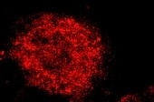
Application: Immunocytochemistry/ImmunofluorescenceSample Tested: Hippocampal neuronsSpecies: MouseVerified Customer | Posted 10/16/2021

There are no reviews that match your criteria.
Protocols
Find general support by application which include: protocols, troubleshooting, illustrated assays, videos and webinars.
- Antigen Retrieval Protocol (PIER)
- Antigen Retrieval for Frozen Sections Protocol
- Appropriate Fixation of IHC/ICC Samples
- Cellular Response to Hypoxia Protocols
- Chromogenic IHC Staining of Formalin-Fixed Paraffin-Embedded (FFPE) Tissue Protocol
- Chromogenic Immunohistochemistry Staining of Frozen Tissue
- ClariTSA™ Fluorophore Kits
- Detection & Visualization of Antibody Binding
- Fluorescent IHC Staining of Frozen Tissue Protocol
- Graphic Protocol for Heat-induced Epitope Retrieval
- Graphic Protocol for the Preparation and Fluorescent IHC Staining of Frozen Tissue Sections
- Graphic Protocol for the Preparation and Fluorescent IHC Staining of Paraffin-embedded Tissue Sections
- Graphic Protocol for the Preparation of Gelatin-coated Slides for Histological Tissue Sections
- IHC Sample Preparation (Frozen sections vs Paraffin)
- Immunofluorescent IHC Staining of Formalin-Fixed Paraffin-Embedded (FFPE) Tissue Protocol
- Immunohistochemistry (IHC) and Immunocytochemistry (ICC) Protocols
- Immunohistochemistry Frozen Troubleshooting
- Immunohistochemistry Paraffin Troubleshooting
- Preparing Samples for IHC/ICC Experiments
- Preventing Non-Specific Staining (Non-Specific Binding)
- Primary Antibody Selection & Optimization
- Protocol for Heat-Induced Epitope Retrieval (HIER)
- Protocol for Making a 4% Formaldehyde Solution in PBS
- Protocol for VisUCyte™ HRP Polymer Detection Reagent
- Protocol for the Preparation & Fixation of Cells on Coverslips
- Protocol for the Preparation and Chromogenic IHC Staining of Frozen Tissue Sections
- Protocol for the Preparation and Chromogenic IHC Staining of Frozen Tissue Sections - Graphic
- Protocol for the Preparation and Chromogenic IHC Staining of Paraffin-embedded Tissue Sections
- Protocol for the Preparation and Chromogenic IHC Staining of Paraffin-embedded Tissue Sections - Graphic
- Protocol for the Preparation and Fluorescent IHC Staining of Frozen Tissue Sections
- Protocol for the Preparation and Fluorescent IHC Staining of Paraffin-embedded Tissue Sections
- Protocol for the Preparation of Gelatin-coated Slides for Histological Tissue Sections
- R&D Systems Quality Control Western Blot Protocol
- TUNEL and Active Caspase-3 Detection by IHC/ICC Protocol
- The Importance of IHC/ICC Controls
- Troubleshooting Guide: Immunohistochemistry
- Troubleshooting Guide: Western Blot Figures
- Western Blot Conditions
- Western Blot Protocol
- Western Blot Protocol for Cell Lysates
- Western Blot Troubleshooting
- Western Blot Troubleshooting Guide
- View all Protocols, Troubleshooting, Illustrated assays and Webinars
Loading...
Associated Pathways

