Human ApoB (Apolipoprotein B-100) is a 550 kDa, secreted, palmitoylated glycoprotein that is part of LDL and VLDL particles. It is made by liver and is 4536 amino acids (aa) in length. It binds LDL to the ApoB/E receptor. Multiple forms of ApoB-100 exist; ApoB-25, 26, 29, 37, 39, 48, and 74. The numbers reflect each molecule’s predicted MW as a percentage of ApoB-100 (512 kDa). Amino acids 1206-1414 of the precursor were used for immunization. Based on likelihood of secretion, this antibody may recognize B26 (1297 aa), B37 (1728 aa), B39 (1799 aa), and B48 (2152 aa). Over the immunization polypeptide range, human ApoB-100 is 80% and 82% aa identical to mouse and dog ApoB-100, respectively.
Human Apolipoprotein B/ApoB Antibody (369717)
R&D Systems | Catalog # MAB4124

Key Product Details
Species Reactivity
Validated:
Human
Cited:
Human
Applications
Validated:
Immunohistochemistry, Western Blot, Simple Western
Cited:
Western Blot, Flow Cytometry, ELISA Capture
Label
Unconjugated
Antibody Source
Monoclonal Mouse IgG2B Clone # 369717
Loading...
Product Specifications
Immunogen
E. coli-derived recombinant human ApoB
Met1206-Asp1413
Accession # NP_000375
Met1206-Asp1413
Accession # NP_000375
Specificity
Detects human Apolipoprotein B in direct ELISAs and Western blots.
Clonality
Monoclonal
Host
Mouse
Isotype
IgG2B
Scientific Data Images for Human Apolipoprotein B/ApoB Antibody (369717)
Detection of Human Apolipoprotein B/ApoB by Western Blot.
Western blot shows human plasma. PVDF membrane was probed with 2 µg/mL of Mouse Anti-Human Apolipoprotein B/ApoB Monoclonal Antibody (Catalog # MAB4124) followed by HRP-conjugated Anti-Mouse IgG Secondary Antibody (Catalog # HAF018). A specific band was detected for Apolipoprotein B/ApoB at approximately 500 kDa (as indicated). This experiment was conducted under reducing conditions and using Immunoblot Buffer Group 1.
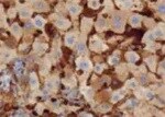
Apolipoprotein B/ApoB in Human Liver.
Apolipoprotein B/ApoB was detected in immersion fixed paraffin-embedded sections of human liver using Mouse Anti-Human Apolipoprotein B/ApoB Monoclonal Antibody (Catalog # MAB4124) at 5 µg/mL for 1 hour at room temperature followed by incubation with the Anti-Mouse IgG VisUCyte™ HRP Polymer Antibody (Catalog # VC001). Tissue was stained using DAB (brown) and counterstained with hematoxylin (blue). Specific staining was localized to plasma membranes in hepatocytes. View our protocol for IHC Staining with VisUCyte HRP Polymer Detection Reagents.
Detection of Human Apolipoprotein B/ApoB by Simple WesternTM.
Simple Western shows lysates of human plasma, loaded at 1:50. A specific band was detected for Apolipoprotein B/ApoB at approximately 333 kDa (as indicated) using 5 µg/mL of Mouse Anti-Human Apolipoprotein B/ApoB Monoclonal Antibody (Catalog # MAB4124). This experiment was conducted under reducing conditions and using the 66-440kDa separation system.Applications for Human Apolipoprotein B/ApoB Antibody (369717)
Application
Recommended Usage
Immunohistochemistry
8-25 µg/mL
Sample: Immersion fixed paraffin-embedded sections of human liver
Sample: Immersion fixed paraffin-embedded sections of human liver
Simple Western
5 µg/mL
Sample: Human plasma
Sample: Human plasma
Western Blot
2 µg/mL
Sample: Human plasma
Sample: Human plasma
Reviewed Applications
Read 1 review rated 5 using MAB4124 in the following applications:
Formulation, Preparation, and Storage
Purification
Protein A or G purified from hybridoma culture supernatant
Reconstitution
Reconstitute at 0.5 mg/mL in sterile PBS. For liquid material, refer to CoA for concentration.
Loading...
Formulation
Lyophilized from a 0.2 μm filtered solution in PBS with Trehalose. *Small pack size (SP) is supplied either lyophilized or as a 0.2 µm filtered solution in PBS.
Shipping
Lyophilized product is shipped at ambient temperature. Liquid small pack size (-SP) is shipped with polar packs. Upon receipt, store immediately at the temperature recommended below.
Stability & Storage
Use a manual defrost freezer and avoid repeated freeze-thaw cycles.
- 12 months from date of receipt, -20 to -70 °C as supplied.
- 1 month, 2 to 8 °C under sterile conditions after reconstitution.
- 6 months, -20 to -70 °C under sterile conditions after reconstitution.
Calculators
Background: Apolipoprotein B/ApoB
Alternate Names
APOB, ApoB-100, ApoB-48, FLDB, LDLCQ4
Gene Symbol
APOB
UniProt
Additional Apolipoprotein B/ApoB Products
Product Documents for Human Apolipoprotein B/ApoB Antibody (369717)
Certificate of Analysis
To download a Certificate of Analysis, please enter a lot or batch number in the search box below.
Note: Certificate of Analysis not available for kit components.
Product Specific Notices for Human Apolipoprotein B/ApoB Antibody (369717)
For research use only
Related Research Areas
Citations for Human Apolipoprotein B/ApoB Antibody (369717)
 Powered by Bioz
Customer Reviews for Human Apolipoprotein B/ApoB Antibody (369717) (1)
5 out of 5
1 Customer Rating
Have you used Human Apolipoprotein B/ApoB Antibody (369717)?
Submit a review and receive an Amazon gift card!
$25/€18/£15/$25CAN/¥2500 Yen for a review with an image
$10/€7/£6/$10CAN/¥1110 Yen for a review without an image
Submit a review
Customer Images
Showing
1
-
1 of
1 review
Showing All
Filter By:
-
Application: ImmunohistochemistrySample Tested: Liver tissueSpecies: HumanVerified Customer | Posted 11/17/2021

There are no reviews that match your criteria.
Protocols
Find general support by application which include: protocols, troubleshooting, illustrated assays, videos and webinars.
- Antigen Retrieval Protocol (PIER)
- Antigen Retrieval for Frozen Sections Protocol
- Appropriate Fixation of IHC/ICC Samples
- Cellular Response to Hypoxia Protocols
- Chromogenic IHC Staining of Formalin-Fixed Paraffin-Embedded (FFPE) Tissue Protocol
- Chromogenic Immunohistochemistry Staining of Frozen Tissue
- ClariTSA™ Fluorophore Kits
- Detection & Visualization of Antibody Binding
- Fluorescent IHC Staining of Frozen Tissue Protocol
- Graphic Protocol for Heat-induced Epitope Retrieval
- Graphic Protocol for the Preparation and Fluorescent IHC Staining of Frozen Tissue Sections
- Graphic Protocol for the Preparation and Fluorescent IHC Staining of Paraffin-embedded Tissue Sections
- Graphic Protocol for the Preparation of Gelatin-coated Slides for Histological Tissue Sections
- IHC Sample Preparation (Frozen sections vs Paraffin)
- Immunofluorescent IHC Staining of Formalin-Fixed Paraffin-Embedded (FFPE) Tissue Protocol
- Immunohistochemistry (IHC) and Immunocytochemistry (ICC) Protocols
- Immunohistochemistry Frozen Troubleshooting
- Immunohistochemistry Paraffin Troubleshooting
- Preparing Samples for IHC/ICC Experiments
- Preventing Non-Specific Staining (Non-Specific Binding)
- Primary Antibody Selection & Optimization
- Protocol for Heat-Induced Epitope Retrieval (HIER)
- Protocol for Making a 4% Formaldehyde Solution in PBS
- Protocol for VisUCyte™ HRP Polymer Detection Reagent
- Protocol for the Preparation & Fixation of Cells on Coverslips
- Protocol for the Preparation and Chromogenic IHC Staining of Frozen Tissue Sections
- Protocol for the Preparation and Chromogenic IHC Staining of Frozen Tissue Sections - Graphic
- Protocol for the Preparation and Chromogenic IHC Staining of Paraffin-embedded Tissue Sections
- Protocol for the Preparation and Chromogenic IHC Staining of Paraffin-embedded Tissue Sections - Graphic
- Protocol for the Preparation and Fluorescent IHC Staining of Frozen Tissue Sections
- Protocol for the Preparation and Fluorescent IHC Staining of Paraffin-embedded Tissue Sections
- Protocol for the Preparation of Gelatin-coated Slides for Histological Tissue Sections
- R&D Systems Quality Control Western Blot Protocol
- TUNEL and Active Caspase-3 Detection by IHC/ICC Protocol
- The Importance of IHC/ICC Controls
- Troubleshooting Guide: Immunohistochemistry
- Troubleshooting Guide: Western Blot Figures
- Western Blot Conditions
- Western Blot Protocol
- Western Blot Protocol for Cell Lysates
- Western Blot Troubleshooting
- Western Blot Troubleshooting Guide
- View all Protocols, Troubleshooting, Illustrated assays and Webinars
Loading...
Associated Pathways
