B7-1 and B7-2, together with their receptors CD28 and CTLA-4, constitute one of the dominant costimulatory pathways that regulate T- and B-cell responses. Although both CTLA-4 and CD28 can bind to the same ligands, CTLA-4 binds to B7-1 and B7-2 with a 20‑100 fold higher affinity than CD28 and is involved in the down‑regulation of the immune response. B7-1 is expressed on activated B cells, activated T cells, and macrophages. B7-2 is constitutively expressed on interdigitating dendritic cells, Langerhans cells, peripheral blood dendritic cells, memory B cells, and germinal center B cells. Additionally, B7-2 is expressed at low levels on monocytes and can be up-regulated through interferon gamma. B7-1 and B7-2 are both members of the immunoglobulin superfamily. Human B7-2 is a 329 amino acid (aa) protein containing a putative 23 aa signal peptide, a 224 aa extracellular domain, a 21 aa transmembrane domain, and a 61 aa cytoplasmic domain. Human B7-2 and B7-1 share 26% amino acid identity. Human and mouse B7-2 share 50% amino acid identity. However, it has been observed that both human and mouse B7‑1 and
B7‑2 can bind to either human or mouse CD28 and CTLA-4, suggesting that there are conserved amino acids which form the B7-1/B7-2/CD28/CTLA-4 critical binding sites.

Key Product Details
Validated by
Knockout/Knockdown
Species Reactivity
Validated:
Human
Cited:
Human, Mouse
Applications
Validated:
Knockout Validated, Immunohistochemistry, Western Blot, Neutralization
Cited:
Immunohistochemistry, Immunohistochemistry-Paraffin, Flow Cytometry, Immunocytochemistry, ELISA Development
Label
Unconjugated
Antibody Source
Polyclonal Goat IgG
Loading...
Product Specifications
Immunogen
S. frugiperda insect ovarian cell line Sf 21-derived recombinant human B7‑2/CD86
Ala23-His244
Accession # P42081
Ala23-His244
Accession # P42081
Specificity
Detects human B7‑2/CD86 in direct ELISAs. In direct ELISAs, less than 10% cross‑reactivity with recombinant mouse B7-2 and recombinant rat B7-2 is observed.
Clonality
Polyclonal
Host
Goat
Isotype
IgG
Endotoxin Level
<0.10 EU per 1 μg of the antibody by the LAL method.
Scientific Data Images for Human B7-2/CD86 Antibody
Detection of Human B7‑2/CD86 by Western Blot.
Western blot shows lysates of Daudi human Burkitt's lymphoma cell line. PVDF membrane was probed with 0.5 µg/mL of Goat Anti-Human B7-2/CD86 Antigen Affinity-purified Polyclonal Antibody (Catalog # AF-141-NA) followed by HRP-conjugated Anti-Goat IgG Secondary Antibody (HAF017). A specific band was detected for B7-2/CD86 at approximately 75 kDa (as indicated). This experiment was conducted under reducing conditions and using Immunoblot Buffer Group 1.
B7‑2/CD86 in Human Tonsil.
B7-2/CD86 was detected in immersion fixed paraffin-embedded sections of human tonsil using Goat Anti-Human B7-2/CD86 Antigen Affinity-purified Polyclonal Antibody (Catalog # AF-141-NA) at 15 µg/mL overnight at 4 °C. Tissue was stained using the Anti-Goat HRP-DAB Cell & Tissue Staining Kit (brown; CTS008) and counterstained with hematoxylin (blue). View our protocol for Chromogenic IHC Staining of immersion fixed paraffin-embedded Tissue Sections.
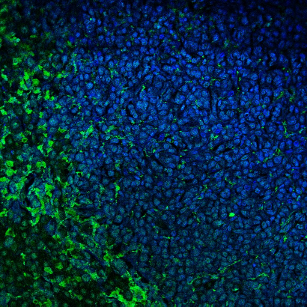
B7‑2/CD86 in Human Tonsil.
B7-2/CD86 was detected in immersion fixed paraffin-embedded sections of human tonsil using Goat Anti-Human B7-2/CD86 Antigen Affinity-purified Polyclonal Antibody (Catalog # AF-141-NA) at 3 µg/mL for 1 hour at room temperature followed by incubation with the Anti-Goat IgG VisUCyte™ HRP Polymer Antibody (Catalog # VC004). Tissue was stained using DAB (brown) and counterstained with hematoxylin (blue). Specific staining was localized to lymphocytes. View our protocol for IHC Staining with VisUCyte HRP Polymer Detection Reagents.
IL‑2 secretion Induced by B7‑2/CD86 and Neutralization by Human B7‑2/CD86 Antibody.
Recombinant Human B7-2/CD86 Fc Chimera (141-B2) co-stimulates IL-2 secretion in the Jurkat human acute T cell leukemia cell line in the presence of PHA in a dose-dependent manner (orange line), as measured by the Human IL-2 Quantikine ELISA Kit (D2050). IL-2 secretion elicited by Recombinant Human B7-2/CD86 Fc Chimera (2 µg/mL) and PHA (10 µg/mL) is neutralized (green line) by increasing concentrations of Goat Anti-Human B7-2/CD86 Antigen Affinity-purified Polyclonal Antibody (Catalog # AF-141-NA). The ND50 is typically 0.25-1.25 µg/mL.
Western Blot Shows Human B7‑2/CD86 Specificity by Using Knockout Cell Line.
Western blot shows lysates of Ramos human Burkitt's lymphoma parental cell line and B7-2/CD86 knockout Ramos cell line (KO). PVDF membrane was probed with 0.5 µg/mL of Goat Anti-Human B7-2/CD86 Antigen Affinity-purified Polyclonal Antibody (Catalog # AF-141-NA) followed by HRP-conjugated Anti-Goat IgG Secondary Antibody (HAF017). A specific band was detected for B7-2/CD86 at approximately 74 kDa (as indicated) in the parental Ramos cell line, but is not detectable in knockout Ramos cell line. GAPDH (Catalog # AF5718) is shown as a loading control. This experiment was conducted under reducing conditions and using Immunoblot Buffer Group 1.Applications for Human B7-2/CD86 Antibody
Application
Recommended Usage
Immunohistochemistry
5-15 µg/mL
Sample: Immersion fixed paraffin-embedded sections of human tonsil
Sample: Immersion fixed paraffin-embedded sections of human tonsil
Knockout Validated
B7-2/CD86 is specifically detected in Ramos human Burkitt's lymphoma parental cell line but is not detectable in B7-2/CD86 knockout Ramos cell line
Western Blot
0.5 µg/mL
Sample: Daudi human Burkitt's lymphoma cell line
Sample: Daudi human Burkitt's lymphoma cell line
Neutralization
Measured by its ability to neutralize B7‑2/CD86-induced IL‑2 secretion in the Jurkat human acute T cell leukemia cell line. Linsley, P. et al. (1990) Proc. Natl. Acad. Sci. 87:5031. The Neutralization Dose (ND50) is typically 0.25-1.25 µg/mL in the presence of 2 µg/mL Recombinant Human B7‑2/CD86 Fc Chimera and 10 µg/mL PHA.
Reviewed Applications
Read 1 review rated 3 using AF-141-NA in the following applications:
Formulation, Preparation, and Storage
Purification
Antigen Affinity-purified
Reconstitution
Reconstitute at 0.2 mg/mL in sterile PBS. For liquid material, refer to CoA for concentration.
Loading...
Formulation
Lyophilized from a 0.2 μm filtered solution in PBS with Trehalose. See Certificate of Analysis for details.
*Small pack size (-SP) is supplied either lyophilized or as a 0.2 µm filtered solution in PBS.
*Small pack size (-SP) is supplied either lyophilized or as a 0.2 µm filtered solution in PBS.
Shipping
Lyophilized product is shipped at ambient temperature. Liquid small pack size (-SP) is shipped with polar packs. Upon receipt, store immediately at the temperature recommended below.
Stability & Storage
Use a manual defrost freezer and avoid repeated freeze-thaw cycles.
- 12 months from date of receipt, -20 to -70 °C as supplied.
- 1 month, 2 to 8 °C under sterile conditions after reconstitution.
- 6 months, -20 to -70 °C under sterile conditions after reconstitution.
Calculators
Background: B7-2/CD86
References
- Azuma, M. et al. (1993) Nature 366:76.
- Freeman, G.J. et al. (1993) Science 262:909.
- Freeman, G. et al. (1991) J. Exp. Med. 174:625.
- Selvakumar, A. et al. (1993) Immunogenetics 38:292.
- Chen, C. et al. (1994) J. Immunol. 152:4929.
- Freeman, G.J. et al. (1993) J. Exp. Med. 178:2185.
Alternate Names
B72, CD86
Gene Symbol
CD86
UniProt
Additional B7-2/CD86 Products
Product Documents for Human B7-2/CD86 Antibody
Certificate of Analysis
To download a Certificate of Analysis, please enter a lot or batch number in the search box below.
Note: Certificate of Analysis not available for kit components.
Product Specific Notices for Human B7-2/CD86 Antibody
For research use only
Citations for Human B7-2/CD86 Antibody
 Powered by Bioz
Customer Reviews for Human B7-2/CD86 Antibody (1)
3 out of 5
1 Customer Rating
Have you used Human B7-2/CD86 Antibody?
Submit a review and receive an Amazon gift card!
$25/€18/£15/$25CAN/¥2500 Yen for a review with an image
$10/€7/£6/$10CAN/¥1110 Yen for a review without an image
Submit a review
Customer Images
Showing
1
-
1 of
1 review
Showing All
Filter By:
-
Application: Immunocytochemistry/ImmunofluorescenceSample Tested: Melanoma tissueSpecies: HumanVerified Customer | Posted 04/11/2022

There are no reviews that match your criteria.
Protocols
Find general support by application which include: protocols, troubleshooting, illustrated assays, videos and webinars.
- Antigen Retrieval Protocol (PIER)
- Antigen Retrieval for Frozen Sections Protocol
- Appropriate Fixation of IHC/ICC Samples
- Cellular Response to Hypoxia Protocols
- Chromogenic IHC Staining of Formalin-Fixed Paraffin-Embedded (FFPE) Tissue Protocol
- Chromogenic Immunohistochemistry Staining of Frozen Tissue
- ClariTSA™ Fluorophore Kits
- Detection & Visualization of Antibody Binding
- Fluorescent IHC Staining of Frozen Tissue Protocol
- Graphic Protocol for Heat-induced Epitope Retrieval
- Graphic Protocol for the Preparation and Fluorescent IHC Staining of Frozen Tissue Sections
- Graphic Protocol for the Preparation and Fluorescent IHC Staining of Paraffin-embedded Tissue Sections
- Graphic Protocol for the Preparation of Gelatin-coated Slides for Histological Tissue Sections
- IHC Sample Preparation (Frozen sections vs Paraffin)
- Immunofluorescent IHC Staining of Formalin-Fixed Paraffin-Embedded (FFPE) Tissue Protocol
- Immunohistochemistry (IHC) and Immunocytochemistry (ICC) Protocols
- Immunohistochemistry Frozen Troubleshooting
- Immunohistochemistry Paraffin Troubleshooting
- Preparing Samples for IHC/ICC Experiments
- Preventing Non-Specific Staining (Non-Specific Binding)
- Primary Antibody Selection & Optimization
- Protocol for Heat-Induced Epitope Retrieval (HIER)
- Protocol for Making a 4% Formaldehyde Solution in PBS
- Protocol for VisUCyte™ HRP Polymer Detection Reagent
- Protocol for the Preparation & Fixation of Cells on Coverslips
- Protocol for the Preparation and Chromogenic IHC Staining of Frozen Tissue Sections
- Protocol for the Preparation and Chromogenic IHC Staining of Frozen Tissue Sections - Graphic
- Protocol for the Preparation and Chromogenic IHC Staining of Paraffin-embedded Tissue Sections
- Protocol for the Preparation and Chromogenic IHC Staining of Paraffin-embedded Tissue Sections - Graphic
- Protocol for the Preparation and Fluorescent IHC Staining of Frozen Tissue Sections
- Protocol for the Preparation and Fluorescent IHC Staining of Paraffin-embedded Tissue Sections
- Protocol for the Preparation of Gelatin-coated Slides for Histological Tissue Sections
- R&D Systems Quality Control Western Blot Protocol
- TUNEL and Active Caspase-3 Detection by IHC/ICC Protocol
- The Importance of IHC/ICC Controls
- Troubleshooting Guide: Immunohistochemistry
- Troubleshooting Guide: Western Blot Figures
- Western Blot Conditions
- Western Blot Protocol
- Western Blot Protocol for Cell Lysates
- Western Blot Troubleshooting
- Western Blot Troubleshooting Guide
- View all Protocols, Troubleshooting, Illustrated assays and Webinars


