
Key Product Details
Species Reactivity
Validated:
Human
Cited:
Human, Mouse
Applications
Validated:
Neutralization, Flow Cytometry, Immunocytochemistry, CyTOF-ready
Cited:
Immunohistochemistry, Immunohistochemistry-Paraffin, Immunohistochemistry-Frozen, Neutralization, Flow Cytometry, Immunocytochemistry, Bioassay, Competitive Binding Assay, Functional Assay
Label
Unconjugated
Antibody Source
Monoclonal Mouse IgG2B Clone # 45531
Loading...
Product Specifications
Immunogen
NS0 mouse myeloma cell line transfected with human CCR5
Met1-Leu352
Accession # P51681
Met1-Leu352
Accession # P51681
Specificity
Detects human CCR5.
Clonality
Monoclonal
Host
Mouse
Isotype
IgG2B
Endotoxin Level
<0.10 EU per 1 μg of the antibody by the LAL method.
Scientific Data Images for Human CCR5 Antibody (45531)
Detection of CCR5 in Human PBMCs by Flow Cytometry.
Human peripheral blood mononuclear cells (PBMCs) were stained with either (A) Mouse Anti-Human CCR5 Monoclonal Antibody (Catalog # MAB182) or (B) Mouse IgG2B Isotype Control (Catalog # MAB0041) followed by Goat anti-Mouse IgG APC-conjugated Secondary Antibody (Catalog # F0101B) and Mouse Anti-Human CD3 epsilon PE-conjugated Monoclonal Antibody (Catalog # FAB100P). View our protocol for Staining Membrane-associated Proteins.
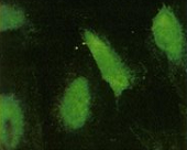
CCR5 in Human Dendritic Cells.
CCR5 was detected in immersion fixed human dendritic cells using Human CCR5 Monoclonal Antibody (Catalog # MAB182) at 10 µg/mL for 3 hours at room temperature. Cells were stained using the NorthernLights™ 557-conjugated Anti-Mouse IgG Secondary Antibody (yellow; Catalog # NL007) and counterstained with DAPI (blue). View our protocol for Fluorescent ICC Staining of Non-adherent Cells.
Chemotaxis Induced by CCL3/MIP‑1 alpha and Neutraliza-tion by Human CCR5 Antibody.
Recombinant Human CCL3/MIP‑1a (Catalog # 270-LD) chemoattracts the BaF3 mouse pro‑B cell line transfected with human CCR5 in a dose-dependent manner (orange line). The amount of cells that migrated through to the lower chemotaxis chamber was measured by Resazurin (Catalog # AR002). Chemotaxis elicited by Recombinant Human CCL3/MIP‑1a (40 ng/mL) is neutralized (green line) by increasing concentrations of Human CCR5 Monoclonal Antibody (Catalog # MAB182). The ND50 is typically 5-20 µg/mL.Applications for Human CCR5 Antibody (45531)
Application
Recommended Usage
CyTOF-ready
Ready to be labeled using established conjugation methods. No BSA or other carrier proteins that could interfere with conjugation.
Flow Cytometry
0.25 µg/106 cells
Sample: Human PBMC
Sample: Human PBMC
Immunocytochemistry
8-25 µg/mL
Sample: Immersion fixed human dendritic cells
Sample: Immersion fixed human dendritic cells
Neutralization
Measured by its ability to neutralize CCL3/MIP‑1 alpha -induced chemotaxis in the BaF3 mouse pro‑B cell line transfected with human CCR5. The Neutralization Dose (ND50) is typically 5-20 µg/mL in the presence of 40 ng/mL Recombinant Human CCL3/MIP‑1 alpha isoform LD78a.
Reviewed Applications
Read 3 reviews rated 3.3 using MAB182 in the following applications:
Flow Cytometry Panel Builder
Bio-Techne Knows Flow Cytometry
Save time and reduce costly mistakes by quickly finding compatible reagents using the Panel Builder Tool.
Advanced Features
- Spectra Viewer - Custom analysis of spectra from multiple fluorochromes
- Spillover Popups - Visualize the spectra of individual fluorochromes
- Antigen Density Selector - Match fluorochrome brightness with antigen density

Formulation, Preparation, and Storage
Purification
Protein A or G purified from ascites
Reconstitution
Reconstitute at 0.5 mg/mL in sterile PBS. For liquid material, refer to CoA for concentration.
Loading...
Formulation
Lyophilized from a 0.2 μm filtered solution in PBS with Trehalose. *Small pack size (SP) is supplied either lyophilized or as a 0.2 µm filtered solution in PBS.
Shipping
Lyophilized product is shipped at ambient temperature. Liquid small pack size (-SP) is shipped with polar packs. Upon receipt, store immediately at the temperature recommended below.
Stability & Storage
Use a manual defrost freezer and avoid repeated freeze-thaw cycles.
- 12 months from date of receipt, -20 to -70 °C as supplied.
- 1 month, 2 to 8 °C under sterile conditions after reconstitution.
- 6 months, -20 to -70 °C under sterile conditions after reconstitution.
Calculators
Background: CCR5
Alternate Names
CCR5, CD195
Gene Symbol
CCR5
UniProt
Additional CCR5 Products
Product Documents for Human CCR5 Antibody (45531)
Certificate of Analysis
To download a Certificate of Analysis, please enter a lot or batch number in the search box below.
Note: Certificate of Analysis not available for kit components.
Product Specific Notices for Human CCR5 Antibody (45531)
For research use only
Citations for Human CCR5 Antibody (45531)
 Powered by Bioz
Customer Reviews for Human CCR5 Antibody (45531) (3)
3.3 out of 5
3 Customer Ratings
Have you used Human CCR5 Antibody (45531)?
Submit a review and receive an Amazon gift card!
$25/€18/£15/$25CAN/¥2500 Yen for a review with an image
$10/€7/£6/$10CAN/¥1110 Yen for a review without an image
Submit a review
Customer Images
Showing
1
-
3 of
3 reviews
Showing All
Filter By:
-
Application: Immunocytochemistry/ImmunofluorescenceSample Tested: Brain endothelial cellsSpecies: HumanVerified Customer | Posted 02/07/2022

-
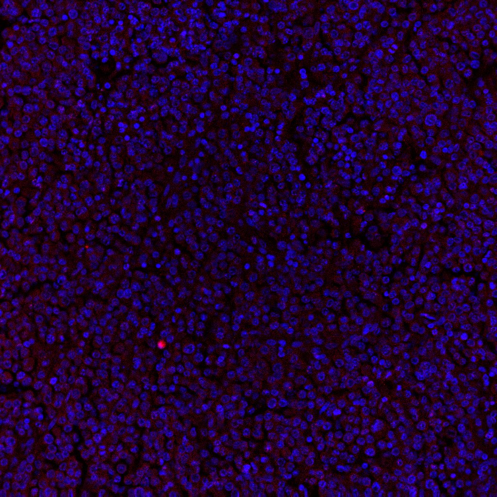
Application: Immunocytochemistry/ImmunofluorescenceSample Tested: Melanoma tissueSpecies: HumanVerified Customer | Posted 07/01/2021
Bio-Techne ResponseThank you for reviewing our product. We are sorry to hear that this product did not perform as expected. We have been in touch with the customer to resolve this issue according to our Product Guarantee and to the customer’s satisfaction.
-
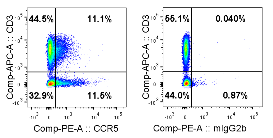
Application: Flow CytometrySample Tested: Peripheral blood mononuclear cells (PBMCs)Species: HumanVerified Customer | Posted 08/28/2018Human PBMCs stained with anti-human CCR5 antibody (MAB182; 2.5 µg/ml) or respective mIgG2b isotype control followed by goat anti-mouse IgG PE and then followed by anti-human CD3 APC. Gated on single live cells.Freshly isolated human PBMCs were stained with mouse anti-human CCR5 monoclonal antibody (MAB182) or mIgG2b isotype control at 2.5 µg/ml for 30 minutes on ice followed by goat anti-mouse IgG PE secondary staining. Additional CD3 APC staining.
There are no reviews that match your criteria.
Protocols
Find general support by application which include: protocols, troubleshooting, illustrated assays, videos and webinars.
- 7-Amino Actinomycin D (7-AAD) Cell Viability Flow Cytometry Protocol
- Appropriate Fixation of IHC/ICC Samples
- Cellular Response to Hypoxia Protocols
- ClariTSA™ Fluorophore Kits
- Detection & Visualization of Antibody Binding
- Extracellular Membrane Flow Cytometry Protocol
- Flow Cytometry Protocol for Cell Surface Markers
- Flow Cytometry Protocol for Staining Membrane Associated Proteins
- Flow Cytometry Staining Protocols
- Flow Cytometry Troubleshooting Guide
- ICC Cell Smear Protocol for Suspension Cells
- ICC Immunocytochemistry Protocol Videos
- ICC for Adherent Cells
- Immunocytochemistry (ICC) Protocol
- Immunocytochemistry Troubleshooting
- Immunofluorescence of Organoids Embedded in Cultrex Basement Membrane Extract
- Immunohistochemistry (IHC) and Immunocytochemistry (ICC) Protocols
- Intracellular Flow Cytometry Protocol Using Alcohol (Methanol)
- Intracellular Flow Cytometry Protocol Using Detergents
- Intracellular Nuclear Staining Flow Cytometry Protocol Using Detergents
- Intracellular Staining Flow Cytometry Protocol Using Alcohol Permeabilization
- Intracellular Staining Flow Cytometry Protocol Using Detergents to Permeabilize Cells
- Preparing Samples for IHC/ICC Experiments
- Preventing Non-Specific Staining (Non-Specific Binding)
- Primary Antibody Selection & Optimization
- Propidium Iodide Cell Viability Flow Cytometry Protocol
- Protocol for Liperfluo
- Protocol for VisUCyte™ HRP Polymer Detection Reagent
- Protocol for the Characterization of Human Th22 Cells
- Protocol for the Characterization of Human Th9 Cells
- Protocol for the Fluorescent ICC Staining of Cell Smears - Graphic
- Protocol for the Fluorescent ICC Staining of Cultured Cells on Coverslips - Graphic
- Protocol for the Preparation and Fluorescent ICC Staining of Cells on Coverslips
- Protocol for the Preparation and Fluorescent ICC Staining of Non-adherent Cells
- Protocol for the Preparation and Fluorescent ICC Staining of Stem Cells on Coverslips
- Protocol for the Preparation of a Cell Smear for Non-adherent Cell ICC - Graphic
- Protocol: Annexin V and PI Staining by Flow Cytometry
- Protocol: Annexin V and PI Staining for Apoptosis by Flow Cytometry
- TUNEL and Active Caspase-3 Detection by IHC/ICC Protocol
- The Importance of IHC/ICC Controls
- Troubleshooting Guide: Fluorokine Flow Cytometry Kits
- View all Protocols, Troubleshooting, Illustrated assays and Webinars
Loading...
Associated Pathways
