Human CD25/IL-2R alpha Antibody (24204)
R&D Systems | Catalog # MAB623

Key Product Details
Species Reactivity
Validated:
Human
Cited:
Human, Cynomolgus Monkey, Xenograft
Applications
Validated:
Immunohistochemistry, Western Blot, ELISA Capture (Matched Antibody Pair), Immunocytochemistry
Cited:
Immunohistochemistry, Western Blot, Antibody Array Development, ELISA Capture, ELISA Development
Label
Unconjugated
Antibody Source
Monoclonal Mouse IgG1 Clone # 24204
Loading...
Product Specifications
Immunogen
S. frugiperda insect ovarian cell line Sf 21-derived recombinant human CD25/IL-2 R alpha
Glu22-Cys213
Accession # P01589
Glu22-Cys213
Accession # P01589
Specificity
Detects human CD25/IL-2 R alpha in ELISAs and Western blots. In Western blots, detects recombinant rat CD25/IL-2 R alpha but no cross-reactivity with recombinant mouse CD25/IL-2 R alpha. In sandwich immunoassays, this antibody does not cross-react or interfere with recombinant human (rh) IL-1 RI, rhIL-1 RII, rhIL-2 R beta, rhIL-3 R alpha, rhIL-4 R, rhIL-5 R alpha, rhIL-5 R beta, rhIL-6 R, or rhgp130.
Clonality
Monoclonal
Host
Mouse
Isotype
IgG1
Scientific Data Images for Human CD25/IL-2R alpha Antibody (24204)
Detection of Recombinant Human and Rat CD25/IL-2 R alpha by Western Blot.
Western blot shows 25 ng of Recombinant Human CD25/IL-2 R alpha (Catalog # 223-2A), Recombinant Mouse CD25/IL-2 R alpha (Catalog # 2438-RM) and Recombinant Rat CD25/IL-2 R alpha Fc Chimera (Catalog # 5156-RM). PVDF Membrane was probed with 1 µg/mL of Mouse Anti-Human CD25/IL-2 R alpha Monoclonal Antibody (Catalog # MAB623) followed by HRP-conjugated Anti-Mouse IgG Secondary Antibody (Catalog # HAF018). Specific bands were detected for CD25/IL-2 R alpha at approximately 30-250 kDa (as indicated). This experiment was conducted under non-reducing conditions and using Immunoblot Buffer Group 3.
CD25/IL‑2 R alpha in Human PBMCs.
CD25/IL-2 Ra was detected in immersion fixed human peripheral blood mononuclear cells (PBMCs) stimulated with PHA using Mouse Anti-Human CD25/IL-2 Ra Monoclonal Antibody (Catalog # MAB623) at 10 µg/mL for 3 hours at room temperature. Cells were stained using the NorthernLights™ 557-conjugated Anti-Mouse IgG Secondary Antibody (yellow; Catalog # NL007) and counterstained with DAPI (blue). Upper panel shows a lack of labeling if primary antibodies are omitted and tissue is stained only with secondary antibody followed by incubation with detection reagents. View our protocol for Fluorescent ICC Staining of Non-adherent Cells.
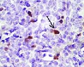
CD25/IL‑2 R alpha in Human Lymph Node.
CD25/IL‑2 R alpha was detected in immersion fixed frozen sections of human lymph node using 15 µg/mL Mouse Anti-Human CD25/IL‑2 R alpha Monoclonal Antibody (Catalog # MAB623) overnight at 4 °C. Tissue was stained (red) and counterstained with hematoxylin (blue). View our protocol for Chromogenic IHC Staining of Frozen Tissue Sections.
Detection of CD25/IL-2R alpha by Western Blot
UBTD1 depletion slows down RTK degradation.(A–E) DU145 cells were transfected for 48 hr with the indicated siRNA (control, siCTRLpool or UBTD1: siUBTD1pool). (A) HGF-alexa647 pulse chase images and quantification. (B) Proximal ligation assay monitoring and quantification of MET associated with ubiquitin in DU145 treated with HGF (40 ng/ml, 10 min). Nuclei were stained with DAPI (blue) on the MERGE image. (C) Immunoblots show MET ubiquitylation in presence of HGF (40 ng/ml, 10 min) in HEK cells in different experimental conditions. Cells were transfected, as indicated, with expression vectors for histidine-tagged ubiquitin (His-Ub) and MET. His-Ub crosslinked forms of MET were purified (IP: His) and the immunoblot of MET showed MET ubiquitylation. The immunoblot of MET (bottom panel) was performed in parallel to verify the amounts of MET protein engaged in His-Ub purifications. The immunoblot of UBTD1 shows the level of siRNA depletion. (D) Immunoblot and quantification of receptors. The immunoblot of UBTD1 shows the level of siRNA depletion. (E) mRNA quantification of receptors. mRNA of UBTD1 shows the level of siRNA depletion. Scale bar = 10 µm. n ≥ 3 independent experiments. ns = non-significant; *p<0.05; **p<0.01; ***p<0.001; ****p<0.0001; (A,B) Bonferroni’s multiple comparison test; (D,E) two-way ANOVA and Bonferroni’s multiple comparisons test; data are mean ± s.e.m. Image collected and cropped by CiteAb from the following open publication (https://pubmed.ncbi.nlm.nih.gov/33884955), licensed under a CC-BY license. Not internally tested by R&D Systems.
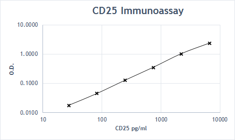
Human CD25 / IL-2 R alpha ELISA Standard Curve
Recombinant Human CD25/IL-2R alpha (Catalog # 223-2A) was serially diluted and captured by Mouse Anti-Human CD25/IL-2R alpha Monoclonal Antibody (Catalog # MAB623) coated on a Clear Polystyrene Microplate (Catalog # DY990). Goat Anti-Human CD25/IL-2R alpha Antigen Affinity-purified Polyclonal Antibody (Catalog # AF-223-NA) was biotinylated and incubated with the protein captured on the plate. Detection of the standard curve was achieved by incubating Streptavidin-HRP (Catalog # DY998)Applications for Human CD25/IL-2R alpha Antibody (24204)
Application
Recommended Usage
Immunocytochemistry
8-25 µg/mL
Sample: Immersion fixed human peripheral blood mononuclear cells stimulated with PHA
Sample: Immersion fixed human peripheral blood mononuclear cells stimulated with PHA
Immunohistochemistry
8-25 µg/mL
Sample: Immersion fixed frozen sections of human lymph node
Sample: Immersion fixed frozen sections of human lymph node
Western Blot
1 µg/mL
Sample: Recombinant Human CD25/IL‑2 R alpha (Catalog # 223-2A) under non-reducing conditions only
Sample: Recombinant Human CD25/IL‑2 R alpha (Catalog # 223-2A) under non-reducing conditions only
Human CD25/IL-2 R alpha Sandwich Immunoassay
Please Note: Optimal dilutions of this antibody should be experimentally determined.
Reviewed Applications
Read 2 reviews rated 5 using MAB623 in the following applications:
Formulation, Preparation, and Storage
Purification
Protein A or G purified from ascites
Reconstitution
Reconstitute at 0.5 mg/mL in sterile PBS. For liquid material, refer to CoA for concentration.
Loading...
Formulation
Lyophilized from a 0.2 μm filtered solution in PBS with Trehalose. *Small pack size (SP) is supplied either lyophilized or as a 0.2 µm filtered solution in PBS.
Shipping
Lyophilized product is shipped at ambient temperature. Liquid small pack size (-SP) is shipped with polar packs. Upon receipt, store immediately at the temperature recommended below.
Stability & Storage
Use a manual defrost freezer and avoid repeated freeze-thaw cycles.
- 12 months from date of receipt, -20 to -70 °C as supplied.
- 1 month, 2 to 8 °C under sterile conditions after reconstitution.
- 6 months, -20 to -70 °C under sterile conditions after reconstitution.
Calculators
Background: CD25/IL-2R alpha
Long Name
Interleukin 2 Receptor alpha
Alternate Names
CD25, IL-2 R alpha, IL-2Ra, IL2R alpha, IL2RA
Entrez Gene IDs
Gene Symbol
IL2RA
UniProt
Additional CD25/IL-2R alpha Products
Product Documents for Human CD25/IL-2R alpha Antibody (24204)
Certificate of Analysis
To download a Certificate of Analysis, please enter a lot or batch number in the search box below.
Note: Certificate of Analysis not available for kit components.
Product Specific Notices for Human CD25/IL-2R alpha Antibody (24204)
For research use only
Related Research Areas
Citations for Human CD25/IL-2R alpha Antibody (24204)
 Powered by Bioz
Customer Reviews for Human CD25/IL-2R alpha Antibody (24204) (2)
5 out of 5
2 Customer Ratings
Have you used Human CD25/IL-2R alpha Antibody (24204)?
Submit a review and receive an Amazon gift card!
$25/€18/£15/$25CAN/¥2500 Yen for a review with an image
$10/€7/£6/$10CAN/¥1110 Yen for a review without an image
Submit a review
Customer Images
Showing
1
-
2 of
2 reviews
Showing All
Filter By:
-
Application: ImmunohistochemistrySample Tested: Tonsil tissueSpecies: HumanVerified Customer | Posted 09/08/2021

-
Application: ELISASample Tested: EDTA PlasmaSpecies: HumanVerified Customer | Posted 07/23/2019
There are no reviews that match your criteria.
Protocols
Find general support by application which include: protocols, troubleshooting, illustrated assays, videos and webinars.
- Antigen Retrieval Protocol (PIER)
- Antigen Retrieval for Frozen Sections Protocol
- Appropriate Fixation of IHC/ICC Samples
- Cellular Response to Hypoxia Protocols
- Chromogenic IHC Staining of Formalin-Fixed Paraffin-Embedded (FFPE) Tissue Protocol
- Chromogenic Immunohistochemistry Staining of Frozen Tissue
- ClariTSA™ Fluorophore Kits
- Detection & Visualization of Antibody Binding
- Fluorescent IHC Staining of Frozen Tissue Protocol
- Graphic Protocol for Heat-induced Epitope Retrieval
- Graphic Protocol for the Preparation and Fluorescent IHC Staining of Frozen Tissue Sections
- Graphic Protocol for the Preparation and Fluorescent IHC Staining of Paraffin-embedded Tissue Sections
- Graphic Protocol for the Preparation of Gelatin-coated Slides for Histological Tissue Sections
- ICC Cell Smear Protocol for Suspension Cells
- ICC Immunocytochemistry Protocol Videos
- ICC for Adherent Cells
- IHC Sample Preparation (Frozen sections vs Paraffin)
- Immunocytochemistry (ICC) Protocol
- Immunocytochemistry Troubleshooting
- Immunofluorescence of Organoids Embedded in Cultrex Basement Membrane Extract
- Immunofluorescent IHC Staining of Formalin-Fixed Paraffin-Embedded (FFPE) Tissue Protocol
- Immunohistochemistry (IHC) and Immunocytochemistry (ICC) Protocols
- Immunohistochemistry Frozen Troubleshooting
- Immunohistochemistry Paraffin Troubleshooting
- Preparing Samples for IHC/ICC Experiments
- Preventing Non-Specific Staining (Non-Specific Binding)
- Primary Antibody Selection & Optimization
- Protocol for Heat-Induced Epitope Retrieval (HIER)
- Protocol for Making a 4% Formaldehyde Solution in PBS
- Protocol for VisUCyte™ HRP Polymer Detection Reagent
- Protocol for the Fluorescent ICC Staining of Cell Smears - Graphic
- Protocol for the Fluorescent ICC Staining of Cultured Cells on Coverslips - Graphic
- Protocol for the Preparation & Fixation of Cells on Coverslips
- Protocol for the Preparation and Chromogenic IHC Staining of Frozen Tissue Sections
- Protocol for the Preparation and Chromogenic IHC Staining of Frozen Tissue Sections - Graphic
- Protocol for the Preparation and Chromogenic IHC Staining of Paraffin-embedded Tissue Sections
- Protocol for the Preparation and Chromogenic IHC Staining of Paraffin-embedded Tissue Sections - Graphic
- Protocol for the Preparation and Fluorescent ICC Staining of Cells on Coverslips
- Protocol for the Preparation and Fluorescent ICC Staining of Non-adherent Cells
- Protocol for the Preparation and Fluorescent ICC Staining of Stem Cells on Coverslips
- Protocol for the Preparation and Fluorescent IHC Staining of Frozen Tissue Sections
- Protocol for the Preparation and Fluorescent IHC Staining of Paraffin-embedded Tissue Sections
- Protocol for the Preparation of Gelatin-coated Slides for Histological Tissue Sections
- Protocol for the Preparation of a Cell Smear for Non-adherent Cell ICC - Graphic
- R&D Systems Quality Control Western Blot Protocol
- TUNEL and Active Caspase-3 Detection by IHC/ICC Protocol
- The Importance of IHC/ICC Controls
- Troubleshooting Guide: Immunohistochemistry
- Troubleshooting Guide: Western Blot Figures
- Western Blot Conditions
- Western Blot Protocol
- Western Blot Protocol for Cell Lysates
- Western Blot Troubleshooting
- Western Blot Troubleshooting Guide
- View all Protocols, Troubleshooting, Illustrated assays and Webinars
Loading...
Associated Pathways


