CD4 is an approximately 55 kDa type I membrane glycoprotein that is expressed predominantly on most thymocytes and a subset of mature T lymphocytes. In humans, CD4 is also expressed to a lesser extent on monocytes and macrophage related cells. Human CD4 cDNA encodes a 458 amino acid (aa) residue precursor protein with a 25 aa residue signal peptide, a 371 aa residue extracellular region containing four immunoglobulin homology domains, a 24 aa residue transmembrane domain and a 38 aa residue cytoplasmic domain. CD4 is a coreceptor required for T cell recognition of antigens that are presented by class II major histocompatibility complexes. CD4 has been shown to be a coreceptor of HIV entry and specifically binds gp120, the external envelope glycoprotein of HIV.

Key Product Details
Species Reactivity
Validated:
Human
Cited:
Human, Mouse, Primate, Primate - Chlorocebus pygerythrus (Vervet Monkey), Xenograft
Applications
Validated:
Immunohistochemistry, Western Blot, Immunocytochemistry, Simple Western
Cited:
Immunohistochemistry, Immunohistochemistry-Paraffin, Immunohistochemistry-Frozen, Western Blot, Neutralization, Immunocytochemistry, Flow Cyomtetry, IF/IHC
Label
Unconjugated
Antibody Source
Polyclonal Goat IgG
Loading...
Product Specifications
Immunogen
S. frugiperda insect ovarian cell line Sf 21-derived recombinant human CD4
Lys26-Trp390
Accession # P01730
Lys26-Trp390
Accession # P01730
Specificity
Detects human CD4 in direct ELISAs and Western blots.
Clonality
Polyclonal
Host
Goat
Isotype
IgG
Scientific Data Images for Human CD4 Antibody
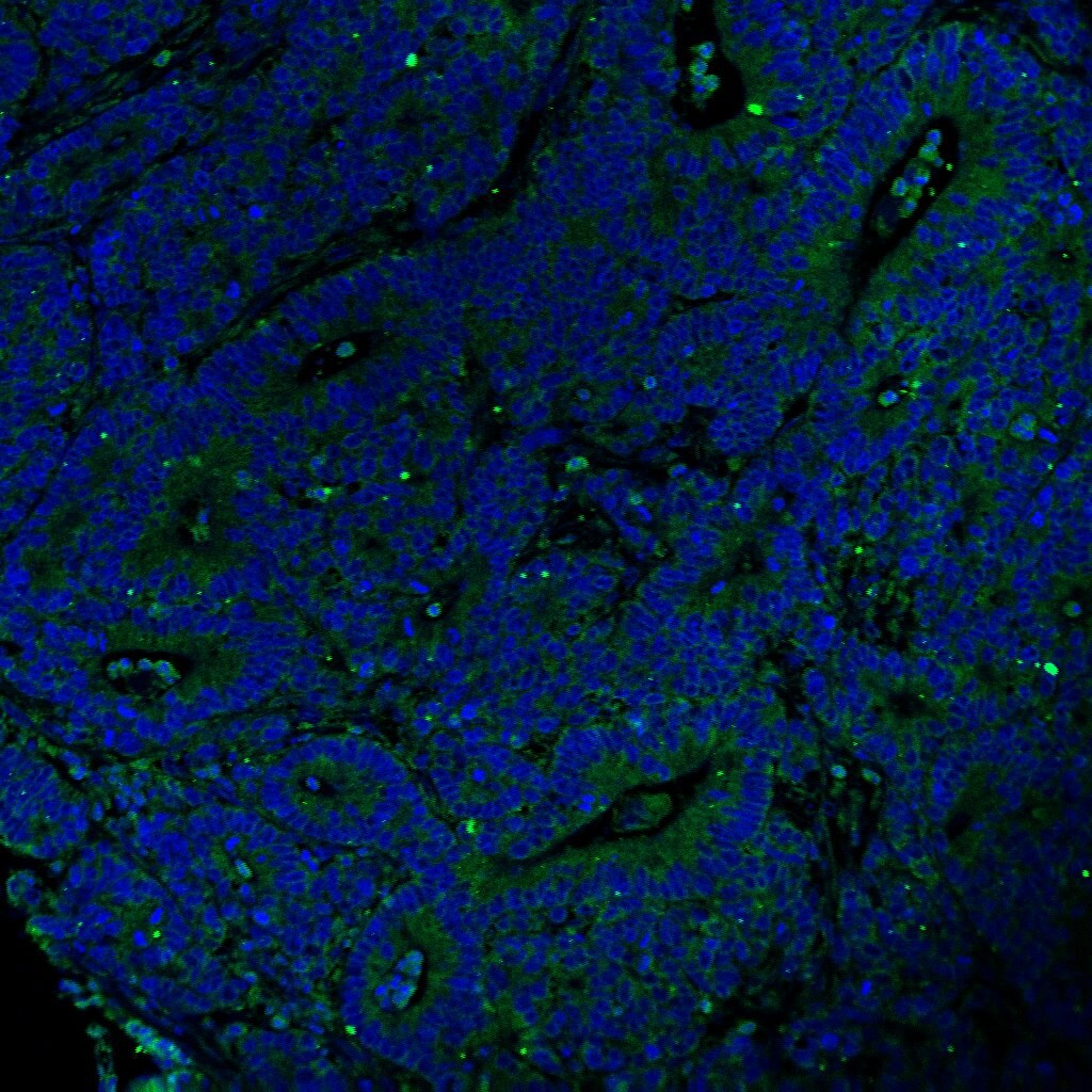
CD4 in Human T Cells.
CD4 was detected in immersion fixed human T cells using 2 µg/mL Goat Anti-Human CD4 Antigen Affinity-purified Polyclonal Antibody (Catalog # AF‑379‑NA) for 3 hours at room temperature. Cells were stained (red) and counterstained (green). View our protocol for Fluorescent ICC Staining of Cells on Coverslips.
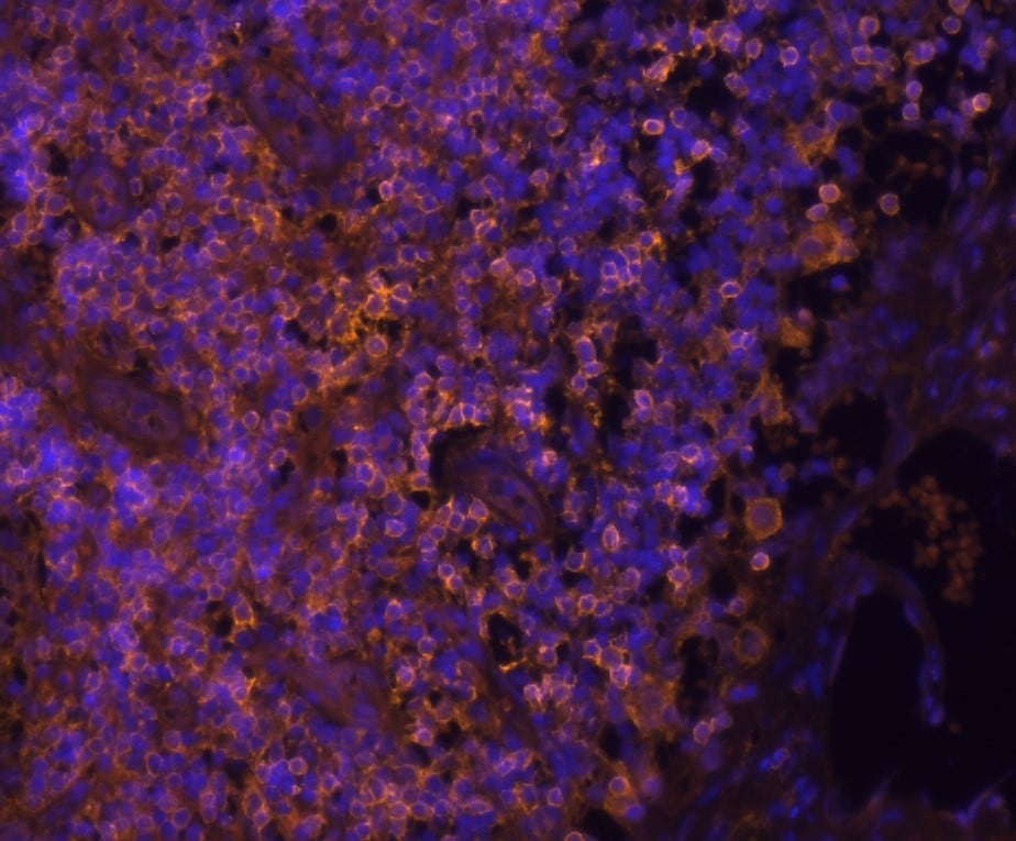
CD4 in Human PBMCs.
CD4 was detected in immersion fixed human peripheral blood mononuclear cells (PBMCs) using Goat Anti-Human CD4 Antigen Affinity-purified Polyclonal Antibody (Catalog # AF-379-NA) at 10 µg/mL for 3 hours at room temperature. Cells were stained using the NorthernLights™ 557-conjugated Anti-Goat IgG Secondary Antibody (yellow; NL001) and counterstained with DAPI (blue). View our protocol for Fluorescent ICC Staining of Non-adherent Cells.
CD4 in Human Tonsil Tissue.
CD4 was detected in immersion fixed paraffin-embedded sections of human tonsil tissue using Goat Anti-Human CD4 Antigen Affinity-purified Polyclonal Antibody (Catalog # AF-379-NA) at 1.7 µg/mL for 1 hour at room temperature followed by incubation with the Anti-Goat IgG VisUCyte™ HRP Polymer Antibody (VC004). Before incubation with the primary antibody, tissue was subjected to heat-induced epitope retrieval using Antigen Retrieval Reagent-Basic (CTS013). Tissue was stained using DAB (brown) and counterstained with hematoxylin (blue). Specific staining was localized to cell surface of lymphocytes. Staining was performed using our IHC Staining with VisUCyte HRP Polymer Detection Reagents protocol.
Detection of Human CD4 by Western Blot.
Western blot shows lysates of PBMC and SUP‑T1 human T cell lymphoblastic lymphoma cells. PVDF membrane was probed with 1 µg/mL of Goat Anti-Human CD4 Antigen Affinity-purified Polyclonal Antibody (Catalog # AF-379-NA) followed by HRP-conjugated Anti-Goat IgG Secondary Antibody (Catalog # HAF017). A specific band was detected for CD4 at approximately ~55kDa kDa (as indicated). This experiment was conducted under reducing conditions and using Western Blot Buffer Group 1.
Detection of Human CD4 by Simple WesternTM.
Simple Western lane view shows lysates of Human PBMC and SUP‑T1 human T cell lymphoblastic lymphoma cell line, loaded at 0.2 mg/mL. A specific band was detected for CD4 at approximately 65 kDa (as indicated) using 10 µg/mL of Goat Anti-Human CD4 Antigen Affinity-purified Polyclonal Antibody (Catalog # AF-379-NA). This experiment was conducted under reducing conditions and using the 12-230 kDa separation system.Applications for Human CD4 Antibody
Application
Recommended Usage
Immunocytochemistry
5-15 µg/mL
Sample: Immersion fixed human T cells and human peripheral blood mononuclear cells (PBMCs)
Sample: Immersion fixed human T cells and human peripheral blood mononuclear cells (PBMCs)
Immunohistochemistry
1-15 µg/mL
Sample: Immersion fixed paraffin-embedded sections of human tonsil tissue
Sample: Immersion fixed paraffin-embedded sections of human tonsil tissue
Simple Western
10 µg/mL
Sample: Human PBMC and SUP‑T1 human T cell lymphoblastic lymphoma cell line
Sample: Human PBMC and SUP‑T1 human T cell lymphoblastic lymphoma cell line
Western Blot
1 µg/mL
Sample: PBMC and SUP‑T1 human T cell lymphoblastic lymphoma cells
Sample: PBMC and SUP‑T1 human T cell lymphoblastic lymphoma cells
Reviewed Applications
Read 3 reviews rated 3.7 using AF-379-NA in the following applications:
Formulation, Preparation, and Storage
Purification
Antigen Affinity-purified
Reconstitution
Reconstitute at 0.2 mg/mL in sterile PBS. For liquid material, refer to CoA for concentration.
Loading...
Formulation
Lyophilized from a 0.2 μm filtered solution in PBS with Trehalose. *Small pack size (SP) is supplied either lyophilized or as a 0.2 µm filtered solution in PBS.
Shipping
Lyophilized product is shipped at ambient temperature. Liquid small pack size (-SP) is shipped with polar packs. Upon receipt, store immediately at the temperature recommended below.
Stability & Storage
Use a manual defrost freezer and avoid repeated freeze-thaw cycles.
- 12 months from date of receipt, -20 to -70 °C as supplied.
- 1 month, 2 to 8 °C under sterile conditions after reconstitution.
- 6 months, -20 to -70 °C under sterile conditions after reconstitution.
Calculators
Background: CD4
References
- Capon, D.I. et al. (1991) Annu. Rev. Immunol. 9:649.
Alternate Names
CD4
Entrez Gene IDs
Gene Symbol
CD4
UniProt
Additional CD4 Products
Product Documents for Human CD4 Antibody
Certificate of Analysis
To download a Certificate of Analysis, please enter a lot or batch number in the search box below.
Note: Certificate of Analysis not available for kit components.
Product Specific Notices for Human CD4 Antibody
For research use only
Citations for Human CD4 Antibody
 Powered by Bioz
Customer Reviews for Human CD4 Antibody (3)
3.7 out of 5
3 Customer Ratings
Have you used Human CD4 Antibody?
Submit a review and receive an Amazon gift card!
$25/€18/£15/$25CAN/¥2500 Yen for a review with an image
$10/€7/£6/$10CAN/¥1110 Yen for a review without an image
Submit a review
Customer Images
Showing
1
-
3 of
3 reviews
Showing All
Filter By:
-
Application: Immunocytochemistry/ImmunofluorescenceSample Tested: Colon cancer tissueSpecies: HumanVerified Customer | Posted 04/06/2021

-
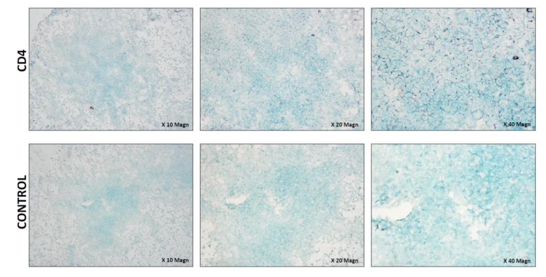
Application: Immunohistochemistry-FrozenSample Tested: Porcine spleenSpecies: PorcineVerified Customer | Posted 12/03/2020Healthy frozen porcine spleen section showing CD4 expression (purple stain).Pig spleen frozen OCT tissues were sectioned at 7µm and fixed in ice cold acetone for 7min. Once dry, blocking buffer 4% FCS/TBS was added to all tissues for 30min to minimize non-specific binding, followed by 10 min incubation with Bloxall, endogenous peroxidase blocking solution at room temperature. Tissues were washed in Tris buffered saline (TBS) for 5 min and incubated for 20min with normal horse serum (Vector laboratories # S-2012-50). Human Polyclonal goat anti-CD4 (Novus biologicals # AF379-NA) was diluted at 1:50 in 0.5% FCS/TBS and added to the arterial tissues for 1 hour incubation at room temperature. Following 5 min wash in TBS, tissues were incubated for 30min with ImmPRESS®HRP horse anti-goat reagent (Vector laboratories # MP-7405). To detect CD4 antigen, tissues were washed in TBS and incubated with VIP HRP substrate (Vector®VIP Substrate kit, Peroxidase (HRP) # SK-4600) for 1 min 30sec then placed immediately in MilliQ water to stop the reaction. Nuclei were counter-stained with methyl green (Sigma-Aldrich # M8884-25G). Control tissues were stained similarly in the absence of the primary antibody. Tissue sections were immediately dehydrated and mounted with ENTELLAN mounting medium (Proscitech, Cat#IA013). Images were captured on Nikon eclipse ME600 microscope and DS-Fi2 camera.
-
Application: Immunocytochemistry/ImmunofluorescenceSample Tested: Tonsil tissueSpecies: Human tonsil and HumanVerified Customer | Posted 12/02/2019
There are no reviews that match your criteria.
Protocols
Find general support by application which include: protocols, troubleshooting, illustrated assays, videos and webinars.
- Antigen Retrieval Protocol (PIER)
- Antigen Retrieval for Frozen Sections Protocol
- Appropriate Fixation of IHC/ICC Samples
- Cellular Response to Hypoxia Protocols
- Chromogenic IHC Staining of Formalin-Fixed Paraffin-Embedded (FFPE) Tissue Protocol
- Chromogenic Immunohistochemistry Staining of Frozen Tissue
- ClariTSA™ Fluorophore Kits
- Detection & Visualization of Antibody Binding
- Fluorescent IHC Staining of Frozen Tissue Protocol
- Graphic Protocol for Heat-induced Epitope Retrieval
- Graphic Protocol for the Preparation and Fluorescent IHC Staining of Frozen Tissue Sections
- Graphic Protocol for the Preparation and Fluorescent IHC Staining of Paraffin-embedded Tissue Sections
- Graphic Protocol for the Preparation of Gelatin-coated Slides for Histological Tissue Sections
- ICC Cell Smear Protocol for Suspension Cells
- ICC Immunocytochemistry Protocol Videos
- ICC for Adherent Cells
- IHC Sample Preparation (Frozen sections vs Paraffin)
- Immunocytochemistry (ICC) Protocol
- Immunocytochemistry Troubleshooting
- Immunofluorescence of Organoids Embedded in Cultrex Basement Membrane Extract
- Immunofluorescent IHC Staining of Formalin-Fixed Paraffin-Embedded (FFPE) Tissue Protocol
- Immunohistochemistry (IHC) and Immunocytochemistry (ICC) Protocols
- Immunohistochemistry Frozen Troubleshooting
- Immunohistochemistry Paraffin Troubleshooting
- Preparing Samples for IHC/ICC Experiments
- Preventing Non-Specific Staining (Non-Specific Binding)
- Primary Antibody Selection & Optimization
- Protocol for Heat-Induced Epitope Retrieval (HIER)
- Protocol for Making a 4% Formaldehyde Solution in PBS
- Protocol for VisUCyte™ HRP Polymer Detection Reagent
- Protocol for the Fluorescent ICC Staining of Cell Smears - Graphic
- Protocol for the Fluorescent ICC Staining of Cultured Cells on Coverslips - Graphic
- Protocol for the Preparation & Fixation of Cells on Coverslips
- Protocol for the Preparation and Chromogenic IHC Staining of Frozen Tissue Sections
- Protocol for the Preparation and Chromogenic IHC Staining of Frozen Tissue Sections - Graphic
- Protocol for the Preparation and Chromogenic IHC Staining of Paraffin-embedded Tissue Sections
- Protocol for the Preparation and Chromogenic IHC Staining of Paraffin-embedded Tissue Sections - Graphic
- Protocol for the Preparation and Fluorescent ICC Staining of Cells on Coverslips
- Protocol for the Preparation and Fluorescent ICC Staining of Non-adherent Cells
- Protocol for the Preparation and Fluorescent ICC Staining of Stem Cells on Coverslips
- Protocol for the Preparation and Fluorescent IHC Staining of Frozen Tissue Sections
- Protocol for the Preparation and Fluorescent IHC Staining of Paraffin-embedded Tissue Sections
- Protocol for the Preparation of Gelatin-coated Slides for Histological Tissue Sections
- Protocol for the Preparation of a Cell Smear for Non-adherent Cell ICC - Graphic
- R&D Systems Quality Control Western Blot Protocol
- TUNEL and Active Caspase-3 Detection by IHC/ICC Protocol
- The Importance of IHC/ICC Controls
- Troubleshooting Guide: Immunohistochemistry
- Troubleshooting Guide: Western Blot Figures
- Western Blot Conditions
- Western Blot Protocol
- Western Blot Protocol for Cell Lysates
- Western Blot Troubleshooting
- Western Blot Troubleshooting Guide
- View all Protocols, Troubleshooting, Illustrated assays and Webinars


