Human IL‑1 beta /IL‑1F2 Antibody
R&D Systems | Catalog # AB-201-NA

Loading...
Key Product Details
Validated by
Biological Validation
Species Reactivity
Validated:
Human
Cited:
Human, Mouse, Porcine
Applications
Validated:
Western Blot, Neutralization, Immunocytochemistry
Cited:
Immunohistochemistry, Western Blot, Neutralization
Label
Unconjugated
Antibody Source
Polyclonal Goat IgG
Loading...
Product Specifications
Immunogen
E. coli-derived recombinant human IL-1 beta /IL-1F2
Ala117-Ser269
Accession # NP_000567
Ala117-Ser269
Accession # NP_000567
Specificity
Detects human IL‑1 beta /IL‑1F2 in ELISAs and Western blots. In direct ELISAs, 100% cross-reactivity with recombinant rhesus monkey is observed, and approximately 50% cross-reactivity with recombinant mouse IL‑1 beta, recombinant rat IL‑1 beta, recombinant canine IL‑1 beta, recombinant equine IL‑1 beta, recombinant feline IL‑1 beta, and approximately 20% cross-reactivity with recombinant porcine IL-1 beta and recombinant cotton rat IL-1 beta is observed.
Clonality
Polyclonal
Host
Goat
Isotype
IgG
Endotoxin Level
<0.10 EU per 1 μg of the antibody by the LAL method.
Scientific Data Images for Human IL‑1 beta /IL‑1F2 Antibody
Detection of Human and Mouse IL‑1 beta /IL‑1F2 by Western Blot.
Western blot shows lysates of THP-1 human acute monocytic leukemia cell line untreated (-) or treated (+) with PMA and LPS and RAW 264.7 mouse monocyte/macrophage cell line untreated (-) or treated (+) with LPS. PVDF membrane was probed with 1 µg/mL of Goat Anti-Human IL-1 beta /IL-1F2 Polyclonal Antibody (Catalog # AB-201-NA) followed by HRP-conjugated Anti-Goat IgG Secondary Antibody (Catalog # HAF017). A specific band was detected for IL-1 beta /IL-1F2 at approximately 35 kDa (as indicated). This experiment was conducted under reducing conditions and using Immunoblot Buffer Group 1.
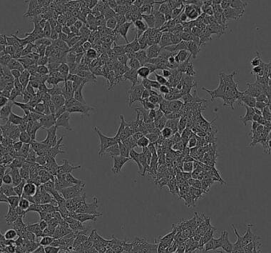
IL‑1 beta /IL‑1F2 in Human Peripheral Blood Mononuclear Cells.
IL-1 beta /IL-1F2 was detected in immersion fixed human peripheral blood mononuclear cells using Goat Anti-Human IL-1 beta /IL-1F2 Polyclonal Antibody (Catalog # AB-201-NA) at 15 µg/mL for 3 hours at room temperature. Cells were stained using the NorthernLights™ 557-conjugated Anti-Goat IgG Secondary Antibody (red; Catalog # NL001) and counterstained with DAPI (blue). Specific staining was localized to cytoplasmic. View our protocol for Fluorescent ICC Staining of Non-adherent Cells.
Cell Proliferation Induced by IL‑1 beta /IL‑1F2 and Neutral-ization by Human IL‑1 beta / IL‑1F2 Antibody.
Recombinant Human IL-1 beta /IL-1F2 (Catalog # 201-LB) stimulates proliferation in the the D10.G4.1 mouse helper T cell line in a dose-dependent manner (orange line). Proliferation elicited by Recombinant Human IL-1 beta / IL-1F2 (50 pg/mL) is neutral-ized (green line) by increasing concentrations of Goat Anti-Human IL-1 beta /IL-1F2 Poly-clonal Antibody (Catalog # AB-201-NA). The ND50 is typically 0.05-0.1 µg/mL.Applications for Human IL‑1 beta /IL‑1F2 Antibody
Application
Recommended Usage
Immunocytochemistry
5-15 µg/mL
Sample: Immersion fixed human peripheral blood mononuclear cells
Sample: Immersion fixed human peripheral blood mononuclear cells
Western Blot
1 µg/mL
Sample: THP‑1 human acute monocytic leukemia cell line -/+ PMA/LPS and RAW 264.7 mouse monocyte/macrophage cell line -/+ LPS.
Sample: THP‑1 human acute monocytic leukemia cell line -/+ PMA/LPS and RAW 264.7 mouse monocyte/macrophage cell line -/+ LPS.
Neutralization
Measured by its ability to neutralize IL‑1 beta /IL‑1F2-induced proliferation in the D10.G4.1 mouse helper T cell line. Symons, J.A. et al. (1987) in Lymphokines and Interferons, a Practical Approach. Clemens, M.J. et al. (eds): IRL Press. 272. The Neutralization Dose (ND50) is typically 0.05-0.1 µg/mL in the presence of 50 pg/mL Recombinant Human IL‑1 beta /IL‑1F2.
Reviewed Applications
Read 1 review rated 5 using AB-201-NA in the following applications:
Formulation, Preparation, and Storage
Purification
Protein A or G purified
Reconstitution
Reconstitute at 1 mg/mL in sterile PBS.
Loading...
Formulation
Lyophilized from a 0.2 μm filtered solution in PBS with Trehalose.
Shipping
The product is shipped at ambient temperature. Upon receipt, store it immediately at the temperature recommended below.
Stability & Storage
Use a manual defrost freezer and avoid repeated freeze-thaw cycles.
- 12 months from date of receipt, -20 to -70 °C as supplied.
- 1 month, 2 to 8 °C under sterile conditions after reconstitution.
- 6 months, -20 to -70 °C under sterile conditions after reconstitution.
Calculators
Background: IL-1 beta/IL-1F2
Long Name
Interleukin 1 beta
Alternate Names
IL-1b, IL-1F2, IL1 beta, IL1B
Entrez Gene IDs
Gene Symbol
IL1B
UniProt
Additional IL-1 beta/IL-1F2 Products
Product Documents for Human IL‑1 beta /IL‑1F2 Antibody
Certificate of Analysis
To download a Certificate of Analysis, please enter a lot or batch number in the search box below.
Note: Certificate of Analysis not available for kit components.
Product Specific Notices for Human IL‑1 beta /IL‑1F2 Antibody
This product is covered by one or more of the following U.S. patents: 4,766,069, 5,510,462, 5,681,933, 4,762,914, 5,474,899, 5,789,185, 5,484,887, 5,122,459, 5,001,057, 5,077,219, 5,286,847.
For research use only
Related Research Areas
Citations for Human IL‑1 beta /IL‑1F2 Antibody
 Powered by Bioz
Customer Reviews for Human IL‑1 beta /IL‑1F2 Antibody (1)
5 out of 5
1 Customer Rating
Have you used Human IL‑1 beta /IL‑1F2 Antibody?
Submit a review and receive an Amazon gift card!
$25/€18/£15/$25CAN/¥2500 Yen for a review with an image
$10/€7/£6/$10CAN/¥1110 Yen for a review without an image
Submit a review
Customer Images
Showing
1
-
1 of
1 review
Showing All
Filter By:
-
Application: Block/NeutralizeSample Tested: 293T human embryonic kidney cell lineSpecies: HumanVerified Customer | Posted 09/02/2022

There are no reviews that match your criteria.
Protocols
Find general support by application which include: protocols, troubleshooting, illustrated assays, videos and webinars.
- Appropriate Fixation of IHC/ICC Samples
- Cellular Response to Hypoxia Protocols
- ClariTSA™ Fluorophore Kits
- Detection & Visualization of Antibody Binding
- ICC Cell Smear Protocol for Suspension Cells
- ICC Immunocytochemistry Protocol Videos
- ICC for Adherent Cells
- Immunocytochemistry (ICC) Protocol
- Immunocytochemistry Troubleshooting
- Immunofluorescence of Organoids Embedded in Cultrex Basement Membrane Extract
- Immunohistochemistry (IHC) and Immunocytochemistry (ICC) Protocols
- Preparing Samples for IHC/ICC Experiments
- Preventing Non-Specific Staining (Non-Specific Binding)
- Primary Antibody Selection & Optimization
- Protocol for VisUCyte™ HRP Polymer Detection Reagent
- Protocol for the Fluorescent ICC Staining of Cell Smears - Graphic
- Protocol for the Fluorescent ICC Staining of Cultured Cells on Coverslips - Graphic
- Protocol for the Preparation and Fluorescent ICC Staining of Cells on Coverslips
- Protocol for the Preparation and Fluorescent ICC Staining of Non-adherent Cells
- Protocol for the Preparation and Fluorescent ICC Staining of Stem Cells on Coverslips
- Protocol for the Preparation of a Cell Smear for Non-adherent Cell ICC - Graphic
- R&D Systems Quality Control Western Blot Protocol
- TUNEL and Active Caspase-3 Detection by IHC/ICC Protocol
- The Importance of IHC/ICC Controls
- Troubleshooting Guide: Western Blot Figures
- Western Blot Conditions
- Western Blot Protocol
- Western Blot Protocol for Cell Lysates
- Western Blot Troubleshooting
- Western Blot Troubleshooting Guide
- View all Protocols, Troubleshooting, Illustrated assays and Webinars
Loading...
Associated Pathways


Innate Lymphoid Cell Differentiation Pathways
 NOD-like Receptor Signaling Pathways
 Th17 Differentiation Pathway
 Toll-Like Receptor Signaling Pathways
