Leptin receptor (OB-R), also named B219, is a type I cytokine receptor family protein with significant amino acid sequence identity with gp130, G-CSF receptor, and the LIF receptor. Multiple isoforms of human and mouse OB-R, including a long form (OB-RL) with a large cytoplasmic domain capable of signal-transduction, and several receptor isoforms with short cytoplasmic domains (OB-Rs) lacking signal-transducing capabilities, have been identified. The extracellular domains of the short and long forms of OB-R are identical. An OB-R transcript, lacking a transmembrane domain and potentially encoding a soluble form of the receptor, has also been described. OB-RL transcripts were reported to be expressed predominantly in regions of the hypothalamus previously thought to be important in body weight regulation. Expression of OB-Rs transcripts have been found in multiple tissues, including the choroid plexus, lung, kidney, and primitive hematopoietic cell populations. OB-R has been shown to be encoded by the mouse diabetes (db) and rat fatty (fa) genes. Rodents homozygous for the db or fa mutations have been known to exhibit an obesity phenotype. Human OB-R long form encodes a 1165 amino acid (aa) precursor protein with a 22 aa signal peptide, an 819 aa extracellular domain, a 21 aa transmembrane domain and a 303 aa cytoplasmic domain. The extracellular domain of OB-R contain two hemopoietin receptor domains, a fibronectin type III domain and the WSXWS domain. Recombinant soluble OB-R has been shown to bind Leptin with high affinity and is a potent Leptin antagonist.

Key Product Details
Species Reactivity
Validated:
Cited:
Applications
Validated:
Cited:
Label
Antibody Source
Product Specifications
Immunogen
Phe22-Asp839 (predicted)
Accession # P48357
Specificity
Clonality
Host
Isotype
Scientific Data Images for Human Leptin R Antibody (52263)
Detection of Recombinant Human Leptin R by Western Blot.
Western blot shows 25 ng of Recombinant Human Leptin R Fc Chimera (Catalog # 389-LR) and Recombinant Mouse Leptin R Fc Chimera (Catalog # 497-LR). PVDF Membrane was probed with 1 µg/mL of Mouse Anti-Human Leptin R Monoclonal Antibody (Catalog # MAB867) followed by HRP-conjugated Anti-Mouse IgG Secondary Antibody (Catalog # HAF007). A specific band was detected for Leptin R at approximately 155-200 kDa (as indicated). This experiment was conducted under reducing conditions and using Immunoblot Buffer Group 3.
Detection of Leptin R in Human Hypothalamus.
Leptin R was detected in immersion fixed paraffin-embedded sections of human hypothalamus using Mouse Anti-Human Leptin R Monoclonal Antibody (Catalog # MAB867) at 1.7 µg/ml for 1 hour at room temperature followed by incubation with the Anti-Mouse IgG VisUCyte™ HRP Polymer Antibody (Catalog # VC001) or the HRP-conjugated Anti-Mouse IgG Secondary Antibody (Catalog # HAF007). Before incubation with the primary antibody, tissue was subjected to heat-induced epitope retrieval using VisUCyte Antigen Retrieval Reagent-Basic (Catalog # VCTS021). Tissue was stained using DAB (brown) and counterstained with hematoxylin (blue). Specific staining was localized to the cell surface of neurons. View our protocol for IHC Staining with VisUCyte HRP Polymer Detection Reagents.
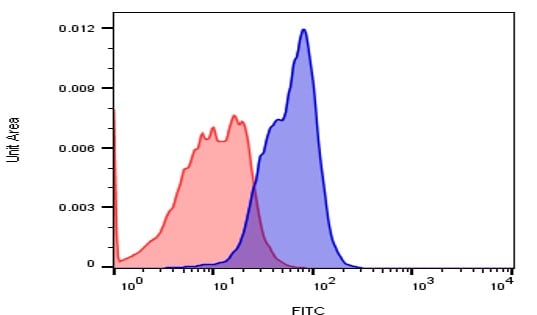
Detection of Leptin R in PBMC by Flow Cytometry
PBMC's were stained with Mouse Anti-Human CD14 PE‑conjugated Monoclonal Antibody (Catalog # FAB3832P) and either (A) Mouse Anti-Human Leptin R Monoclonal Antibody (Catalog # MAB867) or (B) isotype control antibody (Catalog # MAB004) followed by Allophycocyanin-conjugated Anti-Mouse IgG Secondary Antibody (Catalog # F0101B). View our protocol for Staining Membrane-associated Proteins.Applications for Human Leptin R Antibody (52263)
CyTOF-ready
Flow Cytometry
Sample: CD14+ peripheral blood mononuclear cells
Immunohistochemistry
Sample: Immersion fixed paraffin-embedded sections of human hypothalamus
Western Blot
Sample: Recombinant Human Leptin R Fc Chimera (Catalog # 389-LR)
Reviewed Applications
Read 1 review rated 4 using MAB867 in the following applications:
Flow Cytometry Panel Builder
Bio-Techne Knows Flow Cytometry
Save time and reduce costly mistakes by quickly finding compatible reagents using the Panel Builder Tool.
Advanced Features
- Spectra Viewer - Custom analysis of spectra from multiple fluorochromes
- Spillover Popups - Visualize the spectra of individual fluorochromes
- Antigen Density Selector - Match fluorochrome brightness with antigen density

Formulation, Preparation, and Storage
Purification
Reconstitution
Reconstitute at 0.5 mg/mL in sterile PBS. For liquid material, refer to CoA for concentration.
Formulation
*Small pack size (-SP) is supplied either lyophilized or as a 0.2 µm filtered solution in PBS.
Shipping
Stability & Storage
- 12 months from date of receipt, -20 to -70 °C as supplied.
- 1 month, 2 to 8 °C under sterile conditions after reconstitution.
- 6 months, -20 to -70 °C under sterile conditions after reconstitution.
Calculators
Background: Leptin R
References
- Tartaglia, L.A. et al. (1995) Cell 83:1263.
- Cioffi, J.A. et al. (1996) Nature Medicine 2:585.
- Tartaglia, L.A. (1997) J. Biol. Chem. 272:6093.
Long Name
Alternate Names
Gene Symbol
UniProt
Additional Leptin R Products
Product Documents for Human Leptin R Antibody (52263)
Certificate of Analysis
To download a Certificate of Analysis, please enter a lot or batch number in the search box below.
Note: Certificate of Analysis not available for kit components.
Product Specific Notices for Human Leptin R Antibody (52263)
For research use only
Related Research Areas
Citations for Human Leptin R Antibody (52263)
 Powered by Bioz
Customer Reviews for Human Leptin R Antibody (52263) (1)
Have you used Human Leptin R Antibody (52263)?
Submit a review and receive an Amazon gift card!
$25/€18/£15/$25CAN/¥2500 Yen for a review with an image
$10/€7/£6/$10CAN/¥1110 Yen for a review without an image
Submit a review
Customer Images
-
Application: Flow CytometrySample Tested: Human pancreatic islet cellsSpecies: HumanVerified Customer | Posted 12/06/2016Dispersed human islet cells labeled with CD295 and anti-mouse FITC (blue) or anti-mouse FITC only (red).

There are no reviews that match your criteria.
Protocols
Find general support by application which include: protocols, troubleshooting, illustrated assays, videos and webinars.
- 7-Amino Actinomycin D (7-AAD) Cell Viability Flow Cytometry Protocol
- Antigen Retrieval Protocol (PIER)
- Antigen Retrieval for Frozen Sections Protocol
- Appropriate Fixation of IHC/ICC Samples
- Cellular Response to Hypoxia Protocols
- Chromogenic IHC Staining of Formalin-Fixed Paraffin-Embedded (FFPE) Tissue Protocol
- Chromogenic Immunohistochemistry Staining of Frozen Tissue
- ClariTSA™ Fluorophore Kits
- Detection & Visualization of Antibody Binding
- Extracellular Membrane Flow Cytometry Protocol
- Flow Cytometry Protocol for Cell Surface Markers
- Flow Cytometry Protocol for Staining Membrane Associated Proteins
- Flow Cytometry Staining Protocols
- Flow Cytometry Troubleshooting Guide
- Fluorescent IHC Staining of Frozen Tissue Protocol
- Graphic Protocol for Heat-induced Epitope Retrieval
- Graphic Protocol for the Preparation and Fluorescent IHC Staining of Frozen Tissue Sections
- Graphic Protocol for the Preparation and Fluorescent IHC Staining of Paraffin-embedded Tissue Sections
- Graphic Protocol for the Preparation of Gelatin-coated Slides for Histological Tissue Sections
- IHC Sample Preparation (Frozen sections vs Paraffin)
- Immunofluorescent IHC Staining of Formalin-Fixed Paraffin-Embedded (FFPE) Tissue Protocol
- Immunohistochemistry (IHC) and Immunocytochemistry (ICC) Protocols
- Immunohistochemistry Frozen Troubleshooting
- Immunohistochemistry Paraffin Troubleshooting
- Intracellular Flow Cytometry Protocol Using Alcohol (Methanol)
- Intracellular Flow Cytometry Protocol Using Detergents
- Intracellular Nuclear Staining Flow Cytometry Protocol Using Detergents
- Intracellular Staining Flow Cytometry Protocol Using Alcohol Permeabilization
- Intracellular Staining Flow Cytometry Protocol Using Detergents to Permeabilize Cells
- Preparing Samples for IHC/ICC Experiments
- Preventing Non-Specific Staining (Non-Specific Binding)
- Primary Antibody Selection & Optimization
- Propidium Iodide Cell Viability Flow Cytometry Protocol
- Protocol for Heat-Induced Epitope Retrieval (HIER)
- Protocol for Liperfluo
- Protocol for Making a 4% Formaldehyde Solution in PBS
- Protocol for VisUCyte™ HRP Polymer Detection Reagent
- Protocol for the Characterization of Human Th22 Cells
- Protocol for the Characterization of Human Th9 Cells
- Protocol for the Preparation & Fixation of Cells on Coverslips
- Protocol for the Preparation and Chromogenic IHC Staining of Frozen Tissue Sections
- Protocol for the Preparation and Chromogenic IHC Staining of Frozen Tissue Sections - Graphic
- Protocol for the Preparation and Chromogenic IHC Staining of Paraffin-embedded Tissue Sections
- Protocol for the Preparation and Chromogenic IHC Staining of Paraffin-embedded Tissue Sections - Graphic
- Protocol for the Preparation and Fluorescent IHC Staining of Frozen Tissue Sections
- Protocol for the Preparation and Fluorescent IHC Staining of Paraffin-embedded Tissue Sections
- Protocol for the Preparation of Gelatin-coated Slides for Histological Tissue Sections
- Protocol: Annexin V and PI Staining by Flow Cytometry
- Protocol: Annexin V and PI Staining for Apoptosis by Flow Cytometry
- R&D Systems Quality Control Western Blot Protocol
- TUNEL and Active Caspase-3 Detection by IHC/ICC Protocol
- The Importance of IHC/ICC Controls
- Troubleshooting Guide: Fluorokine Flow Cytometry Kits
- Troubleshooting Guide: Immunohistochemistry
- Troubleshooting Guide: Western Blot Figures
- Western Blot Conditions
- Western Blot Protocol
- Western Blot Protocol for Cell Lysates
- Western Blot Troubleshooting
- Western Blot Troubleshooting Guide
- View all Protocols, Troubleshooting, Illustrated assays and Webinars
FAQs for Human Leptin R Antibody (52263)
-
Q: Does this Human Leptin R antibody detect the long form (ObRb) of Leptin R?
A: The immunogen sequence that this antibody was raised against corresponds to the extracellular domain of Leptin R. Because the extracellular domains of the long form and other isoforms of the receptor are identical, we expect that the antibody will detect all forms.