Stem cell factor receptor (CD117, the gene product of the c-kit protooncogene) and its ligand, stem cell factor (also named c-kit ligand, mast cell growth factor), play essential roles in gametogenesis, melanogenesis and hematopoiesis. It is a transmembrane tyrosine kinase that is expressed on endothelial cells, mast cells, megakaryocytes, stem cells and multiple embryonic cells, such as melanoblasts and primordial germ cells.
Best Seller
CD117/c-kit Antibody
R&D Systems | Catalog # AF1356

Key Product Details
Species Reactivity
Validated:
Human, Mouse
Cited:
Human, Mouse, Rat, Porcine, Transgenic Mouse
Applications
Validated:
Immunohistochemistry, Western Blot, Flow Cytometry, Dual RNAscope ISH-IHC Compatible, Simple Western, CyTOF-ready
Cited:
Immunohistochemistry, Immunohistochemistry-Paraffin, Immunohistochemistry-Frozen, Western Blot, Neutralization, Flow Cytometry, Immunofluorescence, Immunocytochemistry, Immunocytochemistry/ Immunofluorescence, Simple Western
Label
Unconjugated
Antibody Source
Polyclonal Goat IgG
Loading...
Product Specifications
Immunogen
Mouse myeloma cell line NS0-derived recombinant mouse CD117/c-kit
Gln25-Thr519 (Ala207Glu)
Accession # P05532
Gln25-Thr519 (Ala207Glu)
Accession # P05532
Specificity
Detects human and mouse CD117/c-kit in direct ELISAs and Western blots.
Clonality
Polyclonal
Host
Goat
Isotype
IgG
Scientific Data Images for CD117/c-kit Antibody
CD117/c-kit in Mouse Embryo.
CD117/c-kit was detected in immersion fixed frozen sections of mouse embryo using Goat Anti-Human/Mouse CD117/c-kit Antigen Affinity-purified Polyclonal Antibody (Catalog # AF1356) at 15 µg/mL overnight at 4 °C. Tissue was stained using the Anti-Goat HRP-DAB Cell & Tissue Staining Kit (brown; CTS008) and counterstained with hematoxylin (blue). View our protocol for Chromogenic IHC Staining of Frozen Tissue Sections.
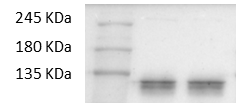
Detection of Human and Mouse CD117/c-kit by Western Blot.
Western blot shows lysates of MO7e human megakaryocytic leukemic cell line, P815 mouse mastocytoma cell line, and MC/9-2 mouse mast cell line. PVDF membrane was probed with 0.1 µg/mL of Goat Anti-Human/Mouse CD117/c-kit Antigen Affinity-purified Polyclonal Antibody (Catalog # AF1356) followed by HRP-conjugated Anti-Goat IgG Secondary Antibody (HAF017). Specific bands were detected for CD117/c-kit at approximately 135, 150 kDa (as indicated). This experiment was conducted under reducing conditions and using Immunoblot Buffer Group 1.
Detection of Human and Mouse CD117/c-kit by Simple WesternTM.
Simple Western lane view shows lysates of MO7e human megakaryocytic leukemic cell line, P815 mouse mastocytoma cell line and MC/9-2 mouse mast cell line, loaded at 0.2 mg/mL. A specific band was detected for CD117/c-kit at approximately 150-165 kDa (as indicated) using 5 µg/mL of Goat Anti-Human/Mouse CD117/c-kit Antigen Affinity-purified Polyclonal Antibody (Catalog # AF1356) followed by 1:50 dilution of HRP-conjugated Anti-Goat IgG Secondary Antibody (HAF109). This experiment was conducted under reducing conditions and using the 12-230 kDa separation system.
Detection of Mouse CD117/c-kit by Immunocytochemistry/Immunofluorescence
Localization of MAFB in mouse testes.(A) Localization of MAFB in E18.5 mouse testes. Double immunostaining of MAFB with E-cadherin (ECAD), GATA4, or STAR is shown. Nuclei were counterstained with DAPI. The color of each marker is indicated above the images. G; germ cells. S; Sertoli cells. L; Leydig cells. MAFB was specifically detected in Leydig cells and Sertoli cells. (B) Localization of MAFB in adult mouse testes. Double immunostaining of MAFB with E-cadherin (ECAD), KIT, SCP3, PNA Lectin, or vimentin is shown. Nuclei were counterstained with DAPI. The color of each marker is indicated above the images. All seminiferous tubules shown represent stage VII. US; undifferentiated spermatogonia. DS; differentiated spermatogonia. P; pachytene spermatocytes. Sp; spermatids. S; Sertoli cells. L; Leydig cells. MAFB was specifically detected in Leydig cells, Sertoli cells, and pachytene spermatocytes. Image collected and cropped by CiteAb from the following publication (https://dx.plos.org/10.1371/journal.pone.0190800), licensed under a CC-BY license. Not internally tested by R&D Systems.
Detection of Mouse CD117/c-kit by Immunocytochemistry/Immunofluorescence
Spermatogenesis development of adult Mafb-cKO mice.Six-week-old mice were injected with tamoxifen and testes of cKO mice at 3- and 8-months of age were examined compared to those of age-matched controls (n = 4 for each group). (A) Testicular sections stained with Periodic acid-Schiff (PAS). (B) Immunostaining with various testicular cell types; undifferentiated spermatogonia (GFRA1 and PLZF), differentiated spermatogonia (KIT+ inside the tubule), spermatocytes (SCP3), spermatids (PNA-Lectin), Sertoli cells (SOX9 and Vimentin), and Leydig cells (KIT+ outside the tubule and STAR). The color of markers is indicated in the left boxes in panels. Colored arrows corresponding to the boxed markers are shown. (C) The expression level of the genes involved in testis function and germ cell development. The marker genes representing undifferentiated spermatogonia (Nanos3), differentiated spermatogonia (c-Kit), undifferentiated and differentiated spermatogonia (Sohlh1), differentiated spermatogonia and preleptotene spermatocytes (Stra8), spermatids (Prm2), Leydig cells (Hsd3b1), and Sertoli cells (Sox9) were analyzed by qRT-PCR. Each reaction was performed in duplicate for each gene. The data represent the means±SEM and are shown as relative mRNA expression after normalization to Hprt. *P<0.05. (D) The proportion of seminiferous stages: I-III, IV-VI, VII-VIII, and IX-XII from each genotype. *P<0.05. M; Month. Image collected and cropped by CiteAb from the following publication (https://dx.plos.org/10.1371/journal.pone.0190800), licensed under a CC-BY license. Not internally tested by R&D Systems.
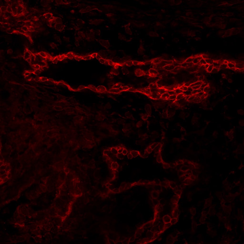
Detection of Mouse CD117/c-kit by Immunocytochemistry/Immunofluorescence
ANXA8+ve/c-kit+ve luminal progenitor cells are mostly Ki67−ve.Co-immunofluorescence staining for ANXA8 (green), c-kit (red), and Ki67 (purple) in mouse mammary gland from virgin and mid-pregnant mice shows that ANXA8+ve cells express c-kit, but not Ki67. The Ki67 staining has been coloured purple for easier visualisation in the triple-merged image. These are representative images of at least three independent mice for each time point. Image collected and cropped by CiteAb from the following publication (https://pubmed.ncbi.nlm.nih.gov/25803307), licensed under a CC-BY license. Not internally tested by R&D Systems.
Detection of CD117/c-kit in Human Mouse Embryos.
Formalin-fixed paraffin-embedded tissue sections of mouse embryo were probed for CD117 mRNA (ACD RNAScope Probe, catalog # 314158; Fast Red chromogen, ACD catalog # 322750). Adjacent tissue section was processed for immunohistochemistry using goat anti-mouse CD117 polyclonal antibody (AF1356) at 5 μg/mL with overnight incubation at 4 degrees Celsius followed by incubation with anti-goat IgG VisUCyte HRP Polymer Antibody (VC004) and DAB chromogen (yellow-brown). Tissue was counterstained with hematoxylin (blue). Specific staining was localized to cell membrane in esophagus in mouse embryo.
Detection of Mouse CD117/c-kit by Flow Cytometry
c-kit-, F4/80- or Ly-6G-positive cell numbers in cornea after alkali injury. Corneal tissues were obtained 4 days after injury from vehicle- or SDF-1 alpha -treated BABL/c mice, and the tissues from 7 to 8 mice were combined and were subjected to analysis using a flow cytometer after being immunostained with anti-c-kit, anti-F4/80, or anti-Ly-6G antibody. Isotype IgG derived from the same species of the test antibody was used as negative control. Representative results from three to four tests of intracorneal infitlration of c-kit- (A), F4/80- (B), or Ly-6G-positive cells (C) from either vehicle- (left plot) or SDF-1 alpha -treated mice (right plot) are shown. Image collected and cropped by CiteAb from the following open publication (https://pubmed.ncbi.nlm.nih.gov/21850188), licensed under a CC-BY license. Not internally tested by R&D Systems.
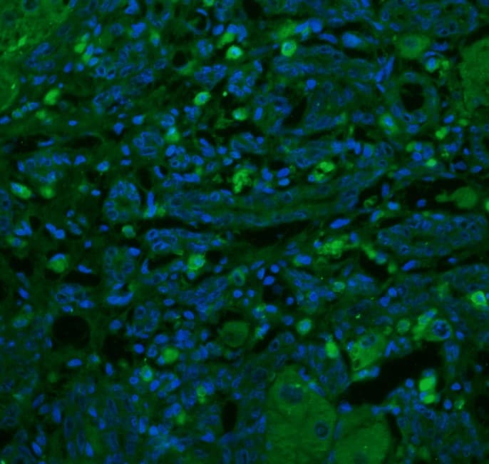
Detection of CD117/c-kit in Human Melanoma
CD117/c-kit was detected in immersion fixed paraffin-embedded sections of human melanoma using Goat Anti-Human/Mouse CD117/c-kit Antigen Affinity-purified Polyclonal Antibody (Catalog # AF1356) at 0.5 µg/ml overnight at 4 °C. Before incubation with the primary antibody, tissue was subjected to heat-induced epitope retrieval using VisUCyte Antigen Retrieval Reagent-Basic (Catalog # VCTS021). Tissue was stained using the HRP-conjugated Anti-Goat IgG Secondary Antibody (Catalog # HAF109) and counterstained with hematoxylin (blue). Specific staining was localized to the cell membrane and cytoplasm. View our protocol for Chromogenic IHC Staining of Paraffin-embedded Tissue Sections.Applications for CD117/c-kit Antibody
Application
Recommended Usage
CyTOF-ready
Ready to be labeled using established conjugation methods. No BSA or other carrier proteins that could interfere with conjugation.
Dual RNAscope ISH-IHC Compatible
5-15 µg/mL
Sample: Immersion fixed paraffin-embedded sections of human mouse embryos
Sample: Immersion fixed paraffin-embedded sections of human mouse embryos
Flow Cytometry
2.5 µg/106 cells
Sample: Lineage depleted mouse bone marrow cells
Sample: Lineage depleted mouse bone marrow cells
Immunohistochemistry
0.5-15 µg/mL
Sample: Immersion fixed frozen sections of mouse embryo (E15), paraffin-embedded human Melanoma
Sample: Immersion fixed frozen sections of mouse embryo (E15), paraffin-embedded human Melanoma
Simple Western
5 µg/mL
Sample: MO7e human megakaryocytic leukemic cell line, P815 mouse mastocytoma cell line, and MC/9‑2 mouse mast cell line
Sample: MO7e human megakaryocytic leukemic cell line, P815 mouse mastocytoma cell line, and MC/9‑2 mouse mast cell line
Western Blot
0.1 µg/mL
Sample: MO7e human megakaryocytic leukemic cell line, P815 mouse mastocytoma cell line, and MC/9‑2 mouse mast cell line
Sample: MO7e human megakaryocytic leukemic cell line, P815 mouse mastocytoma cell line, and MC/9‑2 mouse mast cell line
Reviewed Applications
Read 13 reviews rated 4 using AF1356 in the following applications:
Flow Cytometry Panel Builder
Bio-Techne Knows Flow Cytometry
Save time and reduce costly mistakes by quickly finding compatible reagents using the Panel Builder Tool.
Advanced Features
- Spectra Viewer - Custom analysis of spectra from multiple fluorochromes
- Spillover Popups - Visualize the spectra of individual fluorochromes
- Antigen Density Selector - Match fluorochrome brightness with antigen density

Formulation, Preparation, and Storage
Purification
Antigen Affinity-purified
Reconstitution
Reconstitute at 0.2 mg/mL in sterile PBS. For liquid material, refer to CoA for concentration.
Loading...
Formulation
Lyophilized from a 0.2 μm filtered solution in PBS with Trehalose. See Certificate of Analysis for details.
*Small pack size (-SP) is supplied either lyophilized or as a 0.2 µm filtered solution in PBS.
*Small pack size (-SP) is supplied either lyophilized or as a 0.2 µm filtered solution in PBS.
Shipping
Lyophilized product is shipped at ambient temperature. Liquid small pack size (-SP) is shipped with polar packs. Upon receipt, store immediately at the temperature recommended below.
Stability & Storage
Use a manual defrost freezer and avoid repeated freeze-thaw cycles.
- 12 months from date of receipt, -20 to -70 °C as supplied.
- 1 month, 2 to 8 °C under sterile conditions after reconstitution.
- 6 months, -20 to -70 °C under sterile conditions after reconstitution.
Calculators
Background: CD117/c-kit
Long Name
Stem Cell Factor Receptor
Alternate Names
c-kit, CD117, ckit, KIT, PBT, SCF R, SCFR
Gene Symbol
KIT
UniProt
Additional CD117/c-kit Products
Product Documents for CD117/c-kit Antibody
Certificate of Analysis
To download a Certificate of Analysis, please enter a lot or batch number in the search box below.
Note: Certificate of Analysis not available for kit components.
Product Specific Notices for CD117/c-kit Antibody
For research use only
Related Research Areas
Citations for CD117/c-kit Antibody
 Powered by Bioz
Customer Reviews for CD117/c-kit Antibody (13)
4 out of 5
13 Customer Ratings
Have you used CD117/c-kit Antibody?
Submit a review and receive an Amazon gift card!
$25/€18/£15/$25CAN/¥2500 Yen for a review with an image
$10/€7/£6/$10CAN/¥1110 Yen for a review without an image
Submit a review
Customer Images
Showing
1
-
5 of
13 reviews
Showing All
Filter By:
-
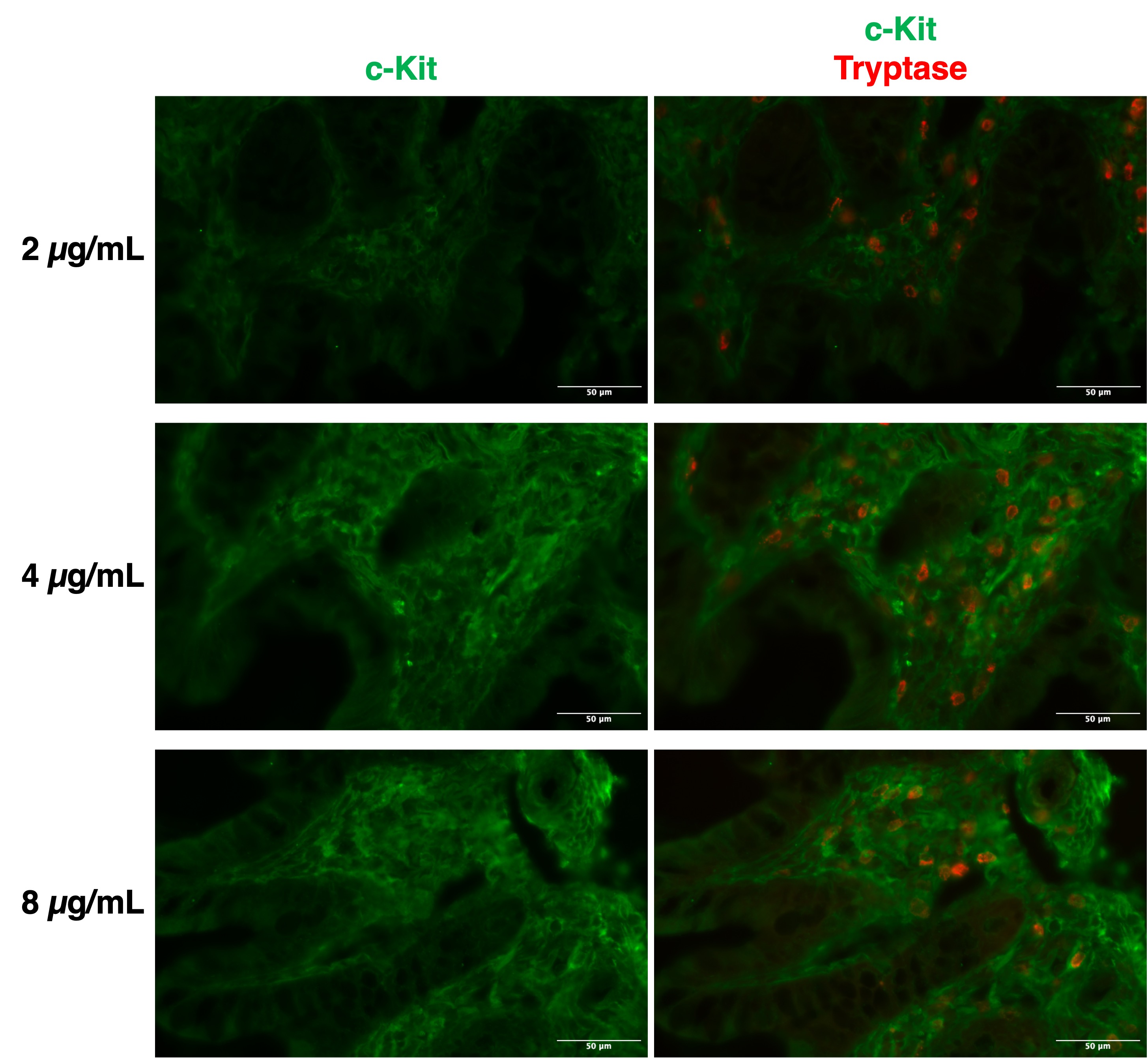
Application: Immunocytochemistry/ImmunofluorescenceSample Tested: Adult small intestineSpecies: HumanVerified Customer | Posted 10/16/2025Human ileum fixed in 4% PFA, cryopreserved in 30% sucrose and frozen. No specific staining in mast cells (tryptase+), which should express c-kit
 Bio-Techne ResponseThank you for reviewing our product. We are sorry to hear that this product did not perform as expected. We have been in touch with the customer to resolve this issue according to our Product Guarantee and to the customer’s satisfaction.
 -
Application: Immunocytochemistry/ImmunofluorescenceSample Tested: Nerve tissueSpecies: MouseVerified Customer | Posted 10/26/2021+4°C overnight incubation 1:200, 2nd anti-goat 647 1:400
-
Application: Immunocytochemistry/ImmunofluorescenceSample Tested: Adult intestineSpecies: MouseVerified Customer | Posted 10/02/2019
-
Application: Western BlotSample Tested: Muscle tissueSpecies: MouseVerified Customer | Posted 06/12/2019
-
Application: Immunocytochemistry/ImmunofluorescenceSample Tested: Liver tissueSpecies: RatVerified Customer | Posted 05/07/20181:100 dilution RT overnight incubation Citrate buffer heat mediated antigen retrival
-
Application: Western BlotSample Tested: ES-E14TG2aSpecies: MouseVerified Customer | Posted 04/05/2018The experiment was performed on ES-E14TG2a cells: two different bands of about 130 KDa (< 135 KDa) were detected.
-
Application: Western BlotSample Tested: mouse es cellsSpecies: MouseVerified Customer | Posted 03/23/2018
-
Application: Western BlotSample Tested: MCF-7 human breast cancer cell line, MCF 10A human breast epithelial cell line and MDA-MB-231 human breast cancer cell lineSpecies: HumanVerified Customer | Posted 07/24/2017
-
Application: Immunocytochemistry/ImmunofluorescenceSample Tested: Embryo tissueSpecies: MouseVerified Customer | Posted 08/08/2016Mouse embryonic yolk sac tissue (E10.5), with 1:100 dilution, Alex Fluro 594 donkey anti-goat 2nd antibody with 1:400 dilution
-
Application: Immunocytochemistry/ImmunofluorescenceSample Tested: Brain (basal ganglia)Species: MouseVerified Customer | Posted 03/22/2016
-
Application: Western BlotSample Tested: See PMID 24375406Species: MouseVerified Customer | Posted 01/05/2015
-
Application: ImmunofluorescenceSample Tested: See PMID 22262798Species: MouseVerified Customer | Posted 01/05/2015
-
Application: ImmunocytochemistrySample Tested: See PMID 24375406Species: MouseVerified Customer | Posted 01/05/2015
There are no reviews that match your criteria.
Protocols
Find general support by application which include: protocols, troubleshooting, illustrated assays, videos and webinars.
- 7-Amino Actinomycin D (7-AAD) Cell Viability Flow Cytometry Protocol
- Antigen Retrieval Protocol (PIER)
- Antigen Retrieval for Frozen Sections Protocol
- Appropriate Fixation of IHC/ICC Samples
- Cellular Response to Hypoxia Protocols
- Chromogenic IHC Staining of Formalin-Fixed Paraffin-Embedded (FFPE) Tissue Protocol
- Chromogenic Immunohistochemistry Staining of Frozen Tissue
- ClariTSA™ Fluorophore Kits
- Detection & Visualization of Antibody Binding
- Extracellular Membrane Flow Cytometry Protocol
- Flow Cytometry Protocol for Cell Surface Markers
- Flow Cytometry Protocol for Staining Membrane Associated Proteins
- Flow Cytometry Staining Protocols
- Flow Cytometry Troubleshooting Guide
- Fluorescent IHC Staining of Frozen Tissue Protocol
- Graphic Protocol for Heat-induced Epitope Retrieval
- Graphic Protocol for the Preparation and Fluorescent IHC Staining of Frozen Tissue Sections
- Graphic Protocol for the Preparation and Fluorescent IHC Staining of Paraffin-embedded Tissue Sections
- Graphic Protocol for the Preparation of Gelatin-coated Slides for Histological Tissue Sections
- IHC Sample Preparation (Frozen sections vs Paraffin)
- ISH-IHC Protocol for Chromogenic Detection on Formalin Fixed Paraffin Embedded (FFPE) Tissue
- Immunofluorescent IHC Staining of Formalin-Fixed Paraffin-Embedded (FFPE) Tissue Protocol
- Immunohistochemistry (IHC) and Immunocytochemistry (ICC) Protocols
- Immunohistochemistry Frozen Troubleshooting
- Immunohistochemistry Paraffin Troubleshooting
- Intracellular Flow Cytometry Protocol Using Alcohol (Methanol)
- Intracellular Flow Cytometry Protocol Using Detergents
- Intracellular Nuclear Staining Flow Cytometry Protocol Using Detergents
- Intracellular Staining Flow Cytometry Protocol Using Alcohol Permeabilization
- Intracellular Staining Flow Cytometry Protocol Using Detergents to Permeabilize Cells
- Preparing Samples for IHC/ICC Experiments
- Preventing Non-Specific Staining (Non-Specific Binding)
- Primary Antibody Selection & Optimization
- Propidium Iodide Cell Viability Flow Cytometry Protocol
- Protocol for Heat-Induced Epitope Retrieval (HIER)
- Protocol for Liperfluo
- Protocol for Making a 4% Formaldehyde Solution in PBS
- Protocol for VisUCyte™ HRP Polymer Detection Reagent
- Protocol for the Characterization of Human Th22 Cells
- Protocol for the Characterization of Human Th9 Cells
- Protocol for the Preparation & Fixation of Cells on Coverslips
- Protocol for the Preparation and Chromogenic IHC Staining of Frozen Tissue Sections
- Protocol for the Preparation and Chromogenic IHC Staining of Frozen Tissue Sections - Graphic
- Protocol for the Preparation and Chromogenic IHC Staining of Paraffin-embedded Tissue Sections
- Protocol for the Preparation and Chromogenic IHC Staining of Paraffin-embedded Tissue Sections - Graphic
- Protocol for the Preparation and Fluorescent IHC Staining of Frozen Tissue Sections
- Protocol for the Preparation and Fluorescent IHC Staining of Paraffin-embedded Tissue Sections
- Protocol for the Preparation of Gelatin-coated Slides for Histological Tissue Sections
- Protocol: Annexin V and PI Staining by Flow Cytometry
- Protocol: Annexin V and PI Staining for Apoptosis by Flow Cytometry
- R&D Systems Quality Control Western Blot Protocol
- TUNEL and Active Caspase-3 Detection by IHC/ICC Protocol
- The Importance of IHC/ICC Controls
- Troubleshooting Guide: Fluorokine Flow Cytometry Kits
- Troubleshooting Guide: Immunohistochemistry
- Troubleshooting Guide: Western Blot Figures
- Western Blot Conditions
- Western Blot Protocol
- Western Blot Protocol for Cell Lysates
- Western Blot Troubleshooting
- Western Blot Troubleshooting Guide
- View all Protocols, Troubleshooting, Illustrated assays and Webinars


