
Key Product Details
Validated by
Knockout/Knockdown
Species Reactivity
Validated:
Human, Mouse
Cited:
Human, Mouse
Applications
Validated:
Immunohistochemistry, Western Blot, Immunocytochemistry, Simple Western
Cited:
Immunohistochemistry, Western Blot, Immunocytochemistry, Chromatin Immunoprecipitation (ChIP), Chromatin Immunoprecipitation Sequencing
Label
Unconjugated
Antibody Source
Polyclonal Goat IgG
Loading...
Product Specifications
Immunogen
E. coli-derived recombinant human GATA-2
Ala15-Thr279
Accession # P23769
Ala15-Thr279
Accession # P23769
Specificity
Detects human and mouse GATA-2 in Western blots.
Clonality
Polyclonal
Host
Goat
Isotype
IgG
Scientific Data Images for GATA-2 Antibody
Detection of Human GATA‑2 by Western Blot.
Western blot shows lysates of NIH-3T3 mouse embryonic fibroblast cell line and KG-1 human acute myelogenous leukemia cell line. PVDF membrane was probed with 0.5 µg/mL of Human/Mouse GATA-2 Antigen Affinity-purified Polyclonal Antibody (Catalog # AF2046) followed by HRP-conjugated Anti-Goat IgG Secondary Antibody (Catalog # HAF017). A specific band was detected for GATA-2 at approximately 51 kDa (as indicated). This experiment was conducted under reducing conditions and using Immunoblot Buffer Group 1.
Detection of Human GATA‑2 by Western Blot.
Western blot shows lysates of LNCaP human prostate cancer cell line and SH-SY5Y human neuroblastoma cell line. PVDF membrane was probed with 0.5 µg/mL of Goat Anti-Human/Mouse GATA-2 Antigen Affinity-purified Polyclonal Antibody (Catalog # AF2046) followed by HRP-conjugated Anti-Goat IgG Secondary Antibody (Catalog # HAF017). A specific band was detected for GATA-2 at approximately 55 kDa (as indicated). This experiment was conducted under reducing conditions and using Immunoblot Buffer Group 1.
GATA‑2 in HUVEC Human Cells.
GATA‑2 was detected in immersion fixed HUVEC human umbilical vein endothelial cells using Goat Anti-Human/Mouse GATA‑2 Antigen Affinity-purified Polyclonal Antibody (Catalog # AF2046) at 5 µg/mL for 3 hours at room temperature. Cells were stained using the NorthernLights™ 557-conjugated Anti-Goat IgG Secondary Antibody (red; NL001) and counterstained with DAPI (blue). Specific staining was localized to cell nuclei. Staining was performed using our protocol for Fluorescent ICC Staining of Non-adherent Cells.
GATA‑2 in SH‑SY5Y Human Cell Line.
GATA‑2 was detected in immersion fixed SH‑SY5Y human neuroblastoma cell line using Goat Anti-Human/Mouse GATA‑2 Antigen Affinity-purified Polyclonal Antibody (Catalog # AF2046) at 5 µg/mL for 3 hours at room temperature. Cells were stained using the NorthernLights™ 557-conjugated Anti-Goat IgG Secondary Antibody (red; NL001) and counterstained with DAPI (blue). Specific staining was localized to cell nuclei. Staining was performed using our protocol for Fluorescent ICC Staining of Non-adherent Cells.
GATA‑2 in Human Duodenum.
GATA-2 was detected in immersion fixed paraffin-embedded sections of human duodenum (blood vessel) using Goat Anti-Human/Mouse GATA-2 Antigen Affinity-purified Polyclonal Antibody (Catalog # AF2046) at 1 µg/mL for 1 hour at room temperature followed by incubation with the Anti-Goat IgG VisUCyte™ HRP Polymer Antibody (Catalog # VC004). Before incubation with the primary antibody, tissue was subjected to heat-induced epitope retrieval using Antigen Retrieval Reagent-Basic (Catalog # CTS013). Tissue was stained using DAB (brown) and counterstained with hematoxylin (blue). Specific staining was localized to nuclei in endothelial cells. View our protocol for IHC Staining with VisUCyte HRP Polymer Detection Reagents.
Detection of Human GATA‑2 by Simple WesternTM.
Simple Western lane view shows lysates of KG-1 human acute myelogenous leukemia cell line, loaded at 0.2 mg/mL. A specific band was detected for GATA-2 at approximately 64 kDa (as indicated) using 5 µg/mL of Goat Anti-Human GATA-2 Antigen Affinity-purified Polyclonal Antibody (Catalog # AF2046) followed by 1:50 dilution of HRP-conjugated Anti-Goat IgG Secondary Antibody (Catalog # HAF109). This experiment was conducted under reducing conditions and using the 12-230 kDa separation system.
Detection of Mouse GATA-2 by Western Blot
RNA-seq identifies the targets of GATA2 in primary human LECs. (A) Principal component analysis (PCA) was performed on RNA-seq data from control shRNA- and shGATA2-infected primary HLECs. A high level of similarity was observed within the groups as indicated by their proximity to each other. (B) Hierarchical clustering shows that approximately 1000 genes were consistently downregulated and 600 genes were upregulated in shGATA2-treated HLECs. (C) GO revealed a list of genes that are likely relevant to the phenotypes observed in mice lacking GATA2. (D) GATA2 was knocked out from a second HLEC line using CRISPR/Cas9. Western blot revealed the lack of GATA2 in the knockout cells (HLEC delta GATA2). In contrast, no obvious differences were observed in the expression of PROX1. Additionally, qRT-PCR revealed the downregulation of miR-126. (A) n=3 independent experiments per shRNA; (D) n=3 independent experiments (antibiotic selection, western blot and qRT-PCR). **P<0.01. Image collected and cropped by CiteAb from the following publication (https://pubmed.ncbi.nlm.nih.gov/31582413), licensed under a CC-BY license. Not internally tested by R&D Systems.
Detection of Human GATA-2 by Western Blot
Changes in KIT gene expression by GATA1 siRNAs. (a) qRT-PCR analysis was performed for K562-WT (left) and one of the K562-G1s clone, C1 #26 (right). The vertical axis represents a relative value with each value of non-target siRNA set at 1. Results are averages of three independent experiments. Error bars represent standard deviation. *P < 0.05, **P < 0.01 (two-side t-test). (b) Representative immunoblots of siRNA experiments with K562-WT (left) and G1s C1 #26 (right). Western blotting was performed with anti-GATA1 antibody (D24E4, Cell signaling technology), anti-GATA2 (AF2046, R&D) anti-c-Kit (SC-17806, Santa Cruz) and anti-beta -actin antibody (Sigma-Aldrich). Images were obtained using ChemiDoc MP. Image collected and cropped by CiteAb from the following open publication (https://pubmed.ncbi.nlm.nih.gov/36447001), licensed under a CC-BY license. Not internally tested by R&D Systems.
Detection of Human GATA-2 by Western Blot
Changes in KIT gene expression by GATA1 siRNAs. (a) qRT-PCR analysis was performed for K562-WT (left) and one of the K562-G1s clone, C1 #26 (right). The vertical axis represents a relative value with each value of non-target siRNA set at 1. Results are averages of three independent experiments. Error bars represent standard deviation. *P < 0.05, **P < 0.01 (two-side t-test). (b) Representative immunoblots of siRNA experiments with K562-WT (left) and G1s C1 #26 (right). Western blotting was performed with anti-GATA1 antibody (D24E4, Cell signaling technology), anti-GATA2 (AF2046, R&D) anti-c-Kit (SC-17806, Santa Cruz) and anti-beta -actin antibody (Sigma-Aldrich). Images were obtained using ChemiDoc MP. Image collected and cropped by CiteAb from the following open publication (https://pubmed.ncbi.nlm.nih.gov/36447001), licensed under a CC-BY license. Not internally tested by R&D Systems.Applications for GATA-2 Antibody
Application
Recommended Usage
Immunocytochemistry
5-15 µg/mL
Sample: Immersion fixed HUVEC human umbilical vein endothelial cells and SH‑SY5Y human neuroblastoma cell line
Sample: Immersion fixed HUVEC human umbilical vein endothelial cells and SH‑SY5Y human neuroblastoma cell line
Immunohistochemistry
1-15 µg/mL
Sample: Immersion fixed paraffin-embedded sections of human duodenum (blood vessel)
Sample: Immersion fixed paraffin-embedded sections of human duodenum (blood vessel)
Simple Western
5 µg/mL
Sample: KG‑1 human acute myelogenous leukemia cell line
Sample: KG‑1 human acute myelogenous leukemia cell line
Western Blot
0.5 µg/mL
Sample: NIH-3T3 mouse embryonic fibroblast cell line, KG-1 human acute myelogenous leukemia cell line, LNCaP human prostate cancer cell line, and SH‑SY5Y human neuroblastoma cell line
Sample: NIH-3T3 mouse embryonic fibroblast cell line, KG-1 human acute myelogenous leukemia cell line, LNCaP human prostate cancer cell line, and SH‑SY5Y human neuroblastoma cell line
Reviewed Applications
Read 1 review rated 5 using AF2046 in the following applications:
Formulation, Preparation, and Storage
Purification
Antigen Affinity-purified
Reconstitution
Reconstitute at 0.2 mg/mL in sterile PBS. For liquid material, refer to CoA for concentration.
Loading...
Formulation
Lyophilized from a 0.2 μm filtered solution in PBS with Trehalose. *Small pack size (SP) is supplied either lyophilized or as a 0.2 µm filtered solution in PBS.
Shipping
Lyophilized product is shipped at ambient temperature. Liquid small pack size (-SP) is shipped with polar packs. Upon receipt, store immediately at the temperature recommended below.
Stability & Storage
Use a manual defrost freezer and avoid repeated freeze-thaw cycles.
- 12 months from date of receipt, -20 to -70 °C as supplied.
- 1 month, 2 to 8 °C under sterile conditions after reconstitution.
- 6 months, -20 to -70 °C under sterile conditions after reconstitution.
Calculators
Background: GATA-2
References
- Tsai, S.F. et al. (1989) Nature 339:446.
- Jiang, Y. and T. Evans (1996) Dev. Biol. 174:258.
Alternate Names
GATA2
Entrez Gene IDs
2624 (Human)
Gene Symbol
GATA2
UniProt
Additional GATA-2 Products
Product Documents for GATA-2 Antibody
Certificate of Analysis
To download a Certificate of Analysis, please enter a lot or batch number in the search box below.
Note: Certificate of Analysis not available for kit components.
Product Specific Notices for GATA-2 Antibody
For research use only
Related Research Areas
Citations for GATA-2 Antibody
 Powered by Bioz
Customer Reviews for GATA-2 Antibody (1)
5 out of 5
1 Customer Rating
Have you used GATA-2 Antibody?
Submit a review and receive an Amazon gift card!
$25/€18/£15/$25CAN/¥2500 Yen for a review with an image
$10/€7/£6/$10CAN/¥1110 Yen for a review without an image
Submit a review
Customer Images
Showing
1
-
1 of
1 review
Showing All
Filter By:
-
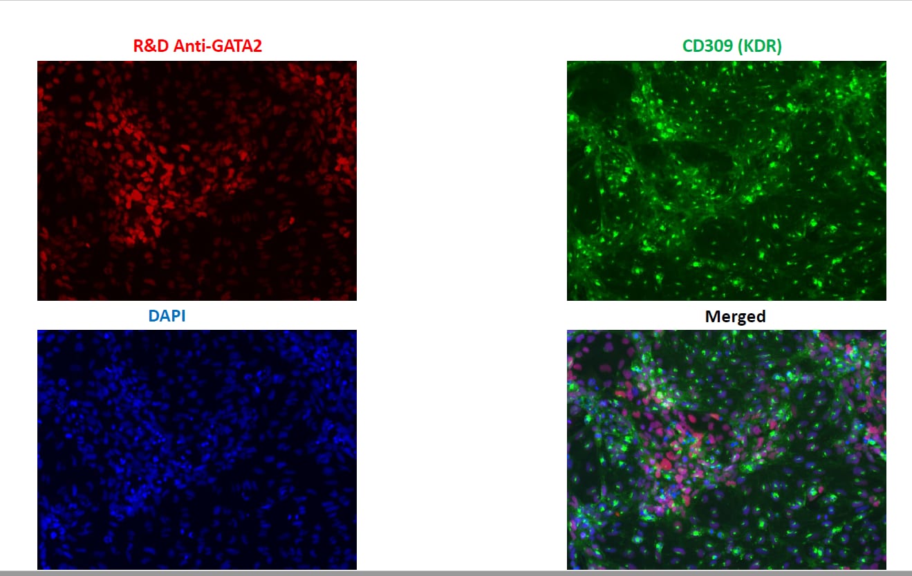
Application: Immunocytochemistry/ImmunofluorescenceSample Tested: HUVEC human umbilical vein endothelial cells and Blood progenitor cellsSpecies: HumanVerified Customer | Posted 05/17/2022Stained with AF+ secondary antibody on endothelial cells undergoing differentiation.

There are no reviews that match your criteria.
Protocols
Find general support by application which include: protocols, troubleshooting, illustrated assays, videos and webinars.
- Antigen Retrieval Protocol (PIER)
- Antigen Retrieval for Frozen Sections Protocol
- Appropriate Fixation of IHC/ICC Samples
- Cellular Response to Hypoxia Protocols
- Chromogenic IHC Staining of Formalin-Fixed Paraffin-Embedded (FFPE) Tissue Protocol
- Chromogenic Immunohistochemistry Staining of Frozen Tissue
- ClariTSA™ Fluorophore Kits
- Detection & Visualization of Antibody Binding
- Fluorescent IHC Staining of Frozen Tissue Protocol
- Graphic Protocol for Heat-induced Epitope Retrieval
- Graphic Protocol for the Preparation and Fluorescent IHC Staining of Frozen Tissue Sections
- Graphic Protocol for the Preparation and Fluorescent IHC Staining of Paraffin-embedded Tissue Sections
- Graphic Protocol for the Preparation of Gelatin-coated Slides for Histological Tissue Sections
- ICC Cell Smear Protocol for Suspension Cells
- ICC Immunocytochemistry Protocol Videos
- ICC for Adherent Cells
- IHC Sample Preparation (Frozen sections vs Paraffin)
- Immunocytochemistry (ICC) Protocol
- Immunocytochemistry Troubleshooting
- Immunofluorescence of Organoids Embedded in Cultrex Basement Membrane Extract
- Immunofluorescent IHC Staining of Formalin-Fixed Paraffin-Embedded (FFPE) Tissue Protocol
- Immunohistochemistry (IHC) and Immunocytochemistry (ICC) Protocols
- Immunohistochemistry Frozen Troubleshooting
- Immunohistochemistry Paraffin Troubleshooting
- Preparing Samples for IHC/ICC Experiments
- Preventing Non-Specific Staining (Non-Specific Binding)
- Primary Antibody Selection & Optimization
- Protocol for Heat-Induced Epitope Retrieval (HIER)
- Protocol for Making a 4% Formaldehyde Solution in PBS
- Protocol for VisUCyte™ HRP Polymer Detection Reagent
- Protocol for the Fluorescent ICC Staining of Cell Smears - Graphic
- Protocol for the Fluorescent ICC Staining of Cultured Cells on Coverslips - Graphic
- Protocol for the Preparation & Fixation of Cells on Coverslips
- Protocol for the Preparation and Chromogenic IHC Staining of Frozen Tissue Sections
- Protocol for the Preparation and Chromogenic IHC Staining of Frozen Tissue Sections - Graphic
- Protocol for the Preparation and Chromogenic IHC Staining of Paraffin-embedded Tissue Sections
- Protocol for the Preparation and Chromogenic IHC Staining of Paraffin-embedded Tissue Sections - Graphic
- Protocol for the Preparation and Fluorescent ICC Staining of Cells on Coverslips
- Protocol for the Preparation and Fluorescent ICC Staining of Non-adherent Cells
- Protocol for the Preparation and Fluorescent ICC Staining of Stem Cells on Coverslips
- Protocol for the Preparation and Fluorescent IHC Staining of Frozen Tissue Sections
- Protocol for the Preparation and Fluorescent IHC Staining of Paraffin-embedded Tissue Sections
- Protocol for the Preparation of Gelatin-coated Slides for Histological Tissue Sections
- Protocol for the Preparation of a Cell Smear for Non-adherent Cell ICC - Graphic
- R&D Systems Quality Control Western Blot Protocol
- TUNEL and Active Caspase-3 Detection by IHC/ICC Protocol
- The Importance of IHC/ICC Controls
- Troubleshooting Guide: Immunohistochemistry
- Troubleshooting Guide: Western Blot Figures
- Western Blot Conditions
- Western Blot Protocol
- Western Blot Protocol for Cell Lysates
- Western Blot Troubleshooting
- Western Blot Troubleshooting Guide
- View all Protocols, Troubleshooting, Illustrated assays and Webinars
Loading...
Associated Pathways
