eIF4E (eukaryotic initiation factor 4E) interacts with the 7-methyl-GTP cap structure to facilitate the initiation and rate of translation of mRNA. Together with eIF4G and eIF4A, it forms the eIF4F complex. eIF4E activity has been shown to play a role in cell cycle progression, tumorigenesis, embryonic development, nuclear export and synaptic plasticity.

Key Product Details
Validated by
Knockout/Knockdown
Species Reactivity
Validated:
Human, Mouse, Rat
Cited:
Human, Mouse
Applications
Validated:
Knockout Validated, Western Blot, Immunocytochemistry
Cited:
Immunohistochemistry, Western Blot
Label
Unconjugated
Antibody Source
Monoclonal Mouse IgG2B Clone # 299910
Loading...
Product Specifications
Immunogen
E. coli-derived recombinant human eIF4E
Met1-Val217
Accession # P06730
Met1-Val217
Accession # P06730
Specificity
Detects human, mouse and rat eIF4E.
Clonality
Monoclonal
Host
Mouse
Isotype
IgG2B
Scientific Data Images for Human/Mouse/Rat eIF4E Antibody
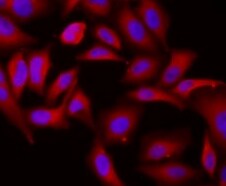
eIF4E in MCF-7 Human Cell Line.
eIF4E was detected in immersion fixed MCF-7 human breast cancer cell line using 10 µg/mL Mouse Anti-Human/Mouse/Rat eIF4E Monoclonal Antibody (Catalog # MAB3228) for 3 hours at room temperature. Cells were stained with the NorthernLights™ 557-conjugated Anti-Mouse IgG Secondary Antibody (red; Catalog # NL007) and counterstained with DAPI (blue). View our protocol for Fluorescent ICC Staining of Cells on Coverslips.
Detection of Human/Mouse/Rat eIF4E by Western Blot.
Western blot shows lysates of MCF-7 human breast cancer cell line, Balb/3T3 mouse embryonic fibroblast cell line, and PC-12 rat adrenal pheochromocytoma cell line. PVDF membrane was probed with 0.1 µg/mL of Mouse Anti-Human/Mouse/Rat eIF4E Monoclonal Antibody (Catalog # MAB3228) followed by HRP-conjugated Anti-Mouse IgG Secondary Antibody (Catalog # HAF007). A specific band was detected for eIF4E at approximately 25 kDa (as indicated). This experiment was conducted under reducing conditions and using Immunoblot Buffer Group 1.
Western Blot Shows Human eIF4E Specificity by Using Knockout Cell Line.
Western blot shows lysates of MCF-7 human breast cancer parental cell line and eIF4E knockout MCF-7 cell line (KO). PVDF membrane was probed with 0.5 µg/mL of Mouse Anti-Human/Mouse/Rat eIF4E Monoclonal Antibody (Catalog # MAB3228) followed by HRP-conjugated Anti-Mouse IgG Secondary Antibody (Catalog # HAF018). A specific band was detected for eIF4E at approximately 25 kDa (as indicated) in the parental MCF-7 cell line, but is not detectable in knockout MCF-7 cell line. GAPDH (Catalog # MAB5718) is shown as a loading control. This experiment was conducted under reducing conditions and using Immunoblot Buffer Group 1.
Detection of eIF4E by Western Blot
mTORC1-mediated upregulation of p21 requires 4E-BP1 phosphorylation.(a) Effect of mTOR inhibitors torin-1 (250 nM) and rapamycin (100 nM) on p21 levels in different cell lines. Cells were treated for 24 h with the indicated drugs and protein levels were subsequently analysed by western blot. (b) Protein levels in MEFs infected simultaneously with lentiviruses expressing either scramble or TSC2 shRNAs together with control or 4E-BP1-4A. (c) Protein levels in U2OS, HCT116 and 293T cells transfected with 4E-BP1-4A mutant alone (U2OS and HCT116 cells) or in combination with pBabe-p21 plasmid (293T cells). (d) Western blot depicting protein levels in MEFs following co-infection with lentiviruses expressing the indicated shRNAs. (e) Primary MEFs were infected with lentiviruses expressing scramble, TSC2 or eIF4E shRNAs in combination with control or 4E-BP1-4A. Protein levels were measured by immunoblotting. For each panel, all the western blots correspond to samples from the same experiment; in some cases, samples were distributed in several electrophoretic gels run in parallel. Image collected and cropped by CiteAb from the following open publication (https://pubmed.ncbi.nlm.nih.gov/26832959), licensed under a CC-BY license. Not internally tested by R&D Systems.
Detection of eIF4E by Western Blot
p38 and ERK1/2 trigger distinct patterns of cytokine release. A, Heatmap of the release of IL-1 alpha, IL-1 beta, IL-6, GM-CSF, and G-CSF by TR146 OECs at 24 h post-candidalysin stimulation in the presence of BIRB796, SP600125, Trametinib, or Gefitinib. The heatmap was generated from the data in fig. S2A. B, Release of GM-CSF from TR146 cells transfected with a pool of Hsp27-targeted siRNAs 72 h prior to candidalysin stimulation for 24 h. Graph shows means of three biological replicates + SD and is expressed as fold change relative to siRNA control + candidalysin. C, Relative expression of IL6 6 h post-candidalysin stimulation in the presence of BIRB796. Graph shows means of three biological replicates + SD and is expressed as fold change relative to DMSO + candidalysin. D and E, IL-6 released into the supernatant D, and present in cell extracts E, before (0 h), after 6 h and after 24 h of candidalysin treatment with or without BIRB796. Graphs show means of three biological replicates + SD and are expressed as fold change relative to DMSO + candidalysin at 24 h. F, Representative immunoblot showing phosphorylated (p-) and total eIF4E 30 min and 2 h post-candidalysin stimulation in the presence of BIRB796. Immunoblots are representative of three biological replicates. GAPDH is a loading control. G, Graphical quantification of immunoblots as in F. Scatter plot shows the mean ± SD of three biological replicates expressed as ratios of p-eIF4E/eIF4E. Statistical significance for B and C was quantified by one sample t test compared to a hypothetical value = 1. Statistical significance in D, E and G was quantified by paired t-tests as indicated in the graphs. *P < 0.05, **P < 0.01, ***P < 0.001. Image collected and cropped by CiteAb from the following open publication (https://pubmed.ncbi.nlm.nih.gov/35380879), licensed under a CC-BY license. Not internally tested by R&D Systems.Applications for Human/Mouse/Rat eIF4E Antibody
Application
Recommended Usage
Immunocytochemistry
8-25 µg/mL
Sample: Immersion fixed MCF-7 human breast cancer cell line
Sample: Immersion fixed MCF-7 human breast cancer cell line
Knockout Validated
eIF4E
is specifically detected in MCF‑7 human breast cancer cell line parental cell line but is not detectable in
eIF4E knockout MCF‑7 cell line.
Western Blot
0.1 µg/mL
Sample: MCF-7 human breast cancer cell line, Balb/3T3 mouse embryonic fibroblast cell line, and PC-12 rat adrenal pheochromocytoma cell line
Sample: MCF-7 human breast cancer cell line, Balb/3T3 mouse embryonic fibroblast cell line, and PC-12 rat adrenal pheochromocytoma cell line
Reviewed Applications
Read 1 review rated 5 using MAB3228 in the following applications:
Formulation, Preparation, and Storage
Purification
Protein A or G purified from hybridoma culture supernatant
Reconstitution
Reconstitute at 0.5 mg/mL in sterile PBS. For liquid material, refer to CoA for concentration.
Loading...
Formulation
Lyophilized from a 0.2 μm filtered solution in PBS with Trehalose. *Small pack size (SP) is supplied either lyophilized or as a 0.2 µm filtered solution in PBS.
Shipping
Lyophilized product is shipped at ambient temperature. Liquid small pack size (-SP) is shipped with polar packs. Upon receipt, store immediately at the temperature recommended below.
Stability & Storage
Use a manual defrost freezer and avoid repeated freeze-thaw cycles.
- 12 months from date of receipt, -20 to -70 °C as supplied.
- 1 month, 2 to 8 °C under sterile conditions after reconstitution.
- 6 months, -20 to -70 °C under sterile conditions after reconstitution.
Calculators
Background: eIF4E
Long Name
Eukaryotic Translation Initiation Factor 4E
Alternate Names
EIF4EL1, EIF4F
Gene Symbol
EIF4E
UniProt
Additional eIF4E Products
Product Documents for Human/Mouse/Rat eIF4E Antibody
Certificate of Analysis
To download a Certificate of Analysis, please enter a lot or batch number in the search box below.
Note: Certificate of Analysis not available for kit components.
Product Specific Notices for Human/Mouse/Rat eIF4E Antibody
For research use only
Related Research Areas
Citations for Human/Mouse/Rat eIF4E Antibody
 Powered by Bioz
Customer Reviews for Human/Mouse/Rat eIF4E Antibody (1)
5 out of 5
1 Customer Rating
Have you used Human/Mouse/Rat eIF4E Antibody?
Submit a review and receive an Amazon gift card!
$25/€18/£15/$25CAN/¥2500 Yen for a review with an image
$10/€7/£6/$10CAN/¥1110 Yen for a review without an image
Submit a review
Customer Images
Showing
1
-
1 of
1 review
Showing All
Filter By:
-
Application: Immunocytochemistry/ImmunofluorescenceSample Tested: MCF-7 Human Cell LineSpecies: HumanVerified Customer | Posted 03/13/2022

There are no reviews that match your criteria.
Protocols
Find general support by application which include: protocols, troubleshooting, illustrated assays, videos and webinars.
- Appropriate Fixation of IHC/ICC Samples
- Cellular Response to Hypoxia Protocols
- ClariTSA™ Fluorophore Kits
- Detection & Visualization of Antibody Binding
- ICC Cell Smear Protocol for Suspension Cells
- ICC Immunocytochemistry Protocol Videos
- ICC for Adherent Cells
- Immunocytochemistry (ICC) Protocol
- Immunocytochemistry Troubleshooting
- Immunofluorescence of Organoids Embedded in Cultrex Basement Membrane Extract
- Immunohistochemistry (IHC) and Immunocytochemistry (ICC) Protocols
- Preparing Samples for IHC/ICC Experiments
- Preventing Non-Specific Staining (Non-Specific Binding)
- Primary Antibody Selection & Optimization
- Protocol for VisUCyte™ HRP Polymer Detection Reagent
- Protocol for the Fluorescent ICC Staining of Cell Smears - Graphic
- Protocol for the Fluorescent ICC Staining of Cultured Cells on Coverslips - Graphic
- Protocol for the Preparation and Fluorescent ICC Staining of Cells on Coverslips
- Protocol for the Preparation and Fluorescent ICC Staining of Non-adherent Cells
- Protocol for the Preparation and Fluorescent ICC Staining of Stem Cells on Coverslips
- Protocol for the Preparation of a Cell Smear for Non-adherent Cell ICC - Graphic
- R&D Systems Quality Control Western Blot Protocol
- TUNEL and Active Caspase-3 Detection by IHC/ICC Protocol
- The Importance of IHC/ICC Controls
- Troubleshooting Guide: Western Blot Figures
- Western Blot Conditions
- Western Blot Protocol
- Western Blot Protocol for Cell Lysates
- Western Blot Troubleshooting
- Western Blot Troubleshooting Guide
- View all Protocols, Troubleshooting, Illustrated assays and Webinars
Loading...
Associated Pathways


