Tyrosine Hydroxylase Antibody (779427)
R&D Systems | Catalog # MAB7566

Key Product Details
Species Reactivity
Validated:
Human, Mouse
Cited:
Human, Mouse, Rat
Applications
Validated:
Immunohistochemistry, Western Blot, Immunocytochemistry, Simple Western
Cited:
Immunohistochemistry, Western Blot, Flow Cytometry, Immunofluorescence, Immunocytochemistry
Label
Unconjugated
Antibody Source
Monoclonal Mouse IgG2B Clone # 779427
Loading...
Product Specifications
Immunogen
E. coli-derived recombinant human Tyrosine Hydroxylase
Ala278-Tyr401
Accession # P07101
Ala278-Tyr401
Accession # P07101
Specificity
Detects human Tyrosine Hydroxylase in direct ELISAs and human, mouse and rat Tyrosine Hydroxylase in Western blots. In direct ELISAs, no cross-reactivity with recombinant human Tryptophan Hydroxylase-1 is observed.
Clonality
Monoclonal
Host
Mouse
Isotype
IgG2B
Scientific Data Images for Tyrosine Hydroxylase Antibody (779427)
Detection of Human, Mouse, and Rat Tyrosine Hydroxylase by Western Blot.
Western blot shows lysates of human adrenal gland tissue, mouse adrenal gland tissue, and rat adrenal gland tissue. PVDF membrane was probed with 0.25 µg/mL of Mouse Anti-Human/Mouse Tyrosine Hydroxylase Monoclonal Antibody (Catalog # MAB7566) followed by HRP-conjugated Anti-Mouse IgG Secondary Antibody (Catalog # HAF018). Specific bands were detected for Tyrosine Hydroxylase at approximately 50-60 kDa (as indicated). This experiment was conducted under reducing conditions and using Immunoblot Buffer Group 1.
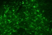
Tyrosine Hydroxylase in Mouse Dopaminergic Neurons.
Tyrosine Hydroxylase was detected in immersion fixed mouse embryonic stem cells differentiated into dopaminergic neurons using Mouse Anti-Human/Mouse Tyrosine Hydroxylase Monoclonal Antibody (Catalog # MAB7566) at 10 µg/mL for 3 hours at room temperature. Cells were stained using the Northern-Lights™ 557-conjugated Anti-Mouse IgG Secondary Antibody (red; Catalog # NL007). Cells were double stained using the Northern-Lights™ 637-conjugated Mouse Anti-Neuron-specific beta-III Tubulin Monoclonal Antibody (white; Catalog # NL1195V). Cells were counterstained with DAPI (blue). Specific staining of Tyrosine Hydroxylase was localized to cytoplasm of dopaminergic neurons. View our protocol for Fluorescent ICC Staining of Cells on Coverslips.
Tyrosine Hydroxylase in Human Brain.
Tyrosine Hydroxylase was detected in immersion fixed paraffin-embedded sections of human brain (medulla) using Mouse Anti-Human/Mouse Tyrosine Hydroxylase Monoclonal Antibody (Catalog # MAB7566) at 25 µg/mL overnight at 4 °C. Tissue was stained using the Anti-Mouse HRP-DAB Cell & Tissue Staining Kit (brown; Catalog # CTS002) and counterstained with hematoxylin (blue). Specific staining was localized to neurons. View our protocol for Chromogenic IHC Staining of Paraffin-embedded Tissue Sections.
Detection of Human and Mouse Tyrosine Hydroxylase by Simple WesternTM.
Simple Western lane view shows lysates of human adrenal gland tissue and mouse adrenal gland tissue, loaded at 0.2 mg/mL. Specific bands were detected for Tyrosine Hydroxylase at approximately 52-61 kDa (as indicated) using 5 µg/mL of Mouse Anti-Human/Mouse Tyrosine Hydroxylase Monoclonal Antibody (Catalog # MAB7566). This experiment was conducted under reducing conditions and using the 12-230 kDa separation system.Non-specific interaction with the 230 kDa Simple Western standard may be seen with this antibody.Applications for Tyrosine Hydroxylase Antibody (779427)
Application
Recommended Usage
Immunocytochemistry
8-25 µg/mL
Sample: Immersion fixed mouse embryonic stem cells differentiated into dopaminergic neurons
Sample: Immersion fixed mouse embryonic stem cells differentiated into dopaminergic neurons
Immunohistochemistry
8-25 µg/mL
Sample: Immersion fixed paraffin-embedded sections of human brain (medulla)
Sample: Immersion fixed paraffin-embedded sections of human brain (medulla)
Simple Western
5 µg/mL
Sample: human adrenal gland tissue and mouse adrenal gland tissue
Sample: human adrenal gland tissue and mouse adrenal gland tissue
Western Blot
0.25 µg/mL
Sample: Human adrenal gland tissue, mouse adrenal gland tissue, and rat adrenal gland tissue
Sample: Human adrenal gland tissue, mouse adrenal gland tissue, and rat adrenal gland tissue
Reviewed Applications
Read 4 reviews rated 4.8 using MAB7566 in the following applications:
Formulation, Preparation, and Storage
Purification
Protein A or G purified from hybridoma culture supernatant
Reconstitution
Sterile PBS to a final concentration of 0.5 mg/mL. For liquid material, refer to CoA for concentration.
Loading...
Formulation
Lyophilized from a 0.2 μm filtered solution in PBS with Trehalose. *Small pack size (SP) is supplied either lyophilized or as a 0.2 µm filtered solution in PBS.
Shipping
Lyophilized product is shipped at ambient temperature. Liquid small pack size (-SP) is shipped with polar packs. Upon receipt, store immediately at the temperature recommended below.
Stability & Storage
Use a manual defrost freezer and avoid repeated freeze-thaw cycles.
- 12 months from date of receipt, -20 to -70 °C as supplied.
- 1 month, 2 to 8 °C under sterile conditions after reconstitution.
- 6 months, -20 to -70 °C under sterile conditions after reconstitution.
Calculators
Background: Tyrosine Hydroxylase
Alternate Names
TH, TYH
Gene Symbol
TH
UniProt
Additional Tyrosine Hydroxylase Products
Product Documents for Tyrosine Hydroxylase Antibody (779427)
Certificate of Analysis
To download a Certificate of Analysis, please enter a lot or batch number in the search box below.
Note: Certificate of Analysis not available for kit components.
Product Specific Notices for Tyrosine Hydroxylase Antibody (779427)
For research use only
Citations for Tyrosine Hydroxylase Antibody (779427)
 Powered by Bioz
Customer Reviews for Tyrosine Hydroxylase Antibody (779427) (4)
4.8 out of 5
4 Customer Ratings
Have you used Tyrosine Hydroxylase Antibody (779427)?
Submit a review and receive an Amazon gift card!
$25/€18/£15/$25CAN/¥2500 Yen for a review with an image
$10/€7/£6/$10CAN/¥1110 Yen for a review without an image
Submit a review
Customer Images
Showing
1
-
4 of
4 reviews
Showing All
Filter By:
-
Application: ImmunohistochemistrySample Tested: Brain tissueSpecies: MouseVerified Customer | Posted 09/13/2021

-
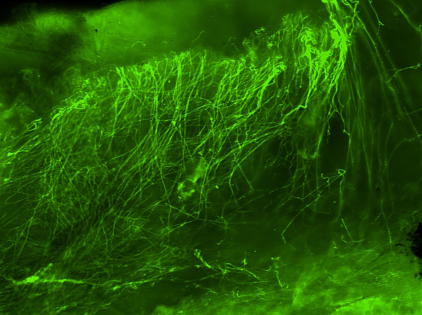
Application: Immunocytochemistry/ImmunofluorescenceSample Tested: Sympathetic nerve and conjunctival sympathetic nerveSpecies: MouseVerified Customer | Posted 10/27/2017
-
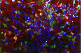
Application: Immunocytochemistry/ImmunofluorescenceSample Tested: iPS2 human induced pluripotent stem cellsSpecies: HumanVerified Customer | Posted 10/24/2016TH positive neurons in red, co-stained with TUJ1 to mark neurons.
-
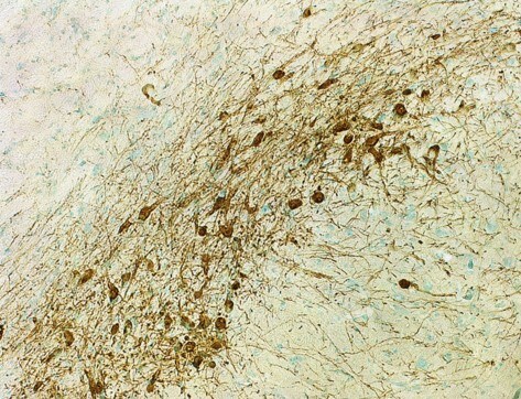
Application: Immunohistochemistry-FrozenSample Tested: Frozen tissue sections or rat cerebrumSpecies: RatVerified Customer | Posted 09/02/2015TH positive cells, IHC-Fr 1:500 dilution
There are no reviews that match your criteria.
Protocols
Find general support by application which include: protocols, troubleshooting, illustrated assays, videos and webinars.
- Antigen Retrieval Protocol (PIER)
- Antigen Retrieval for Frozen Sections Protocol
- Appropriate Fixation of IHC/ICC Samples
- Cellular Response to Hypoxia Protocols
- Chromogenic IHC Staining of Formalin-Fixed Paraffin-Embedded (FFPE) Tissue Protocol
- Chromogenic Immunohistochemistry Staining of Frozen Tissue
- ClariTSA™ Fluorophore Kits
- Detection & Visualization of Antibody Binding
- Fluorescent IHC Staining of Frozen Tissue Protocol
- Graphic Protocol for Heat-induced Epitope Retrieval
- Graphic Protocol for the Preparation and Fluorescent IHC Staining of Frozen Tissue Sections
- Graphic Protocol for the Preparation and Fluorescent IHC Staining of Paraffin-embedded Tissue Sections
- Graphic Protocol for the Preparation of Gelatin-coated Slides for Histological Tissue Sections
- ICC Cell Smear Protocol for Suspension Cells
- ICC Immunocytochemistry Protocol Videos
- ICC for Adherent Cells
- IHC Sample Preparation (Frozen sections vs Paraffin)
- Immunocytochemistry (ICC) Protocol
- Immunocytochemistry Troubleshooting
- Immunofluorescence of Organoids Embedded in Cultrex Basement Membrane Extract
- Immunofluorescent IHC Staining of Formalin-Fixed Paraffin-Embedded (FFPE) Tissue Protocol
- Immunohistochemistry (IHC) and Immunocytochemistry (ICC) Protocols
- Immunohistochemistry Frozen Troubleshooting
- Immunohistochemistry Paraffin Troubleshooting
- Preparing Samples for IHC/ICC Experiments
- Preventing Non-Specific Staining (Non-Specific Binding)
- Primary Antibody Selection & Optimization
- Protocol for Heat-Induced Epitope Retrieval (HIER)
- Protocol for Making a 4% Formaldehyde Solution in PBS
- Protocol for VisUCyte™ HRP Polymer Detection Reagent
- Protocol for the Fluorescent ICC Staining of Cell Smears - Graphic
- Protocol for the Fluorescent ICC Staining of Cultured Cells on Coverslips - Graphic
- Protocol for the Preparation & Fixation of Cells on Coverslips
- Protocol for the Preparation and Chromogenic IHC Staining of Frozen Tissue Sections
- Protocol for the Preparation and Chromogenic IHC Staining of Frozen Tissue Sections - Graphic
- Protocol for the Preparation and Chromogenic IHC Staining of Paraffin-embedded Tissue Sections
- Protocol for the Preparation and Chromogenic IHC Staining of Paraffin-embedded Tissue Sections - Graphic
- Protocol for the Preparation and Fluorescent ICC Staining of Cells on Coverslips
- Protocol for the Preparation and Fluorescent ICC Staining of Non-adherent Cells
- Protocol for the Preparation and Fluorescent ICC Staining of Stem Cells on Coverslips
- Protocol for the Preparation and Fluorescent IHC Staining of Frozen Tissue Sections
- Protocol for the Preparation and Fluorescent IHC Staining of Paraffin-embedded Tissue Sections
- Protocol for the Preparation of Gelatin-coated Slides for Histological Tissue Sections
- Protocol for the Preparation of a Cell Smear for Non-adherent Cell ICC - Graphic
- R&D Systems Quality Control Western Blot Protocol
- TUNEL and Active Caspase-3 Detection by IHC/ICC Protocol
- The Importance of IHC/ICC Controls
- Troubleshooting Guide: Immunohistochemistry
- Troubleshooting Guide: Western Blot Figures
- Western Blot Conditions
- Western Blot Protocol
- Western Blot Protocol for Cell Lysates
- Western Blot Troubleshooting
- Western Blot Troubleshooting Guide
- View all Protocols, Troubleshooting, Illustrated assays and Webinars
Loading...
Associated Pathways
