Human Pref-1/DLK1/FA1 Antibody (211309)
R&D Systems | Catalog # MAB1144

Key Product Details
Species Reactivity
Validated:
Human
Cited:
Human
Applications
Validated:
Western Blot, Flow Cytometry, Immunocytochemistry, CyTOF-ready
Cited:
Immunohistochemistry, Western Blot, Immunocytochemistry, Immunoprecipitation
Label
Unconjugated
Antibody Source
Monoclonal Mouse IgG2B Clone # 211309
Loading...
Product Specifications
Immunogen
Mouse myeloma cell line NS0-derived recombinant human Pref-1 long isoform
Ala24-Pro297 (with Arg248Pro and Lys295Ser substitutions)
Accession # P80370
Ala24-Pro297 (with Arg248Pro and Lys295Ser substitutions)
Accession # P80370
Specificity
Detects human Pref‑1/DLK‑1/FA1 in direct ELISAs and Western blots. Shows approximately 50% cross-reactivity with recombinant mouse Pref‑1.
Clonality
Monoclonal
Host
Mouse
Isotype
IgG2B
Scientific Data Images for Human Pref-1/DLK1/FA1 Antibody (211309)
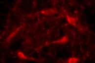
Pref‑1/DLK1/FA1 in HepG2 Human Cell Line.
Pref-1/DLK1/FA1 was detected in immersion fixed HepG2 human hepatocellular carcinoma cell line using Mouse Anti-Human Pref-1/DLK1/FA1 Monoclonal Antibody (Catalog # MAB1144) at 10 µg/mL for 3 hours at room temperature. Cells were stained using the NorthernLights™ 557-conjugated Anti-Mouse IgG Secondary Antibody (red; Catalog # NL007) and counterstained with DAPI (blue). Specific staining was localized to cell membranes. View our protocol for Fluorescent ICC Staining of Cells on Coverslips.
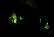
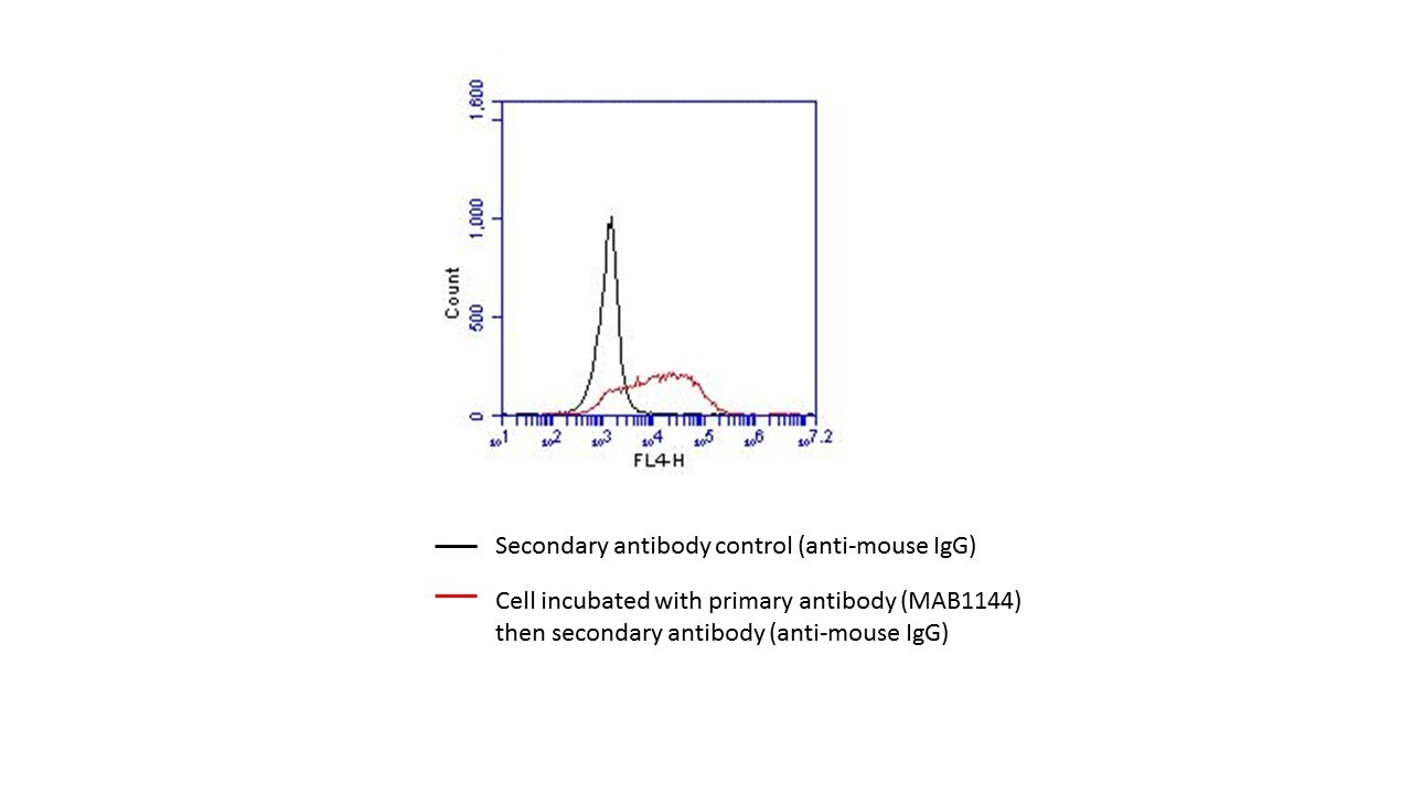
Detection of Pref‑1/DLK1/FA1 in HEK293/Pref-1/eGFP vs Irrelevant transfectants by Flow Cytometry
HEK293/Pref-1/eGFP (A) vs Irrelevant transfectants (B) were stained with Mouse Anti-Human Pref‑1/DLK1/FA1 Monoclonal Antibody (Catalog # MAB1144) followed by Allophycocyanin-conjugated Anti-Mouse IgG Secondary Antibody (Catalog # F0101B). View our protocol for Staining Membrane-associated Proteins.Applications for Human Pref-1/DLK1/FA1 Antibody (211309)
Application
Recommended Usage
CyTOF-ready
Ready to be labeled using established conjugation methods. No BSA or other carrier proteins that could interfere with conjugation.
Flow Cytometry
0.25 µg/106 cells
Sample: HEK293/hPref-1/eGFP transfectant cell line vs irrelevant
Sample: HEK293/hPref-1/eGFP transfectant cell line vs irrelevant
Immunocytochemistry
8-25 µg/mL
Sample: Immersion fixed HepG2 human hepatocellular carcinoma cell line
Sample: Immersion fixed HepG2 human hepatocellular carcinoma cell line
Western Blot
1 µg/mL
Sample: Recombinant Human Pref‑1/DLK‑1/FA1 under non-reducing conditions only
Sample: Recombinant Human Pref‑1/DLK‑1/FA1 under non-reducing conditions only
Reviewed Applications
Read 4 reviews rated 4.8 using MAB1144 in the following applications:
Flow Cytometry Panel Builder
Bio-Techne Knows Flow Cytometry
Save time and reduce costly mistakes by quickly finding compatible reagents using the Panel Builder Tool.
Advanced Features
- Spectra Viewer - Custom analysis of spectra from multiple fluorochromes
- Spillover Popups - Visualize the spectra of individual fluorochromes
- Antigen Density Selector - Match fluorochrome brightness with antigen density

Formulation, Preparation, and Storage
Purification
Protein A or G purified from hybridoma culture supernatant
Reconstitution
Reconstitute at 0.5 mg/mL in sterile PBS. For liquid material, refer to CoA for concentration.
Loading...
Formulation
Lyophilized from a 0.2 μm filtered solution in PBS with Trehalose. See Certificate of Analysis for details.
*Small pack size (-SP) is supplied either lyophilized or as a 0.2 µm filtered solution in PBS.
*Small pack size (-SP) is supplied either lyophilized or as a 0.2 µm filtered solution in PBS.
Shipping
Lyophilized product is shipped at ambient temperature. Liquid small pack size (-SP) is shipped with polar packs. Upon receipt, store immediately at the temperature recommended below.
Stability & Storage
Use a manual defrost freezer and avoid repeated freeze-thaw cycles.
- 12 months from date of receipt, -20 to -70 °C as supplied.
- 1 month, 2 to 8 °C under sterile conditions after reconstitution.
- 6 months, -20 to -70 °C under sterile conditions after reconstitution.
Calculators
Background: Pref-1/DLK1/FA1
Long Name
Preadipocyte Factor-1/Protein delta Homolog 1/Fetal Antigen 1
Alternate Names
DLK-1, DLK1, FA1, pG2, Pref1, ZOG
Gene Symbol
DLK1
UniProt
Additional Pref-1/DLK1/FA1 Products
Product Documents for Human Pref-1/DLK1/FA1 Antibody (211309)
Certificate of Analysis
To download a Certificate of Analysis, please enter a lot or batch number in the search box below.
Note: Certificate of Analysis not available for kit components.
Product Specific Notices for Human Pref-1/DLK1/FA1 Antibody (211309)
For research use only
Related Research Areas
Citations for Human Pref-1/DLK1/FA1 Antibody (211309)
 Powered by Bioz
Customer Reviews for Human Pref-1/DLK1/FA1 Antibody (211309) (4)
4.8 out of 5
4 Customer Ratings
Have you used Human Pref-1/DLK1/FA1 Antibody (211309)?
Submit a review and receive an Amazon gift card!
$25/€18/£15/$25CAN/¥2500 Yen for a review with an image
$10/€7/£6/$10CAN/¥1110 Yen for a review without an image
Submit a review
Customer Images
Showing
1
-
4 of
4 reviews
Showing All
Filter By:
-
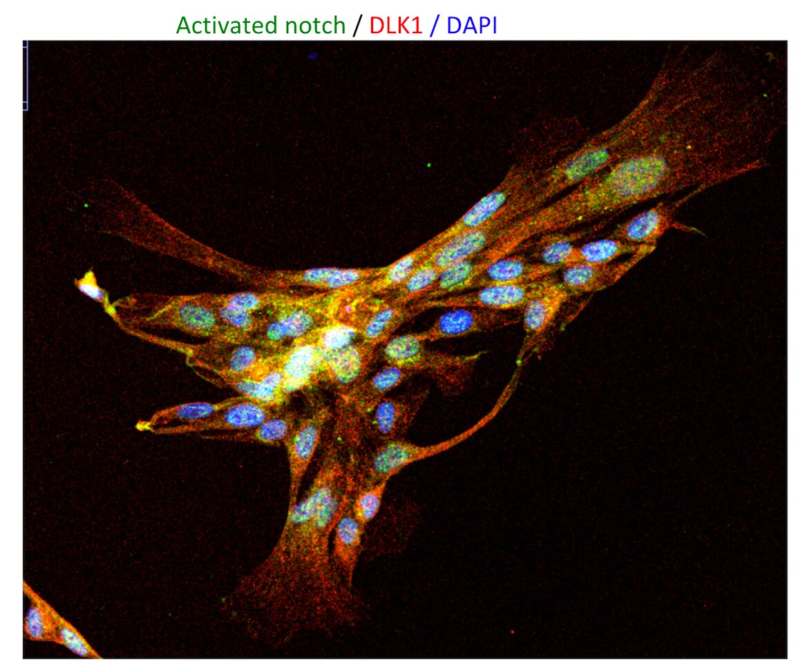
Application: Immunocytochemistry/ImmunofluorescenceSample Tested: SVF-cellsSpecies: HumanVerified Customer | Posted 10/18/2021

-
Application: ImmunohistochemistrySample Tested: Brain (substantia nigra)Species: HumanVerified Customer | Posted 10/13/2021
-
Application: Flow CytometrySample Tested: NIH3T3 cells overxpressing human DLK1Species: HumanVerified Customer | Posted 01/09/2018
-
Application: Immunocytochemistry/ImmunofluorescenceSample Tested: mesenchymal cellsSpecies: HumanVerified Customer | Posted 10/18/2016
There are no reviews that match your criteria.
Protocols
Find general support by application which include: protocols, troubleshooting, illustrated assays, videos and webinars.
- 7-Amino Actinomycin D (7-AAD) Cell Viability Flow Cytometry Protocol
- Appropriate Fixation of IHC/ICC Samples
- Cellular Response to Hypoxia Protocols
- ClariTSA™ Fluorophore Kits
- Detection & Visualization of Antibody Binding
- Extracellular Membrane Flow Cytometry Protocol
- Flow Cytometry Protocol for Cell Surface Markers
- Flow Cytometry Protocol for Staining Membrane Associated Proteins
- Flow Cytometry Staining Protocols
- Flow Cytometry Troubleshooting Guide
- ICC Cell Smear Protocol for Suspension Cells
- ICC Immunocytochemistry Protocol Videos
- ICC for Adherent Cells
- Immunocytochemistry (ICC) Protocol
- Immunocytochemistry Troubleshooting
- Immunofluorescence of Organoids Embedded in Cultrex Basement Membrane Extract
- Immunohistochemistry (IHC) and Immunocytochemistry (ICC) Protocols
- Intracellular Flow Cytometry Protocol Using Alcohol (Methanol)
- Intracellular Flow Cytometry Protocol Using Detergents
- Intracellular Nuclear Staining Flow Cytometry Protocol Using Detergents
- Intracellular Staining Flow Cytometry Protocol Using Alcohol Permeabilization
- Intracellular Staining Flow Cytometry Protocol Using Detergents to Permeabilize Cells
- Preparing Samples for IHC/ICC Experiments
- Preventing Non-Specific Staining (Non-Specific Binding)
- Primary Antibody Selection & Optimization
- Propidium Iodide Cell Viability Flow Cytometry Protocol
- Protocol for Liperfluo
- Protocol for VisUCyte™ HRP Polymer Detection Reagent
- Protocol for the Characterization of Human Th22 Cells
- Protocol for the Characterization of Human Th9 Cells
- Protocol for the Fluorescent ICC Staining of Cell Smears - Graphic
- Protocol for the Fluorescent ICC Staining of Cultured Cells on Coverslips - Graphic
- Protocol for the Preparation and Fluorescent ICC Staining of Cells on Coverslips
- Protocol for the Preparation and Fluorescent ICC Staining of Non-adherent Cells
- Protocol for the Preparation and Fluorescent ICC Staining of Stem Cells on Coverslips
- Protocol for the Preparation of a Cell Smear for Non-adherent Cell ICC - Graphic
- Protocol: Annexin V and PI Staining by Flow Cytometry
- Protocol: Annexin V and PI Staining for Apoptosis by Flow Cytometry
- R&D Systems Quality Control Western Blot Protocol
- TUNEL and Active Caspase-3 Detection by IHC/ICC Protocol
- The Importance of IHC/ICC Controls
- Troubleshooting Guide: Fluorokine Flow Cytometry Kits
- Troubleshooting Guide: Western Blot Figures
- Western Blot Conditions
- Western Blot Protocol
- Western Blot Protocol for Cell Lysates
- Western Blot Troubleshooting
- Western Blot Troubleshooting Guide
- View all Protocols, Troubleshooting, Illustrated assays and Webinars
Loading...
Associated Pathways

