Matrix metalloproteinases are a family of zinc and calcium dependent endopeptidases with the combined ability to degrade all the components of the extracellular matrix. MMP-13 (Collagenase-3) has been demonstrated to degrade a range of extracellular matrix proteins, including collagen types I, II, III, IV, IX, X and XIV, gelatin, aggrecan, perlecan and fibronectin. MMP-13 is distinguished from the other human collagenases by its effecient degradation of type II collagen. MMP-13 is expressed by fibroblasts, chrondrocytes and squamous epithelial cells. Structurally, MMP-13 may be divided into several distinct domains; a pro-domain which is cleaved upon activation; a catalytic domain containing the zinc binding site; a short hinge region and a carboxyl terminal (hemopexin-like) domain.
Human Pro MMP-13 Antibody (87518)
R&D Systems | Catalog # MAB913

Key Product Details
Species Reactivity
Validated:
Cited:
Applications
Validated:
Cited:
Label
Antibody Source
Product Specifications
Immunogen
Leu20-Cys471
Accession # P45452
Specificity
Clonality
Host
Isotype
Scientific Data Images for Human Pro MMP-13 Antibody (87518)
Human Pro- MMP‑13 ELISA Standard Curve.
Recombinant Human Pro-MMP-13 protein was serially diluted 2-fold and captured by Mouse Anti-Human MMP-13 Monoclonal Antibody (Catalog # MAB5111) coated on a Clear Polystyrene Microplate (Catalog # DY990). Mouse Anti-Human Pro MMP-13 Monoclonal Antibody (Catalog # MAB913) was biotinylated and incubated with the protein captured on the plate. Detection of the standard curve was achieved by incubating Streptavidin-HRP (Catalog # DY998) followed by Substrate Solution (Catalog # DY999) and stopping the enzymatic reaction with Stop Solution (Catalog # DY994).Applications for Human Pro MMP-13 Antibody (87518)
ELISA
This antibody functions as a Pro-MMP-13 ELISA ELISA detection antibody when paired with Mouse Anti-Human MMP‑13 Monoclonal Antibody (Catalog # MAB5111).
This product is intended for assay development on various assay platforms requiring antibody pairs. We recommend the Human Pro-MMP-13 DuoSet ELISA Kit (Catalog # DY913) for convenient development of a sandwich ELISA or the Human Pro-MMP-13 Quantikine ELISA Kit (Catalog # DM1300) for a complete optimized ELISA.
Immunoaffinity Purification
Immunohistochemistry
Sample: Immersion fixed paraffin-embedded sections of human breast cancer tissue subjected to Antigen Retrieval Reagent-Basic (Catalog # CTS013)
Immunoprecipitation
Sample: Conditioned cell culture medium spiked with Recombinant Human MMP‑13 (Catalog # 511‑MM), see our available Western blot detection antibodies
Western Blot
Sample: Recombinant Human MMP-13 Western Blot Standard (Catalog # WBC020) under non‑reducing conditions only
Reviewed Applications
Read 1 review rated 4 using MAB913 in the following applications:
Formulation, Preparation, and Storage
Purification
Reconstitution
Reconstitute at 0.5 mg/mL in sterile PBS. For liquid material, refer to CoA for concentration.
Formulation
Shipping
Stability & Storage
- 12 months from date of receipt, -20 to -70 °C as supplied.
- 1 month, 2 to 8 °C under sterile conditions after reconstitution.
- 6 months, -20 to -70 °C under sterile conditions after reconstitution.
Calculators
Background: MMP-13
References
- Jeffery, J.J. (1998) in Collagenase 3. A.J. Barrett, et al. (eds): Handbook of Proteolytic Enzymes, San Diego: Academic Press, p. 1167.
Long Name
Alternate Names
Entrez Gene IDs
Gene Symbol
UniProt
Additional MMP-13 Products
Product Documents for Human Pro MMP-13 Antibody (87518)
Certificate of Analysis
To download a Certificate of Analysis, please enter a lot or batch number in the search box below.
Note: Certificate of Analysis not available for kit components.
Product Specific Notices for Human Pro MMP-13 Antibody (87518)
For research use only
Related Research Areas
Citations for Human Pro MMP-13 Antibody (87518)
 Powered by Bioz
Customer Reviews for Human Pro MMP-13 Antibody (87518) (1)
Have you used Human Pro MMP-13 Antibody (87518)?
Submit a review and receive an Amazon gift card!
$25/€18/£15/$25CAN/¥2500 Yen for a review with an image
$10/€7/£6/$10CAN/¥1110 Yen for a review without an image
Submit a review
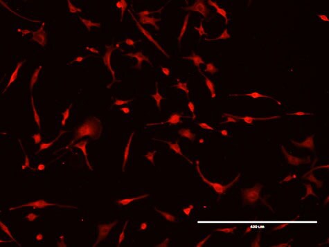
Customer Images
-
Application: Immunocytochemistry/ImmunofluorescenceSample Tested: C20A4 Chondrocyte cell lineSpecies: HumanVerified Customer | Posted 08/28/2018

There are no reviews that match your criteria.
Protocols
Find general support by application which include: protocols, troubleshooting, illustrated assays, videos and webinars.
- Antigen Retrieval Protocol (PIER)
- Antigen Retrieval for Frozen Sections Protocol
- Appropriate Fixation of IHC/ICC Samples
- Cellular Response to Hypoxia Protocols
- Chromogenic IHC Staining of Formalin-Fixed Paraffin-Embedded (FFPE) Tissue Protocol
- Chromogenic Immunohistochemistry Staining of Frozen Tissue
- ClariTSA™ Fluorophore Kits
- Detection & Visualization of Antibody Binding
- ELISA Sample Preparation & Collection Guide
- ELISA Troubleshooting Guide
- Fluorescent IHC Staining of Frozen Tissue Protocol
- Graphic Protocol for Heat-induced Epitope Retrieval
- Graphic Protocol for the Preparation and Fluorescent IHC Staining of Frozen Tissue Sections
- Graphic Protocol for the Preparation and Fluorescent IHC Staining of Paraffin-embedded Tissue Sections
- Graphic Protocol for the Preparation of Gelatin-coated Slides for Histological Tissue Sections
- How to Run an R&D Systems DuoSet ELISA
- How to Run an R&D Systems Quantikine ELISA
- How to Run an R&D Systems Quantikine™ QuicKit™ ELISA
- IHC Sample Preparation (Frozen sections vs Paraffin)
- Immunofluorescent IHC Staining of Formalin-Fixed Paraffin-Embedded (FFPE) Tissue Protocol
- Immunohistochemistry (IHC) and Immunocytochemistry (ICC) Protocols
- Immunohistochemistry Frozen Troubleshooting
- Immunohistochemistry Paraffin Troubleshooting
- Immunoprecipitation Protocol
- Preparing Samples for IHC/ICC Experiments
- Preventing Non-Specific Staining (Non-Specific Binding)
- Primary Antibody Selection & Optimization
- Protocol for Heat-Induced Epitope Retrieval (HIER)
- Protocol for Making a 4% Formaldehyde Solution in PBS
- Protocol for VisUCyte™ HRP Polymer Detection Reagent
- Protocol for the Preparation & Fixation of Cells on Coverslips
- Protocol for the Preparation and Chromogenic IHC Staining of Frozen Tissue Sections
- Protocol for the Preparation and Chromogenic IHC Staining of Frozen Tissue Sections - Graphic
- Protocol for the Preparation and Chromogenic IHC Staining of Paraffin-embedded Tissue Sections
- Protocol for the Preparation and Chromogenic IHC Staining of Paraffin-embedded Tissue Sections - Graphic
- Protocol for the Preparation and Fluorescent IHC Staining of Frozen Tissue Sections
- Protocol for the Preparation and Fluorescent IHC Staining of Paraffin-embedded Tissue Sections
- Protocol for the Preparation of Gelatin-coated Slides for Histological Tissue Sections
- Quantikine HS ELISA Kit Assay Principle, Alkaline Phosphatase
- Quantikine HS ELISA Kit Principle, Streptavidin-HRP Polymer
- R&D Systems Quality Control Western Blot Protocol
- Sandwich ELISA (Colorimetric) – Biotin/Streptavidin Detection Protocol
- Sandwich ELISA (Colorimetric) – Direct Detection Protocol
- TUNEL and Active Caspase-3 Detection by IHC/ICC Protocol
- The Importance of IHC/ICC Controls
- Troubleshooting Guide: ELISA
- Troubleshooting Guide: Immunohistochemistry
- Troubleshooting Guide: Western Blot Figures
- Western Blot Conditions
- Western Blot Protocol
- Western Blot Protocol for Cell Lysates
- Western Blot Troubleshooting
- Western Blot Troubleshooting Guide
- View all Protocols, Troubleshooting, Illustrated assays and Webinars