
Key Product Details
Species Reactivity
Validated:
Human
Cited:
Human, Mouse
Applications
Validated:
Immunohistochemistry, Western Blot, Neutralization, Simple Western
Cited:
Immunohistochemistry, Immunohistochemistry-Paraffin, Immunohistochemistry-Frozen, Western Blot, Neutralization, Flow Cytometry, Immunocytochemistry, Immunoprecipitation, Cell Culture
Label
Unconjugated
Antibody Source
Polyclonal Goat IgG
Loading...
Product Specifications
Immunogen
S. frugiperda insect ovarian cell line Sf 21-derived recombinant human ST2/IL-33 R
Lys19-Phe328
Accession # BAA02233
Lys19-Phe328
Accession # BAA02233
Specificity
Detects human ST2/IL-33 R in direct ELISAs and Western blots.
Clonality
Polyclonal
Host
Goat
Isotype
IgG
Endotoxin Level
<0.10 EU per 1 μg of the antibody by the LAL method.
Scientific Data Images for Human ST2/IL-33R Antibody
Detection of Human ST2/IL-33 R by Western Blot.
Western blot shows lysates of human kidney tissue and human lung tissue. PVDF membrane was probed with 0.2 µg/mL of Goat Anti-Human ST2/IL-33 R Antigen Affinity-purified Polyclonal Antibody (Catalog # AF523) followed by HRP-conjugated Anti-Goat IgG Secondary Antibody (HAF017). A specific band was detected for ST2/IL-33 R at approximately 50 kDa (as indicated). This experiment was conducted under reducing conditions and using Immunoblot Buffer Group 1.
ST2/IL-33 R in Human Liver.
ST2/IL-33 R was detected in immersion fixed paraffin-embedded sections of human liver using Goat Anti-Human ST2/IL-33 R Antigen Affinity-purified Polyclonal Antibody (Catalog # AF523) at 1.7 µg/mL for 1 hour at room temperature followed by incubation with the Anti-Goat IgG VisUCyte™ HRP Polymer Antibody (Catalog # (VC004). Before incubation with the primary antibody, tissue was subjected to heat-induced epitope retrieval using Antigen Retrieval Reagent-Basic (CTS013). Tissue was stained using DAB (brown) and counterstained with hematoxylin (blue). Specific staining was localized to cell membranes. View our protocol for IHC Staining with VisUCyte HRP Polymer Detection Reagents.
Detection of Human ST2/IL-33 R by Simple WesternTM.
Simple Western lane view shows lysates of human kidney tissue and human lung tissue, loaded at 0.2 mg/mL. A specific band was detected for ST2/IL-33 R at approximately 60 kDa (as indicated) using 2 µg/mL of Goat Anti-Human ST2/IL-33 R Antigen Affinity-purified Polyclonal Antibody (Catalog # AF523) followed by 1:50 dilution of HRP-conjugated Anti-Goat IgG Secondary Antibody (HAF109). This experiment was conducted under reducing conditions and using the 12-230 kDa separation system.
IFN-gamma Secretion Induced by IL‑33 and Neutralization by Human ST2/IL-33 R Antibody.
Recombinant Human IL-33 induces IFN-gamma secretion in Human Peripheral Blood Mononuclear cells (PBMC) in the presence of 0.25 ng/mL Recombinant Human IL-12 (219-IL) in a dose-dependent manner (orange line), as measured by the Human IFN-gamma Quantikine ELISA Kit (DIF50C). Under these conditions, IFN-gamma secretion elicited by IL-33 is neutralized (green line) by increasing concentrations of Goat Anti-Human ST2/IL-33 R Antigen Affinity-purified Polyclonal Antibody (Catalog # AF523). The ND50 is typically 0.1-0.6 µg/mL.
Detection of ST2/IL-33R by Western Blot
IL‐33 protein levels in whole lungs of mice exposed to SUHX (a) Western Blot representation of IL‐33, ST2, and MYD88 protein in lungs of C57BL/6J, ST2−/−, and MYD88−/− mice exposed to either SUHX or DMSO/RA conditions (all n = 8). Equal loading of protein was confirmed using alpha ‐Tubulin. (b) Quantitative analysis of IL‐33 in whole lungs. Values are presented as the means ± SD Image collected and cropped by CiteAb from the following open publication (https://pubmed.ncbi.nlm.nih.gov/35150208), licensed under a CC-BY license. Not internally tested by R&D Systems.
Detection of ST2/IL-33R by Western Blot
The effect of IL-33 on the pro-fibrotic activity of fibroblasts. Non-IPF (n = 2) and IPF (n = 2) HLFs were stimulated with 2 ng/ml of TGF beta or 10 ng/ml of IL-33 for 4, 8 and 24 h and ACTA2 (A), COL1A1 (B), IL6 (C) and CXCL8 (D) gene expression measured by qPCR. For each donor, all data was normalised to the unstimulated media alone control at 4 h. Non-IPF and IPF donors are shown in black and grey respectively. Bars indicate median values and each data point represents a single donor with statistical analysis performed by Wilcoxon signed-rank test. Whole cell lysates from resting (rest.) M1 and M2 MDMs, activated (act.) M1 (IFN gamma stimulated) and M2 (IL-4 stimulated) MDMs, HPAECs stimulated with 5 ng/ml of TNF alpha, 5 ng/ml of TGF beta and 0.1 ng/ml of IL-1 beta (TTI) and a single non-IPF HLF donor were separated by SDS-PAGE (20 μg protein/lane) and expression of ST2 and GAPDH measured by western blot (E). Whole cell lysates from HUVECs, non-IPF (n = 3) and IPF (n = 4) HLFs were separated by SDS-PAGE (20 μg protein/lane) and the expression of ST2 and GAPDH determined by western blot (F). Representative cropped western blots are shown Image collected and cropped by CiteAb from the following open publication (https://pubmed.ncbi.nlm.nih.gov/36949463), licensed under a CC-BY license. Not internally tested by R&D Systems.
Detection of ST2/IL-33R by Western Blot
The effect of IL-33 on the pro-fibrotic activity of fibroblasts. Non-IPF (n = 2) and IPF (n = 2) HLFs were stimulated with 2 ng/ml of TGF beta or 10 ng/ml of IL-33 for 4, 8 and 24 h and ACTA2 (A), COL1A1 (B), IL6 (C) and CXCL8 (D) gene expression measured by qPCR. For each donor, all data was normalised to the unstimulated media alone control at 4 h. Non-IPF and IPF donors are shown in black and grey respectively. Bars indicate median values and each data point represents a single donor with statistical analysis performed by Wilcoxon signed-rank test. Whole cell lysates from resting (rest.) M1 and M2 MDMs, activated (act.) M1 (IFN gamma stimulated) and M2 (IL-4 stimulated) MDMs, HPAECs stimulated with 5 ng/ml of TNF alpha, 5 ng/ml of TGF beta and 0.1 ng/ml of IL-1 beta (TTI) and a single non-IPF HLF donor were separated by SDS-PAGE (20 μg protein/lane) and expression of ST2 and GAPDH measured by western blot (E). Whole cell lysates from HUVECs, non-IPF (n = 3) and IPF (n = 4) HLFs were separated by SDS-PAGE (20 μg protein/lane) and the expression of ST2 and GAPDH determined by western blot (F). Representative cropped western blots are shown Image collected and cropped by CiteAb from the following open publication (https://pubmed.ncbi.nlm.nih.gov/36949463), licensed under a CC-BY license. Not internally tested by R&D Systems.Applications for Human ST2/IL-33R Antibody
Application
Recommended Usage
Immunohistochemistry
1-15 µg/mL
Sample: Immersion fixed paraffin-embedded sections of human liver
Sample: Immersion fixed paraffin-embedded sections of human liver
Simple Western
2 µg/mL
Sample: Human kidney tissue and human lung tissue
Sample: Human kidney tissue and human lung tissue
Western Blot
0.2 µg/mL
Sample: Human kidney tissue and human lung tissue
Sample: Human kidney tissue and human lung tissue
Neutralization
Measured by its ability to neutralize IL‑33-induced IFN-gamma secretion in Human Peripheral Blood Mononuclear cells (PBMC). Blom, L and Poulsen, LK (2012) JI 189:4331. The Neutralization Dose (ND50) is typically 0.1-0.6 μg/mL in the presence of 1 ng/mL Recombinant Human IL‑33.
Reviewed Applications
Read 1 review rated 5 using AF523 in the following applications:
Formulation, Preparation, and Storage
Purification
Antigen Affinity-purified
Reconstitution
Reconstitute at 0.2 mg/mL in sterile PBS. For liquid material, refer to CoA for concentration.
Loading...
Formulation
Lyophilized from a 0.2 μm filtered solution in PBS with Trehalose. See Certificate of Analysis for details.
*Small pack size (-SP) is supplied either lyophilized or as a 0.2 µm filtered solution in PBS.
*Small pack size (-SP) is supplied either lyophilized or as a 0.2 µm filtered solution in PBS.
Shipping
Lyophilized product is shipped at ambient temperature. Liquid small pack size (-SP) is shipped with polar packs. Upon receipt, store immediately at the temperature recommended below.
Stability & Storage
Use a manual defrost freezer and avoid repeated freeze-thaw cycles.
- 12 months from date of receipt, -20 to -70 °C as supplied.
- 1 month, 2 to 8 °C under sterile conditions after reconstitution.
- 6 months, -20 to -70 °C under sterile conditions after reconstitution.
Calculators
Background: ST2/IL-33R
References
-
Barksby, H.E. et al. (2007) Clin. Exp. Immunol. 149:217.
-
Gadina, M. and C.A. Jefferies (2007) Science STKE 2007:pe31.
-
Tominaga, S. et al. (1992) Biochim. Biophys. Acta 1171:215.
-
Li, H. et al. (2000) Genomics 67:284.
-
Schmitz, J. et al. (2005) Immunity 23:479.
-
Lecart, S. et al. (2002) Eur. J. Immunol. 32:2979.
-
Brint, E.K. et al. (2004) Nat. Immunol. 5:373.
-
Weinberg, E.O. et al. (2002) Circulation 106:2961.
-
Sanada S. et al. (2007) J. Clin. Invest. 117:1538.
-
Palmer, G. et al. (2008) Cytokine 42:358.
-
Chackerian, A.A. et al. (2007) J. Immunol. 179:2551.
-
Allakhverdi, Z. et al. (2007) J. Immunol. 179:2051.
-
Verri Jr., W.A. et al. (2008) Proc. Natl. Acad. Sci. 105:2723.
-
Miller, A.M. et al. (2008) J. Exp. Med. 205:339.
-
Hayakawa, H. et al. (2007) J. Biol. Chem. 282:26369.
Long Name
Interleukin 33 Receptor
Alternate Names
DER4, Fit-1, IL-1 R4, IL-1R4, IL-1RL1, IL-33 R, IL-33R, IL1R4, IL33R, Ly84, ST2L, ST2V, T1
Gene Symbol
IL1RL1
UniProt
Additional ST2/IL-33R Products
Product Documents for Human ST2/IL-33R Antibody
Certificate of Analysis
To download a Certificate of Analysis, please enter a lot or batch number in the search box below.
Note: Certificate of Analysis not available for kit components.
Product Specific Notices for Human ST2/IL-33R Antibody
For research use only
Related Research Areas
Citations for Human ST2/IL-33R Antibody
 Powered by Bioz
Customer Reviews for Human ST2/IL-33R Antibody (1)
5 out of 5
1 Customer Rating
Have you used Human ST2/IL-33R Antibody?
Submit a review and receive an Amazon gift card!
$25/€18/£15/$25CAN/¥2500 Yen for a review with an image
$10/€7/£6/$10CAN/¥1110 Yen for a review without an image
Submit a review
Customer Images
Showing
1
-
1 of
1 review
Showing All
Filter By:
-
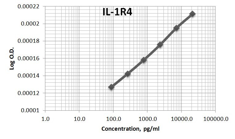
Application: ELISASample Tested: Serum and Plasma and Cell culture supernatantSpecies: HumanVerified Customer | Posted 11/09/2017AF523 was used as both the capture and the detection antibody for the sandwich ELISA for sIL-1R4. The immunoassay standard was 523-ST. Assay had sensitivity of ~90pg/ml.

There are no reviews that match your criteria.
Protocols
Find general support by application which include: protocols, troubleshooting, illustrated assays, videos and webinars.
- Antigen Retrieval Protocol (PIER)
- Antigen Retrieval for Frozen Sections Protocol
- Appropriate Fixation of IHC/ICC Samples
- Cellular Response to Hypoxia Protocols
- Chromogenic IHC Staining of Formalin-Fixed Paraffin-Embedded (FFPE) Tissue Protocol
- Chromogenic Immunohistochemistry Staining of Frozen Tissue
- ClariTSA™ Fluorophore Kits
- Detection & Visualization of Antibody Binding
- Fluorescent IHC Staining of Frozen Tissue Protocol
- Graphic Protocol for Heat-induced Epitope Retrieval
- Graphic Protocol for the Preparation and Fluorescent IHC Staining of Frozen Tissue Sections
- Graphic Protocol for the Preparation and Fluorescent IHC Staining of Paraffin-embedded Tissue Sections
- Graphic Protocol for the Preparation of Gelatin-coated Slides for Histological Tissue Sections
- IHC Sample Preparation (Frozen sections vs Paraffin)
- Immunofluorescent IHC Staining of Formalin-Fixed Paraffin-Embedded (FFPE) Tissue Protocol
- Immunohistochemistry (IHC) and Immunocytochemistry (ICC) Protocols
- Immunohistochemistry Frozen Troubleshooting
- Immunohistochemistry Paraffin Troubleshooting
- Preparing Samples for IHC/ICC Experiments
- Preventing Non-Specific Staining (Non-Specific Binding)
- Primary Antibody Selection & Optimization
- Protocol for Heat-Induced Epitope Retrieval (HIER)
- Protocol for Making a 4% Formaldehyde Solution in PBS
- Protocol for VisUCyte™ HRP Polymer Detection Reagent
- Protocol for the Preparation & Fixation of Cells on Coverslips
- Protocol for the Preparation and Chromogenic IHC Staining of Frozen Tissue Sections
- Protocol for the Preparation and Chromogenic IHC Staining of Frozen Tissue Sections - Graphic
- Protocol for the Preparation and Chromogenic IHC Staining of Paraffin-embedded Tissue Sections
- Protocol for the Preparation and Chromogenic IHC Staining of Paraffin-embedded Tissue Sections - Graphic
- Protocol for the Preparation and Fluorescent IHC Staining of Frozen Tissue Sections
- Protocol for the Preparation and Fluorescent IHC Staining of Paraffin-embedded Tissue Sections
- Protocol for the Preparation of Gelatin-coated Slides for Histological Tissue Sections
- R&D Systems Quality Control Western Blot Protocol
- TUNEL and Active Caspase-3 Detection by IHC/ICC Protocol
- The Importance of IHC/ICC Controls
- Troubleshooting Guide: Immunohistochemistry
- Troubleshooting Guide: Western Blot Figures
- Western Blot Conditions
- Western Blot Protocol
- Western Blot Protocol for Cell Lysates
- Western Blot Troubleshooting
- Western Blot Troubleshooting Guide
- View all Protocols, Troubleshooting, Illustrated assays and Webinars
Loading...
Associated Pathways


