NKp46, along with NKp30 and NKp44, are activating receptors that have been collectively termed the natural cytotoxicity receptors (NCR) (1). These receptors are expressed almost exclusively by NK cells and play a major role in triggering some of the key lytic activities of NK cells. In human systems, the CD56dimCD16+ subpopulation that makes up the majority of NK cells in the peripheral blood and spleen expresses NKp46 in both resting and activated states (2). The main NK cell population of the lymph node (CD56brightCD16-) expresses low levels of NKp46 in resting cells, but expression is upregulated by IL-2. Mouse NKp46, also known as MAR-1 (3), is a type I transmembrane protein with two extracellular Ig-like domains. It has a positive charge in its transmembrane domain that permits association with the ITAM-bearing signal adapter proteins, CD3 zeta and Fc epsilon RI gamma (4). Studies with neutralizing antibodies indicate that the three NCR are primarily responsible for triggering the NK-mediated lysis of many human tumor cell lines. Blocking any of the NCRs individually resulted in partial inhibition of tumor cell lysis, but nearly complete inhibition of lysis was observed if all three receptors were blocked simultaneously (5). NKp46 has also been implicated in recognition of virus-infected cells through its capacity to bind to viral hemagglutinins (6-8).
Mouse NKp46/NCR1 Antibody
R&D Systems | Catalog # AF2225

Key Product Details
Validated by
Knockout/Knockdown
Species Reactivity
Validated:
Mouse
Cited:
Human, Mouse, Rat, Transgenic Mouse, Xenograft
Applications
Validated:
Immunohistochemistry, Western Blot, Flow Cytometry, Agonist Activity, CyTOF-ready
Cited:
Immunohistochemistry, Immunohistochemistry-Paraffin, Immunohistochemistry-Frozen, Western Blot, Neutralization, Flow Cytometry, Immunocytochemistry, Bioassay, Functional Assay
Label
Unconjugated
Antibody Source
Polyclonal Goat IgG
Loading...
Product Specifications
Immunogen
Mouse myeloma cell line NS0-derived recombinant mouse NKp46/NCR1
Glu22-Asn255
Accession # Q8C567
Glu22-Asn255
Accession # Q8C567
Specificity
Detects mouse NKp46/NCR1 in direct ELISAs and Western blots. In direct ELISAs, less than 15% cross-reactivity with recombinant human (rh) NKp46 is observed.
Clonality
Polyclonal
Host
Goat
Isotype
IgG
Endotoxin Level
<0.10 EU per 1 μg of the antibody by the LAL method.
Scientific Data Images for Mouse NKp46/NCR1 Antibody
Mouse NKp46/NCR1 Antibody Induces IFN-gamma Secretion in Activated Mouse NK Cells.
Mouse NKp46/NCR1 Antigen Affinity-purified Polyclonal Antibody induces IFN-? secretion in mouse natural killer (NK) cells activated with 25 ng/mL Recombinant Mouse IL-2 (Catalog # 402-ML) and 25 ng/mL Recombinant Mouse IL-12 (Catalog # 419-ML), in a dose-dependent manner, as measured using the Quantikine Mouse IFN-? ELISA Kit (Catalog # MIF00). The ED50 for this effect is typically 0.4-2.4 µg/mL.
Detection of NKp46/NCR1 in Mouse DX5/CD49b+Splenocytes by Flow Cytometry.
Mouse DX5/CD49b+splenocytes were stained with Goat Anti-Mouse NKp46/NCR1 Antigen Affinity-purified Polyclonal Antibody (Catalog # AF2225, filled histogram) or control antibody (Catalog # AB-108-C, open histogram), followed by Allophycocyanin-conjugated Anti-Goat IgG Secondary Antibody (Catalog # F0108).
NKp46/NCR1 in Mouse Spleen.
NKp46/NCR1 was detected in perfusion fixed frozen sections of mouse spleen using Goat Anti-Mouse NKp46/NCR1 Antigen Affinity-purified Polyclonal Antibody (Catalog # AF2225) at 3 µg/mL for 1 hour at room temperature followed by incubation with the Anti-Goat IgG VisUCyte™ HRP Polymer Antibody (Catalog # VC004). Tissue was stained using DAB (brown) and counterstained with hematoxylin (blue). Specific staining was localized to cytoplasm in lymphocytes. View our protocol for IHC Staining with VisUCyte HRP Polymer Detection Reagents.
Detection of Mouse NKp46/NCR1 by Immunocytochemistry/Immunofluorescence
Acutely Cultured Embryonic but Not Adult DRG Neurons Reveal Susceptibility to NK-Mediated Cytotoxicity by RAE1(A) Immunolabeling of co-culture (4 h) between embryonic (top) or adult (bottom) DRG neurons ( beta -tubulin, magenta) and either freshly isolated (control) or IL-2-stimulated natural killer (NK) cells (NKp46, green). The inset shows a high-magnification image of NK cell in contact with embryonic DRG neurite.(B) LDH-release cytotoxicity assay of acutely cultured (1 day in vitro) embryonic (top) and adult (bottom) DRG at various Effector (NK):Target (DRG) (E:T) ratios. Matched two-way ANOVA: embryonic DRG, F(1,10) = 100.01, p < 0.0001); adult DRG, F(1,10) = 1.25, p = 0.2982). Three replicate co-cultures for each DRG group.(C) Still images of in vitro time-lapse confocal Ca2+ imaging of rhodamine 3 AM-loaded embryonic (top) and adult (bottom) DRG (magenta) co-cultured with IL-2-stimulated NK cells (green) isolated from adult male NKp46-YFP mice.(D) Frequency histogram (30 s time bins) of neurite Ca2+ events in embryonic (top) and adult (bottom) DRG during NK co-culture. Cumulative area under the curve (right). Student’s paired t test; t = 2.290, p = 0.045. n = 6 fields of view from two repeat co-cultures per group.(E) RT-PCR of mRNA transcripts in freshly isolated splenic NK cells and embryonic and adult DRG.(F) qRT-PCR shows higher Raet1 mRNA expression in embryonic compared to adult DRG tissue. Student’s paired t test; t = 16.16, p < 0.0001. n = 5 mice, or replicates per group.(G) Western blot of embryonic and adult mouse DRG tissue (40 μg loading) with pan-RAE1 antibody and beta -actin control. Images are representative of three independent experiments.(H) Selective siRNA knockdown reduces RAE1 protein (top) and Raet1 mRNA (bottom) expression in embryonic DRG (2 d culture). Student’s unpaired t test; t = 9.060, p = 0.0008. n = 3 mice, or replicates per group.(I) LDH-release cytotoxicity assay of negative control or Raet1-selective siRNA knockdown embryonic DRG. Thre
Detection of NKp46/NCR1 by Immunohistochemistry
mNemvaleukin alone and combination treatments expand tumor-infiltrating NK cells in SCLC. (A) Schematic showing experimental design for immune profiling. After confirming tumor burden by MRI, mice were randomized into vehicle, chemotherapy, mNemvaleukin, and combo (chemotherapy+mNemvaleukin) treatment groups. Tumor nodules were collected and tumor-infiltrating lymphocytes were analyzed by flow cytometry at day 7 after treatment initiation. (B) Representative plots of infiltrating NK cells (CD3−CD49b+) are shown on indicated treatment. (C, D) Frequencies of total infiltrating NK cells (C), activated NK cells (D) and expression of Ki-67+ (E) in NK cells are presented. Two independent cohorts were performed (n=4 in total for each group). (F) IHC staining of NKp46 on tumor samples treated with vehicle, chemotherapy, mNemvaleukin, and combo regimens. Data shown as means±SEM. *P<0.05, **P<0.01, ***P<0.001. IHC, Immunohistochemistry; mNemvaleukin, mouse version of nemvaleukin; NK, natural killer; ns, not significant (unpaired two-tailed t test); SCLC, small cell lung cancer. Image collected and cropped by CiteAb from the following open publication (https://pubmed.ncbi.nlm.nih.gov/36472839), licensed under a CC-BY license. Not internally tested by R&D Systems.
Detection of NKp46/NCR1 by Immunohistochemistry
mNemvaleukin alone and combination treatments expand tumor-infiltrating NK cells in SCLC. (A) Schematic showing experimental design for immune profiling. After confirming tumor burden by MRI, mice were randomized into vehicle, chemotherapy, mNemvaleukin, and combo (chemotherapy+mNemvaleukin) treatment groups. Tumor nodules were collected and tumor-infiltrating lymphocytes were analyzed by flow cytometry at day 7 after treatment initiation. (B) Representative plots of infiltrating NK cells (CD3−CD49b+) are shown on indicated treatment. (C, D) Frequencies of total infiltrating NK cells (C), activated NK cells (D) and expression of Ki-67+ (E) in NK cells are presented. Two independent cohorts were performed (n=4 in total for each group). (F) IHC staining of NKp46 on tumor samples treated with vehicle, chemotherapy, mNemvaleukin, and combo regimens. Data shown as means±SEM. *P<0.05, **P<0.01, ***P<0.001. IHC, Immunohistochemistry; mNemvaleukin, mouse version of nemvaleukin; NK, natural killer; ns, not significant (unpaired two-tailed t test); SCLC, small cell lung cancer. Image collected and cropped by CiteAb from the following open publication (https://pubmed.ncbi.nlm.nih.gov/36472839), licensed under a CC-BY license. Not internally tested by R&D Systems.Applications for Mouse NKp46/NCR1 Antibody
Application
Recommended Usage
Agonist Activity
0.4-2.4 µg/mL
Sample: Mouse natural killer (NK) cells activated with Recombinant Mouse IL-2 (Catalog # 402-ML) and Recombinant Mouse IL-12 (Catalog # 419-ML)
Sample: Mouse natural killer (NK) cells activated with Recombinant Mouse IL-2 (Catalog # 402-ML) and Recombinant Mouse IL-12 (Catalog # 419-ML)
CyTOF-ready
Ready to be labeled using established conjugation methods. No BSA or other carrier proteins that could interfere with conjugation.
Flow Cytometry
2.5 µg/106 cells
Sample: Mouse DX5/CD49b+ splenocytes
Sample: Mouse DX5/CD49b+ splenocytes
Immunohistochemistry
3-15 µg/mL
Sample: Perfusion fixed frozen sections of mouse spleen
Sample: Perfusion fixed frozen sections of mouse spleen
Western Blot
0.1 µg/mL
Sample: Recombinant Mouse NKp46/NCR1 Fc Chimera (Catalog # 2225-NK)
Sample: Recombinant Mouse NKp46/NCR1 Fc Chimera (Catalog # 2225-NK)
Reviewed Applications
Read 2 reviews rated 5 using AF2225 in the following applications:
Flow Cytometry Panel Builder
Bio-Techne Knows Flow Cytometry
Save time and reduce costly mistakes by quickly finding compatible reagents using the Panel Builder Tool.
Advanced Features
- Spectra Viewer - Custom analysis of spectra from multiple fluorochromes
- Spillover Popups - Visualize the spectra of individual fluorochromes
- Antigen Density Selector - Match fluorochrome brightness with antigen density

Formulation, Preparation, and Storage
Purification
Antigen Affinity-purified
Reconstitution
Reconstitute at 0.2 mg/mL in sterile PBS. For liquid material, refer to CoA for concentration.
Loading...
Formulation
Lyophilized from a 0.2 μm filtered solution in PBS with Trehalose. *Small pack size (SP) is supplied either lyophilized or as a 0.2 µm filtered solution in PBS.
Shipping
Lyophilized product is shipped at ambient temperature. Liquid small pack size (-SP) is shipped with polar packs. Upon receipt, store immediately at the temperature recommended below.
Stability & Storage
Use a manual defrost freezer and avoid repeated freeze-thaw cycles.
- 12 months from date of receipt, -20 to -70 °C as supplied.
- 1 month, 2 to 8 °C under sterile conditions after reconstitution.
- 6 months, -20 to -70 °C under sterile conditions after reconstitution.
Calculators
Background: NKp46/NCR1
References
- Moretta, L. and A. Moretta (2004) EMBO J. 23:255.
- Ferlazzo, G. et al. (2004) J. Immunol. 172:1455.
- Biassoni, R. et al. (1999) Eur. J. Immunol. 29:1014.
- Westgaard, I. et al. (2004) J. Leukoc. Biol. PMID 15356098.
- Pende, D. et al. (1999) J. Exp. Med. 190:1505.
- Arnon, T. et al. (2004) Blood 103:664.
- Arnon, T. et al. (2001) Eur. J. Immunol. 31:2680.
- Mandelboim, O. et al. (2001) Nature 409:1055.
Alternate Names
CD335, Ly94, MAR-1, NCR1
Gene Symbol
NCR1
UniProt
Additional NKp46/NCR1 Products
Product Documents for Mouse NKp46/NCR1 Antibody
Certificate of Analysis
To download a Certificate of Analysis, please enter a lot or batch number in the search box below.
Note: Certificate of Analysis not available for kit components.
Product Specific Notices for Mouse NKp46/NCR1 Antibody
For research use only
Citations for Mouse NKp46/NCR1 Antibody
 Powered by Bioz
Customer Reviews for Mouse NKp46/NCR1 Antibody (2)
5 out of 5
2 Customer Ratings
Have you used Mouse NKp46/NCR1 Antibody?
Submit a review and receive an Amazon gift card!
$25/€18/£15/$25CAN/¥2500 Yen for a review with an image
$10/€7/£6/$10CAN/¥1110 Yen for a review without an image
Submit a review
Customer Images
Showing
1
-
2 of
2 reviews
Showing All
Filter By:
-
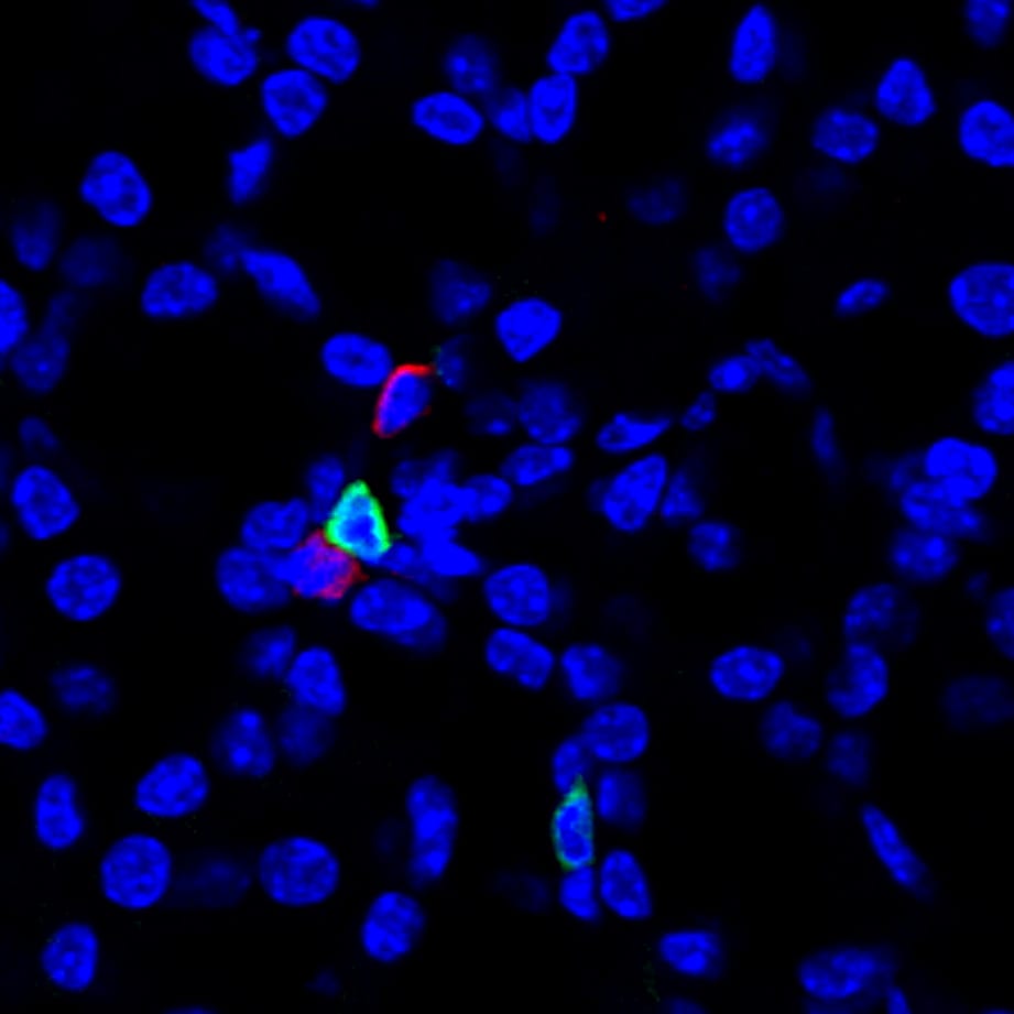
Application: Immunocytochemistry/ImmunofluorescenceSample Tested: Salivary gland tissueSpecies: MouseVerified Customer | Posted 07/10/2019PLP-fixed salivary gland cryosections were stained with anti NKp46/NCR1 for 4h at room temperature. After washing, the samples were stained with Donkey anti-goat alexa546 2ndary antibody for 1h at room temperature. NKp46 in red, CD3 in green & Hoechst in blue.

-
Application: Immunohistochemistry-FrozenSample Tested: See PMID 23902341Species: MouseVerified Customer | Posted 01/06/2015
There are no reviews that match your criteria.
Protocols
Find general support by application which include: protocols, troubleshooting, illustrated assays, videos and webinars.
- 7-Amino Actinomycin D (7-AAD) Cell Viability Flow Cytometry Protocol
- Antigen Retrieval Protocol (PIER)
- Antigen Retrieval for Frozen Sections Protocol
- Appropriate Fixation of IHC/ICC Samples
- Cellular Response to Hypoxia Protocols
- Chromogenic IHC Staining of Formalin-Fixed Paraffin-Embedded (FFPE) Tissue Protocol
- Chromogenic Immunohistochemistry Staining of Frozen Tissue
- ClariTSA™ Fluorophore Kits
- Detection & Visualization of Antibody Binding
- Extracellular Membrane Flow Cytometry Protocol
- Flow Cytometry Protocol for Cell Surface Markers
- Flow Cytometry Protocol for Staining Membrane Associated Proteins
- Flow Cytometry Staining Protocols
- Flow Cytometry Troubleshooting Guide
- Fluorescent IHC Staining of Frozen Tissue Protocol
- Graphic Protocol for Heat-induced Epitope Retrieval
- Graphic Protocol for the Preparation and Fluorescent IHC Staining of Frozen Tissue Sections
- Graphic Protocol for the Preparation and Fluorescent IHC Staining of Paraffin-embedded Tissue Sections
- Graphic Protocol for the Preparation of Gelatin-coated Slides for Histological Tissue Sections
- IHC Sample Preparation (Frozen sections vs Paraffin)
- Immunofluorescent IHC Staining of Formalin-Fixed Paraffin-Embedded (FFPE) Tissue Protocol
- Immunohistochemistry (IHC) and Immunocytochemistry (ICC) Protocols
- Immunohistochemistry Frozen Troubleshooting
- Immunohistochemistry Paraffin Troubleshooting
- Intracellular Flow Cytometry Protocol Using Alcohol (Methanol)
- Intracellular Flow Cytometry Protocol Using Detergents
- Intracellular Nuclear Staining Flow Cytometry Protocol Using Detergents
- Intracellular Staining Flow Cytometry Protocol Using Alcohol Permeabilization
- Intracellular Staining Flow Cytometry Protocol Using Detergents to Permeabilize Cells
- Preparing Samples for IHC/ICC Experiments
- Preventing Non-Specific Staining (Non-Specific Binding)
- Primary Antibody Selection & Optimization
- Propidium Iodide Cell Viability Flow Cytometry Protocol
- Protocol for Heat-Induced Epitope Retrieval (HIER)
- Protocol for Liperfluo
- Protocol for Making a 4% Formaldehyde Solution in PBS
- Protocol for VisUCyte™ HRP Polymer Detection Reagent
- Protocol for the Characterization of Human Th22 Cells
- Protocol for the Characterization of Human Th9 Cells
- Protocol for the Preparation & Fixation of Cells on Coverslips
- Protocol for the Preparation and Chromogenic IHC Staining of Frozen Tissue Sections
- Protocol for the Preparation and Chromogenic IHC Staining of Frozen Tissue Sections - Graphic
- Protocol for the Preparation and Chromogenic IHC Staining of Paraffin-embedded Tissue Sections
- Protocol for the Preparation and Chromogenic IHC Staining of Paraffin-embedded Tissue Sections - Graphic
- Protocol for the Preparation and Fluorescent IHC Staining of Frozen Tissue Sections
- Protocol for the Preparation and Fluorescent IHC Staining of Paraffin-embedded Tissue Sections
- Protocol for the Preparation of Gelatin-coated Slides for Histological Tissue Sections
- Protocol: Annexin V and PI Staining by Flow Cytometry
- Protocol: Annexin V and PI Staining for Apoptosis by Flow Cytometry
- R&D Systems Quality Control Western Blot Protocol
- TUNEL and Active Caspase-3 Detection by IHC/ICC Protocol
- The Importance of IHC/ICC Controls
- Troubleshooting Guide: Fluorokine Flow Cytometry Kits
- Troubleshooting Guide: Immunohistochemistry
- Troubleshooting Guide: Western Blot Figures
- Western Blot Conditions
- Western Blot Protocol
- Western Blot Protocol for Cell Lysates
- Western Blot Troubleshooting
- Western Blot Troubleshooting Guide
- View all Protocols, Troubleshooting, Illustrated assays and Webinars
Loading...
Associated Pathways

