Mouse Serum Amyloid A1/A2 Antibody
R&D Systems | Catalog # AF2948

Key Product Details
Species Reactivity
Validated:
Cited:
Applications
Validated:
Cited:
Label
Antibody Source
Product Specifications
Immunogen
Gly20-Tyr122
Accession # P05366
Specificity
Clonality
Host
Isotype
Scientific Data Images for Mouse Serum Amyloid A1/A2 Antibody
Detection of Mouse Serum Amyloid A1/A2 by Western Blot.
Western blot shows mouse serum and mouse plasma. PVDF membrane was probed with 1 µg/mL of Goat Anti-Mouse Serum Amyloid A1/A2 Antigen Affinity-purified Polyclonal Antibody (Catalog # AF2948) followed by HRP-conjugated Anti-Goat IgG Secondary Antibody (Catalog # HAF017). A specific band was detected for Serum Amyloid A1/A2 at approximately 12 kDa (as indicated). This experiment was conducted under reducing conditions and using Immunoblot Buffer Group 1.
Detection of Mouse Serum Amyloid A1/A2 by Western Blot.
Western blot shows recombinant mouse Serum Amyloid A1 and recombinant mouse Serum Amyloid A2. PVDF membrane was probed with 1 µg/mL of Goat Anti-Mouse Serum Amyloid A1/A2 Antigen Affinity-purified Polyclonal Antibody (Catalog # AF2948) followed by HRP-conjugated Anti-Goat IgG Secondary Antibody (Catalog # HAF017). A specific band was detected for Serum Amyloid A1/A2 at approximately 12 kDa (as indicated). This experiment was conducted under reducing conditions and using Immunoblot Buffer Group 1.
Serum Amyloid A1/A2 in Mouse Liver.
Serum Amyloid A1/A2 was detected in perfusion fixed frozen sections of mouse liver using Goat Anti-Mouse Serum Amyloid A1/A2 Antigen Affinity-purified Polyclonal Antibody (Catalog # AF2948) at 15 µg/mL overnight at 4 °C. Tissue was stained using the Anti-Goat HRP-DAB Cell & Tissue Staining Kit (brown; Catalog # CTS008) and counterstained with hematoxylin (blue). Lower panel shows a lack of labeling when primary antibodies are omitted and tissue is stained only with secondary antibody followed by incubation with detection reagents. Specific staining was localized to cytoplasm. View our protocol for Chromogenic IHC Staining of Frozen Tissue Sections.
Detection of Mouse Serum Amyloid A1/A2 by Simple WesternTM.
Simple Western lane view shows mouse serum, loaded at a 1:100 dilution. A specific band was detected for Serum Amyloid A1/A2 at approximately 14 kDa (as indicated) using 50 µg/mL of (Catalog # AF2948) followed by 1:50 dilution of HRP-conjugated Anti-Goat IgG Secondary Antibody (Catalog # HAF109). This experiment was conducted under reducing conditions and using the 12-230 kDa separation system.
Mouse Serum Amyloid A1/A2 ELISA Standard Curve.
Recombinant Mouse Serum Amyloid A1/A2 protein was serially diluted 2-fold and captured by Rat Anti-Mouse Serum Amyloid A1/A2 Monoclonal Antibody (Catalog # MAB2948) coated on a Clear Polystyrene Microplate (Catalog # DY990). Goat Anti-Mouse Serum Amyloid A1/A2 Antigen Affinity-purified Polyclonal Antibody (Catalog # AF2948) was biotinylated and incubated with the protein captured on the plate. Detection of the standard curve was achieved by incubating Streptavidin-HRP (Catalog # DY998) followed by Substrate Solution (Catalog # DY999) and stopping the enzymatic reaction with Stop Solution (Catalog # DY994).
Detection of Serum Amyloid A1/A2 by Western Blot
Robust expression of acute phase response proteins in skeletal muscle versus liver in C26 cachexia.A. Western blotting and quantitation of fibrinogen levels in control and C26 quadriceps and liver. Data (mean ± SEM) are expressed as relative densitometry value. **P<0.01, ***P<0.001. B, Western blotting analysis of fibrinogen standard proteins and quadriceps and liver extracts for control, CHO-IL6 injected nude mice and C26 injected CD2F1 mice. Quantitation was performed on the band indicated by the arrow. Data (means ± SEM) are expressed as ng fibrinogen / µg protein. *P<0.05, **P<0.01, ***P<0.001. C, Western blotting analysis demonstrates significantly increased fibrinogen and SAA1 protein levels in quadriceps and gastrocnemius in moderate and severe C26 cachexia. *P<0.05, **P<0.01, ***P<0.001. Image collected and cropped by CiteAb from the following open publication (https://pubmed.ncbi.nlm.nih.gov/21799891), licensed under a CC-BY license. Not internally tested by R&D Systems.
Detection of Serum Amyloid A1/A2 by Western Blot
FMT-AD neither induced peripheral inflammation nor caused intestinal damage to recipient mice. (a) Western blot showing protein Serum Amyloid A (SAA) levels in the serum from FMT-AD and FMT-young (FMT-y) mice at 3 d post-TBI. (b) Quantification of the western blot shows a not statistically significant increase in SAA in the serum of males after FMT-AD compared to FMT-y. (c) Representative histology images from intestinal tissue do not show any abnormality between the FMT-y and FMT-AD groups in male or female mice. Left, Hematoxylin and eosin (H.E.) staining; right (×10 Objective), Alcian blue staining (×20 Objective). (d) Densiometric quantification of the Alcian blue positive area and counting the number of goblet cells per villus are not different between FMT-y and FMT-AD groups. Male and female data were combined, since two-way ANOVA showed no sex differences (Alcian blue area, p = 0.429; goblet cells, p = 0.471). Image collected and cropped by CiteAb from the following open publication (https://pubmed.ncbi.nlm.nih.gov/35562867), licensed under a CC-BY license. Not internally tested by R&D Systems.
Mouse Serum Amyloid A ELISA Standard Curve
Recombinant Mouse Serum Amyloid A1 (Catalog # 2948-SA) was serially diluted and captured by Rat Anti-Mouse Serum Amyloid A1/A2 Monoclonal Antibody (Catalog # MAB2948) coated on a Clear Polystyrene Microplate (Catalog # DY990). Goat Anti-Mouse Serum Amyloid A1/A2 Antigen Affinity-purified Polyclonal Antibody (Catalog # AF2948) was biotinylated and incubated with the protein captured on the plate. Detection of the standard curve was achieved by incubating Streptavidin-HRP (Catalog # DY998)Applications for Mouse Serum Amyloid A1/A2 Antibody
ELISA
This antibody functions as an ELISA detection antibody when paired with Rat Anti-Mouse Serum Amyloid A1/A2 Monoclonal Antibody (Catalog # MAB2948).
This product is intended for assay development on various assay platforms requiring antibody pairs. We recommend the Mouse Serum Amyloid A DuoSet ELISA Kit (Catalog # DY2948-05) for convenient development of a sandwich ELISA or the Mouse Serum Amyloid A Quantikine ELISA Kit (Catalog # MSAA00) for a complete optimized ELISA.
Immunohistochemistry
Sample: Perfusion fixed frozen sections of mouse liver
Simple Western
Sample: Mouse serum
Western Blot
Sample: Mouse serum and mouse plasma
Reviewed Applications
Read 3 reviews rated 3.7 using AF2948 in the following applications:
Formulation, Preparation, and Storage
Purification
Reconstitution
Reconstitute at 0.2 mg/mL in sterile PBS. For liquid material, refer to CoA for concentration.
Formulation
Shipping
Stability & Storage
- 12 months from date of receipt, -20 to -70 °C as supplied.
- 1 month, 2 to 8 °C under sterile conditions after reconstitution.
- 6 months, -20 to -70 °C under sterile conditions after reconstitution.
Calculators
Background: Serum Amyloid A1/A2
Alternate Names
UniProt
Additional Serum Amyloid A1/A2 Products
Product Documents for Mouse Serum Amyloid A1/A2 Antibody
Certificate of Analysis
To download a Certificate of Analysis, please enter a lot or batch number in the search box below.
Note: Certificate of Analysis not available for kit components.
Product Specific Notices for Mouse Serum Amyloid A1/A2 Antibody
For research use only
Related Research Areas
Citations for Mouse Serum Amyloid A1/A2 Antibody
 Powered by Bioz
Customer Reviews for Mouse Serum Amyloid A1/A2 Antibody (3)
Have you used Mouse Serum Amyloid A1/A2 Antibody?
Submit a review and receive an Amazon gift card!
$25/€18/£15/$25CAN/¥2500 Yen for a review with an image
$10/€7/£6/$10CAN/¥1110 Yen for a review without an image
Submit a review
Customer Images
-
Application: Immunocytochemistry/ImmunofluorescenceSample Tested: Spinal cordSpecies: MouseVerified Customer | Posted 12/24/2021

-
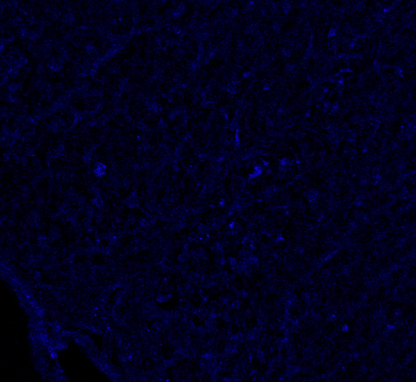
Application: Immunocytochemistry/ImmunofluorescenceSample Tested: Brain tissueSpecies: MouseVerified Customer | Posted 10/17/2018
-
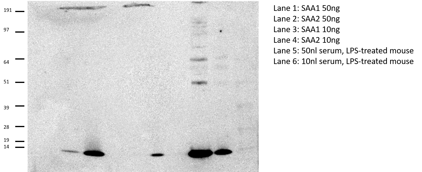
Application: Western BlotSample Tested: Serum and Recombinant proteinSpecies: MouseVerified Customer | Posted 10/14/2016Protein loaded: Lane 1: SAA1 50ng Lane 2: SAA2 50ng Lane 3: SAA1 10ng Lane 4: SAA2 10ng Lane 5: 50nl serum, LPS-treated mouse Lane 6: 10nl serum, LPS-treated mouse Recombinant proteins were from R and D systems Prepared in Invitrogen 1x LDS sample buffer plus 1x reducing agent. Run on 4-12% Bis-tris gel under reducing conditions in MOPS buffer. Transferred using iBlot 20V 7 min Blocked 1 hour 5% BSA/TBS-T Incubated overnight 4 C, antibody prepared 1/1000 in 5% BSA/TBS-T Secondary antibody was anti-goat from Jackson Immuno 1:5000 for 30 minutes. West Pico Chemiluminescent Reagent ImageQuant LAS4000 for imaging *This antibody gives great signal, but predominantly detects SAA2 over SAA1.
There are no reviews that match your criteria.
Protocols
Find general support by application which include: protocols, troubleshooting, illustrated assays, videos and webinars.
- Antigen Retrieval Protocol (PIER)
- Antigen Retrieval for Frozen Sections Protocol
- Appropriate Fixation of IHC/ICC Samples
- Cellular Response to Hypoxia Protocols
- Chromogenic IHC Staining of Formalin-Fixed Paraffin-Embedded (FFPE) Tissue Protocol
- Chromogenic Immunohistochemistry Staining of Frozen Tissue
- ClariTSA™ Fluorophore Kits
- Detection & Visualization of Antibody Binding
- ELISA Sample Preparation & Collection Guide
- ELISA Troubleshooting Guide
- Fluorescent IHC Staining of Frozen Tissue Protocol
- Graphic Protocol for Heat-induced Epitope Retrieval
- Graphic Protocol for the Preparation and Fluorescent IHC Staining of Frozen Tissue Sections
- Graphic Protocol for the Preparation and Fluorescent IHC Staining of Paraffin-embedded Tissue Sections
- Graphic Protocol for the Preparation of Gelatin-coated Slides for Histological Tissue Sections
- How to Run an R&D Systems DuoSet ELISA
- How to Run an R&D Systems Quantikine ELISA
- How to Run an R&D Systems Quantikine™ QuicKit™ ELISA
- IHC Sample Preparation (Frozen sections vs Paraffin)
- Immunofluorescent IHC Staining of Formalin-Fixed Paraffin-Embedded (FFPE) Tissue Protocol
- Immunohistochemistry (IHC) and Immunocytochemistry (ICC) Protocols
- Immunohistochemistry Frozen Troubleshooting
- Immunohistochemistry Paraffin Troubleshooting
- Preparing Samples for IHC/ICC Experiments
- Preventing Non-Specific Staining (Non-Specific Binding)
- Primary Antibody Selection & Optimization
- Protocol for Heat-Induced Epitope Retrieval (HIER)
- Protocol for Making a 4% Formaldehyde Solution in PBS
- Protocol for VisUCyte™ HRP Polymer Detection Reagent
- Protocol for the Preparation & Fixation of Cells on Coverslips
- Protocol for the Preparation and Chromogenic IHC Staining of Frozen Tissue Sections
- Protocol for the Preparation and Chromogenic IHC Staining of Frozen Tissue Sections - Graphic
- Protocol for the Preparation and Chromogenic IHC Staining of Paraffin-embedded Tissue Sections
- Protocol for the Preparation and Chromogenic IHC Staining of Paraffin-embedded Tissue Sections - Graphic
- Protocol for the Preparation and Fluorescent IHC Staining of Frozen Tissue Sections
- Protocol for the Preparation and Fluorescent IHC Staining of Paraffin-embedded Tissue Sections
- Protocol for the Preparation of Gelatin-coated Slides for Histological Tissue Sections
- Quantikine HS ELISA Kit Assay Principle, Alkaline Phosphatase
- Quantikine HS ELISA Kit Principle, Streptavidin-HRP Polymer
- R&D Systems Quality Control Western Blot Protocol
- Sandwich ELISA (Colorimetric) – Biotin/Streptavidin Detection Protocol
- Sandwich ELISA (Colorimetric) – Direct Detection Protocol
- TUNEL and Active Caspase-3 Detection by IHC/ICC Protocol
- The Importance of IHC/ICC Controls
- Troubleshooting Guide: ELISA
- Troubleshooting Guide: Immunohistochemistry
- Troubleshooting Guide: Western Blot Figures
- Western Blot Conditions
- Western Blot Protocol
- Western Blot Protocol for Cell Lysates
- Western Blot Troubleshooting
- Western Blot Troubleshooting Guide
- View all Protocols, Troubleshooting, Illustrated assays and Webinars