B Cell Culture and Analysis

How Do B Cells Regulate Immune Responses?
B cells play an integral role in the adaptive immune response due to their abilities to:
- Produce antibodies
- Recognize and present antigens to T cells
- Secrete regulatory cytokines that affect the activities of other immune cell types
- Establish a state of immunological memory
This page outlines the tools that we offer for studying B cells, including cytokines and antibodies for B cell culture and activation, antibodies for B cell characterization, and single and multianalyte immunoassays for B cell analysis.
B Cell Culture
B cells represent a small portion of the peripheral blood lymphocyte population. Therefore, it is essential to expand B cells in vitro for many downstream applications, including functional studies, infectious disease research, and the development of vaccines and therapeutic antibodies.
Mouse B cells can be purified from splenocytes using magnetic bead-based negative selection, while human B cells are isolated from peripheral blood mononuclear cells by either negative selection, or CD19+ positive selection. B cells are then commonly cultured in vitro on a stromal feeder cell layer expressing CD40 Ligand in media supplemented with specific cytokines, which can include IL-4, IL-2, IL-21, and/or BAFF. Additional cytokines can be used to promote B cell differentiation, antibody production, and the generation of specific antibody isotypes such as IgA, IgG, IgE, or IgM.
Cytokines for B Cell Culture and Differentiation
R&D Systems large selection of cytokines can help optimize your B cell cultures. Our proteins are rigorously tested to ensure exceptional performance and lot-to-lot consistency, so you can have confidence in their ability to promote robust B cell expansion and differentiation with minimal variability between cultures. From basic research to clinical applications, we offer research-grade, Animal-free preclinical, and GMP-grade proteins to meet all research needs.
| BAFF/BLyS/TNFSF13B | CD40 Ligand | IFN-gamma* | IL-2*† | IL-4* | IL-5 |
| IL-6* | IL-10* | IL-15*† | IL-21*† | RANK Ligand | TNF-alpha* |
* GMP-grade proteins are available for these molecules.
† Liquid formulations of the GMP and Animal-free Preclinical grades of these proteins are available.
CellXVivo™ Human B Cell Expansion Kit
CellXVivo Immune Cell Expansion and Differentiation Kits contain optimized cytokine combinations and protocols for the expansion and differentiation of immune cell types. The CellXVivo Human B Cell Expansion Kit provides enough reagents for the 3-5-fold expansion of a starting population of 107 human B cells.
| Product Name |
| CellXVivo Human B Cell Expansion Kit |
ExCellerate™ Media for B Cell Expansion
ExCellerate B Cell Media is designed to promote robust B cell expansion, while ensuring consistent culture conditions. This media is specially formulated and optimized for the ex vivo culture of B cells under xeno-free conditions. It supports target antigen-specific B cell clonogenicity and the culture of mouse B cell hybridoma cell lines for antibody production, and it is compatible with B cells from multiple species.
| Product Name |
| ExCellerate B Cell Media, Xeno-Free |
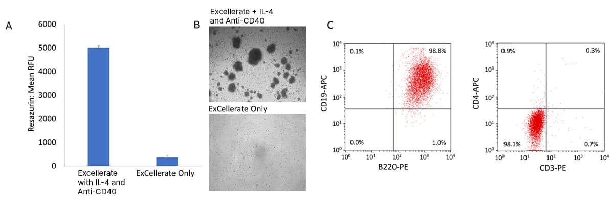
Expansion of Mouse B Cells in ExCellerate™ Human B Cell Expansion Media Supplemented with Recombinant Mouse IL-4 and an Anti-Mouse CD40 Antibody. Mouse B cells were isolated from splenocytes using the MagCellect™ Mouse B Cell Isolation Kit (Catalog # MAGM204) and expanded using ExCellerate B Cell Media (Catalog # CCM031) with or without Recombinant Mouse IL-4 (Catalog # 404-ML) and a Rat Anti-Mouse CD40/TNFRSF5 Monoclonal Antibody (Catalog # MAB440). (A) Expansion of mouse B cells was monitored using Rezasurin (Catalog # AR002). Data show that ExCellerate B Cell Media supplemented with IL-4 and an Anti-Mouse CD40 Antibody results in robust mouse B cell expansion. (B) Representative brightfield images of mouse B cell colonies. (C) Expanded mouse B cells are B220+CD19+ (>98%) and negative for both CD3 and CD4.
B Cell Activation
B cell activation requires two signals:
- The first signal is provided by an antigen binding to the B cell receptor (BCR), which is followed by endocytosis of the BCR-antigen complex, antigen degradation, and antigen presentation by MHC class II molecules on the B cell surface.
- The second signal is a co-stimulatory signal that is provided by the antigen itself or by interactions between the B cell and a T cell. The T cell interacts with the B cell through recognition of the antigen-MHC II complex on the B cell. This interaction promotes the binding of co-stimulatory molecules such as CD40 on the B cell with CD40L on the T cell and stimulates T cell activation, resulting in the secretion of cytokines such as IL-4, IL-5, IL-6, and IL-21 that drive B cell activation. Once B cells are activated, they proliferate and form germinal centers, where they differentiate into antibody-producing plasma cells and memory B cells.
In cell culture, anti-IgD or IgM antibodies can be used to mimic BCR activation, and CD40 Ligand or agonistic anti-CD40 antibodies can be used with cytokines such as IL-2, IL-4, IL-21, and/or BAFF to mimic the T cell-dependent co-stimulatory signal that promotes B cell activation.
Antibodies for B Cell Activation
| CD40 | IgD | IgM |
B Cell Blocking and Neutralizing Antibodies
Blocking and neutralizing antibodies are often used to investigate the functions of proteins expressed on the surface of a cell. We offer a wide selection of blocking/neutralization antibodies for key ligand-receptor pairs that regulate B cell activity. These antibodies are validated using the same bioassays that we use to test the bioactivity of our proteins, ensuring that they block/neutralize the appropriate cellular function.
| APRIL | BAFF | BCMA | CD22 |
| CD40 | CD40L | TACI |
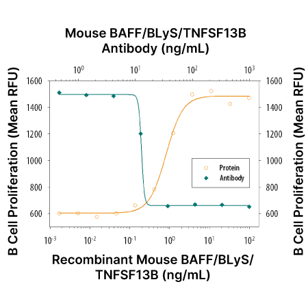
BAFF-induced Mouse B Cell Proliferation and Neutralization using an Anti-Mouse BAFF Antibody. In the presence of Goat F(ab’)2 Anti-Mouse IgM, Recombinant Mouse BAFF (Catalog # 8876-BF) stimulates proliferation of mouse B cells in a dose-dependent manner (orange line). Under these conditions, proliferation elicited by 3 ng/mL Recombinant Mouse BAFF is neutralized by increasing concentrations of a Goat Anti-Mouse BAFF Antigen Affinity-purified Polyclonal Antibody (Catalog # AF2106; green line). The ND50 is typically 0.01-0.04 µg/mL.
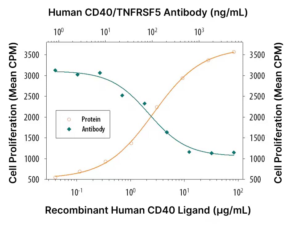
CD40 Ligand-induced B Cell Proliferation and Neutralization Using an Anti-Human CD40 Antibody. In the presence of Recombinant Human IL-4 (Catalog # 204-IL), Recombinant Human CD40 Ligand (Catalog # 6245-CL) stimulates proliferation of human B cell enriched peripheral blood mononuclear cells in a dose-dependent manner (orange line). Proliferation elicited by 10 µg/mL is neutralized by increasing concentrations of a Mouse Anti-Human CD40 Monoclonal Antibody (Catalog # MAB6322; green line). At 5 µg/mL, this anti-CD40 antibody will neutralize approximately 80% of Recombinant Human CD40 Ligand-induced proliferation in the presence of Recombinant Human IL-4.
B Cell Markers
Different B cell subsets are commonly identified based on the expression or lack of expression of specific cell surface and intracellular markers. Our selection of primary antibodies conjugated to one of over 30 fluorescent labels allows direct detection of cell surface and intracellular targets with the highest level of flexibility for multiplex experiments. Our antibodies are validated for specificity and reproducibility in multiple applications, including flow cytometry, immunocytochemistry/immunofluorescence (ICC/IF), immunohistochemistry (IHC), and Western blot. The tables below list the markers that are commonly used to identify different B cell subsets.
When an R&D Systems antibody is not available for a specific target molecule, we recommend using an antibody from our sister brand, Novus Biologicals™, whose products can be found at novusbio.com. Links provided in the tables below will show you to a list of antibodies from Novus Biologicals when an R&D Systems antibody is not available for a specific target.
Follicular B Cells
Follicular B cells are the largest subset of mature B cells in mice and humans and are found in the follicles of the lymph nodes and spleen. They recirculate in the secondary lymphoid organs and can differentiate into short-lived, antibody-secreting plasma cells and memory B cells following activation.
| Human Markers | Expression | Mouse Markers | Expression |
| CD10/Neprilysin | Negative | B220/CD45 R | Positive |
| CD19 | Positive | CD1d | Mid |
| CD20/MS4A1 | Positive | CD19 | Mid |
| CD21 | Positive | CD21 | Low |
| CD22/Siglec-2 | Positive | CD23/Fcε RII | Positive |
| CD23/Fcε RII | Positive | CD43 | Negative |
| CD24 | Low | CXCR5 | Positive |
| CD27 | Negative | IgD | High |
| CD38 | Low | IgM | Low |
| CXCR5 | Positive | L-Selectin/CD62L | Positive |
| HLA-DR | Positive | MHC class II | Positive |
| TACI | Positive | ||
| IgD | High | ||
| IgM | Low |
Marginal Zone B Cells
Marginal zone B cells are a subset of innate-like B cells located in between the white and red pulp in the marginal zone of the spleen, where they play a key role in protecting against blood borne pathogens. Marginal zone B cells have a poly-reactive BCR that can bind to a variety of antigens. Following activation, they produce low-affinity antibodies aimed at rapidly clearing pathogens and apoptotic debris.
| Human Markers | Expression | Mouse Markers | Expression |
| CD1c/BDCA-1 | Positive | B220/CD45 R | Positive |
| CD19 | Positive | C1q R1/CD93 | Negative |
| CD20/MS4A1 | Positive | CD1d | Positive |
| CD21 | Positive | CD19 | Mid |
| CD23/Fcε RII | Negative/Low | CD21 | High |
| CD27 | Positive | CD22/Siglec-2 | Positive |
| FCRL3/FcRH3 | Positive | CD23/Fcε RII | Negative |
| IgD | Low | CD43 | Negative |
| IgM | Positive | IgD | Low |
| TACI | Positive | IgM | High |
Memory B Cells
Once B cells are activated, they differentiate into memory B cells in germinal centers that form within the B cell follicles in the lymph nodes and spleen. Memory B cells circulate in the blood and have BCRs that are specific to the antigens that triggered their initial formation. Upon re-exposure to the antigen, memory B cells quickly respond by producing high affinity, antigen-specific antibodies to protect the host from re-infection.
| Human Markers | Expression | Mouse Markers | Expression |
| B7-1/CD80 | Positive | B220/CD45 R | Positive |
| B7-2/CD86 | Positive | B7-1/CD80 | Positive |
| C1q R1/CD93 | Negative | CD19 | Positive |
| CD19 | Positive | CD21 | Positive |
| CD20/MS4A1 | Positive | CD27 | Mid/Positive |
| CD21 | Positive | CD40 | Positive |
| CD27 | Mid/Positive | MHC class II | Positive |
| HLA-DR | Positive | ||
| TACI | Positive |
Plasma Cells
Plasma cells are antibody-secreting cells required for both short-lived and long-term antibody responses following antigen exposure. They are terminally-differentiated, non-proliferative cells, whose primary function is to secrete antibodies at a high rate to defend the host against an invading pathogen.
| Human Markers | Expression | Mouse Markers | Expression |
| BCMA | Positive | B220/CD45 R | Low |
| BLIMP1 | Positive | BCMA | Positive |
| CD10/Neprilysin | Negative | BLIMP1 | Positive |
| CD19 | Low | CD19 | Negative |
| CD20/MS4A1 | Negative/Low | CD27 | High |
| CD27 | Positive | CD38 | Low |
| CD38 | High | CXCR4 | High |
| CXCR4 | Positive | IgD | Negative |
| HLA-DR | Low | MHC class II | Negative/Low |
| IgD | Negative | Syndecan-1/CD138 | Positive |
| Syndecan-1/CD138 | High |
Regulatory B Cells
Regulatory B cells are a small subset of B cells with immunosuppressive properties that are involved in dampening inflammation and restoring immune homeostasis. These cells produce cytokines such as IL-10, IL-35, and TGF-β, which inhibit the differentiation, proliferation, and activation of pro-inflammatory T effector cells.
| Human Markers | Expression | Mouse Markers | Expression |
| CD1d | Positive | CD1d | High |
| CD5 | Positive | CD5 | Positive |
| CD19 | Positive | CD19 | Positive |
| CD21 | Positive | CD23/Fcε RII | Negative/Low |
| CD24 | Positive | CD24 | Positive |
| IL-10 | Positive | TIM-1 | Positive |
| IL-35 | Positive | IL-10 | Positive |
| TGF-β | Positive | IL-35 | Positive |
| TGF-β | Positive |
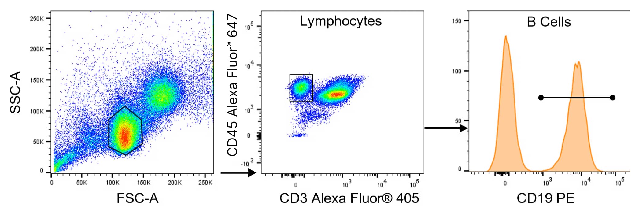
Detection of B Cells Using Multicolor Flow Cytometry. B cells were detected in human peripheral blood mononuclear cells (PBMCs) by staining with antibodies in the CD19 B Cell Panel (Catalog # FMC-P-003), which includes an Alexa Fluor® 405-conjugated Mouse Anti-Human CD3 Monoclonal Antibody, an Alexa Fluor® 647-conjugated Mouse Anti-Human CD45 Monoclonal Antibody, and a PE-conjugated Mouse Anti-Human CD19 Monoclonal Antibody. Alexa Fluor® is a registered trademark of Molecular Probes, Inc., Eugene, OR.
B Cell Cytokine Analysis – Single Analyte ELISAs
Following activation, B cells primarily secrete CCL17 and CCL22, which both bind to the CCR4 receptor that is highly expressed on Th2 cells. Further B cell-mediated cytokine secretion is regulated by extrinsic stimuli and cytokines secreted by other immune cells. Depending on these signals, activated B cells can produce both pro-inflammatory cytokines such as IL-1β, IL-6, and TNF-α, and anti-inflammatory cytokines such as IL-10.
As a result, B cells play an important role in immune responses not only due to their abilities to produce antibodies and generate immunological memory, but also because they secrete cytokines that regulate the activities of other immune cell types.
To characterize B cells based on cytokine secretion analysis, we offer a broad range of immunoassays for quantifying soluble proteins. This includes R&D Systems complete, ready-to-use Quantikine™ ELISA Kits, our 90-minute QuicKit™ ELISAs, and the more versatile DuoSet™ ELISA Development Systems that provide all the components necessary for a customer to develop their own working assay.
Quantikine ELISA Kits
- Extensively tested for long-term consistency and reproducibility
- Fully validated sample types
- Optimized reagents and diluents for accurate and sensitive sample values
Quantikine QuicKit ELISA Kits
- Data generated in 90 minutes
- Quantikine quality with lot-to-lot consistency and reproducibility
- Simplified protocol – only one wash step
DuoSet ELISA Development Systems
- Carefully selected with validated matched antibody pairs for optimal performance
- Large menu – over 1,000 targets with novel targets across multiple species
- Adaptable for use across multiple platforms and 384 well
Single Analyte ELISAs for B Cell Characterization – Products by Molecule
| CCL17 | CCL22 | GM-CSF | IFN-gamma | IL-1 beta | IL-2 |
| IL-4 | IL-6 | IL-10 | TGF-beta 1 | TNF-alpha |
ELISpot Kits and ELISpot Development Modules
ELISpot Kits are highly sensitive, microplate-based assays for the detection of cytokine-secreting cells. Complete ELISpot Kits are ready-to-run and require no assay development or refinement. As an alternative to our complete kits, we also offer ELISpot Development Modules, which provide a flexible, do-it-yourself format for ELISpot development.
- High sensitivity – ELISpot Assays can measure responses with frequencies below 1 in 100,000 cells
- No in vitro expansion of cells required
- High-throughput – ELISpot Assays use only a small number of primary cells
| IFN-gamma | IgG | IgM | IL-2 | IL-4 | IL-5 |
| IL-6 | IL-10 | IL-13 | IL-17 | TNF-alpha |
Learn more about ELISpot Assays.
Multiplex Assays for Cytokine Secretion Analysis
Proteome Profiler™ Antibody Arrays
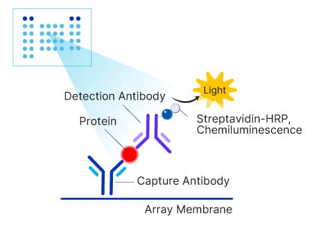
Proteome Profiler Antibody Arrays are membrane-based assays designed to simultaneous measure up to 119 proteins in a single sample. They provide a simple, economical, semi-quantitative multiplex assay for early stage discovery research. The arrays utilize chemiluminescence for detection, so membranes can be assessed for protein levels in the same manner as traditional Western blots.
| Proteome Profiler Antibody Arrays | No. of Analytes | Analytes |
| Human Chemokine Antibody Array | 31-plex | CCL1 · CCL2 · CCL3/CCL4 · CCL5 · CCL7 · CCL14 · CCL15 · CCL17 · CCL18 · CCL19 · CCL20 · CCL21 · CCL22 · CCL26 · CCL28 · Chemerin · CX3CL1 · CXCL1 · CXCL4 · CXCL5 · CXCL7 · CXCL8 · CXCL9 · CXCL10 · CXCL11 · CXCL12 · CXCL16 · CXCL17 · IL-16 · Midkine · XCL1 |
| Human Cytokine Antibody Array | 36-plex | C5a · CCL1 · CCL2 · CCL5 · CXCL1 · CXCL10 · CXCL11 · CXCL12 · CD40 Ligand · G-CSF · GM-CSF · ICAM-1 · IFN-γ · IL-1α · IL-1β · IL-1ra · IL-2 · IL-4 · IL-5 · IL-6 · IL-8 · IL-10 · IL-12 p70 · IL-13 · IL-16 · IL-17 · IL-17E · IL-18 · IL-21 · IL-27 · IL-32α · MIF · MIP-1α/MIP-1β · Serpin E1 · TNF-α · TREM-1 |
| Human XL Cytokine Antibody Array | 105-plex | Adiponectin · Angiogenin · Angiopoietin-1 · Angiopoietin-2 · Apolipoprotein A1 · BAFF · BDNF · C5a· CCL2 · CCL3/CCL4/MIP-1α/β · CCL5 · CCL7 · CCL17 · CCL19 · CCL20 · CD14 · CD30 · CD31 · CD40 Ligand · Chitinase 3-like · Complement Factor D · C-Reactive Protein · Cripto-1 · CXCL1 · CXCL4 · CXCL5 · CXCL9 · CXCL10 · CXCL11 · CXCL12 · Cystatin C · Dkk-1 · DPPIV · EGF · Endoglin · EMMPRIN · Fas Ligand · FGF-basic · FGF-7/KGF · FGF-19 · Flt-3 Ligand · G-CSF · GDF-15 · GM-CSF · Growth Hormone · HGF · ICAM-1 · IFN-γ · IGFBP-2 · IGFBP-3 · IL-1α · IL-1β · IL-1ra · IL-2 · IL-3 · IL-4 · IL-5 · IL-6 · IL-8 · IL-10 · IL-11· IL-12 p70 · IL-13 · IL-15 · IL-16 · IL-17A · IL-18 BPa · IL-19 · IL-22 · IL-23 · IL-24 · IL-27 · IL-31 · IL-32α/β/γ · IL-33 · IL-34 · Kallikrein 3 · Leptin · LIF · Lipocalin-2 · M-CSF · MIF · MMP-9 · Myeloperoxidase · Osteopontin · PDGF-AA ·PDGF-AB/BB · Pentraxin 3 · RAGE · RBP4 · Relaxin-2 · Resistin · Serpin E1 · SHBG · ST2 · TFF3 ·TfR · TGF-α · Thrombospondin-1 · TIM-1 · TNF-α · uPAR · VCAM-1 · VEGF · Vitamin D BP |
| Mouse Chemokine Antibody Array | 25-plex | C5a · CCL2 · CCL3/CCL4 · CCL5 · CCL6/C10 · CCL8 · CCL9/10/MIP-1γ · CCL11 · CCL12 · CCL21 · CCL22 · CCL27 · CCL28 · Chemerin · CX3CL1 · CXCL1 · CXCL2 · CXCL9 · CXCL10 · CXCL11 · CXCL12 · CXCL13 · CXCL16 · IL-16 · LIX |
| Mouse Cytokine Antibody Array | 40-plex | C5a · CCL1 · CCL2 · CCL3 · CCL4 · CCL5 · CCL11 · CCL12 · CCL17 · CXCL1 · CXCL2 · CXCL9 · CXCL10 · CXCL11 · CXCL12 · CXCL13 · G-CSF · GM-CSF · ICAM-1 · IFN-γ · IL-1α · IL-1β · IL-1ra · IL-2 · IL-3 · IL-4 · IL-5 · IL-6 · IL-7 · IL-10 · IL-12 p70 · IL-13 · IL-16 · IL-17 · IL-23 · IL-27 · M-CSF · TIMP-1 · TNF-α · TREM-1 |
| Mouse XL Cytokine Antibody Array | 111-plex | Adiponectin · Amphiregulin · Angiopoietin-1 · Angiopoietin-2 · Angiopoietin-like 3 · BAFF · C1q R1/CD93 · C5a · CCL2 · CCL3/CCL4/MIP-1α/β · CCL5 · CCL6/C10 · CCL11 · CCL12 · CCL17 · CCL19 · CCL20 · CCL21 · CCL22 · CD14 · CD40 · CD160 · Chemerin · Chitinase 3-like · Coagulation Factor III/TF · Complement Factor D · C-Reactive Protein · CX3CL1 · CXCL1 · CXCL2 · CXCL9 · CXCL10 · CXCL11 · CXCL13 · CXCL16 · Cystatin C · Dkk-1 · DPPIV · EGF · Endoglin · Endostatin · Fetuin A · FGF acidic · FGF-21 · Flt-3 Ligand · Gas6 · G-CSF · GDF-15 · GM-CSF · HGF · ICAM-1 · IFN-γ · IGFBP-1 · IGFBP-2 · IGFBP-3 · IGFBP-5 · IGFBP-6 · IL-1α · IL-1β · IL-1ra · IL-2 · IL-3 · IL-4 · IL-5 · IL-6 · IL-7 · IL-10 · IL-11· IL-12 p40 · IL-13 · IL-15 · IL-17A · IL-22 · IL-23 · IL-27 · IL-28 · IL-33 · LDL R · Leptin · LIF · Lipocalin-2 · LIX · M-CSF · MMP-2 · MMP-3 · MMP-9 · Myeloperoxidase · Osteopontin · Osteoprotegerin · PD-ECGF · PDGF-BB · Pentraxin 2 · Pentraxin 3 · Periostin · Pref-1 · Proliferin · Proprotein Convertase 9 · RAGE · RBP4 · Reg3G · Resistin · E-Selectin/CD62E · P-Selectin/CD62P · Serpin E1 · Serpin F1 · Thrombopoietin · TIM-1 · TNF-α · VCAM-1 · VEGF · WISP-1 |
Learn more about Proteome Profiler Antibody Arrays.
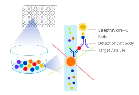
Luminex® Panels
Luminex panels are bead-based multiplex assays that enable you to multiplex up to 50 analytes, conserving on sample, time, and cost. Select from more than 490 targets spanning human, mouse, rat, porcine, and non-human primate species. Luminex Assays will deliver data that is reliable and reproducible because every panel is QC tested.
Luminex Assays are available in 3 flexible formats:
- Discovery Assays: Mix and match analyte customization
- Fixed High Performance Panels: Fully stocked and ready to ship
- Configurable High Performance Panels: Choose from a selection of the analytes or the entire panel
| Luminex High Performance Panels - Fixed | Analytes |
| Human Cytokine Panel 15-plex | IFN-α2 · IFN-γ · IL-1α · IL-1β · IL-1ra · IL-2 · IL-3 · IL-4 · IL-6 · IL-7 · IL-9 · IL-10 · IL-15 · IL-33 · VEGF |
| Human Immunotherapy Panel 25-plex | CD40 Ligand · GM-CSF · Granzyme B · IFN-α2 · IFN-γ · IL-1α · IL-1β · IL-1ra · IL-2 · IL-4 · IL-6 · IL-8 · IL-9 · IL-10 · IL-12 p70 · IL-13 · IL-15 · IL-17A · IL-33 · IP-10 · MCP-1 · MIP-1α · MIP-1β· PD-L1 · TNF-α |
| Human XL Cytokine Panel 46-plex | CD40 Ligand · EGF · Eotaxin · FGF basic · Flt-3 Ligand · G-CSF · GM-CSF · Granzyme B · GRO α · GRO β· IFN-α2 · IFN-β · IFN-γ · IL-1α · IL-1β · IL-1ra · IL-2 · IL-3 · IL-4 · IL-5 · IL-6 · IL-7 · IL-8 · IL-9 · IL-10 · IL-12 p70 · IL-13 · IL-15 · IL-17A · IL-17E · IL-33 · IP-10 · MCP-1 · MIP-1α · MIP-1β · MIP-3α · MIP-3β · PDGF-AA · PDGF-AB/BB · PD-L1 · RANTES · TGF-α · TNF-α · TNF-β · TRAIL · VEGF |
| Mouse XL Cytokine Panel 45-plex | BAFF · CCL2 · CCL3 · CCL4 · CCL5 · CCL11 · CCL19 · Chitinase 3-like 1 · CXCL1 · CXCL10 · EGF · FGF basic · Flt-3 Ligand · G-CSF · GDF-15 · GM-CSF · ICAM-1 · IFN-γ · IL-1α · IL-1β · IL-2 · IL-3 · IL-4 · IL-5 · IL-6 · IL-7 · IL-9 · IL-10 · IL-11 · IL-12 p70 · IL-13 · IL-16 · IL-17A · IL-18 · IL-21 · IL-27 · IL-31 · LDL R · LIF · LIX · M-CSF · TIMP-1 · TNF-α · VEGF |
| Luminex High Performance Panels - Configurable | Analytes |
| Human Cytokine Panel A | CCL2 · CCL3 · CCL4 · CCL5 · CXCL5 · FGF basic · G-CSF · GM-CSF · IFN-γ · IL-1α · IL-1β · IL-1ra · IL-2 · IL-4 · IL-5 · IL-6 · IL-8 · IL-10 · IL-17A · Thrombopoietin · TNF-α · VEGF |
| Human High Sensitivity Cytokine Panel A | GM-CSF · IFN-γ · IL-1β · IL-2 · IL-4 · IL-5 · IL-6 · IL-8 · IL-10 · IL-12 · TNF-α · VEGF |
| Human High Sensitivity Cytokine Panel B | GM-CSF · IFN-γ · IL-1β · IL-2 · IL-5 · IL-6 · IL-7 · IL-13 · IL-15 · IL-17A · IL-17F · IL-22 · IL-23 ·IL-31 · IL-33 · IL-36β · TNF-α |
| Human XL Cytokine Panel | CCL2 · CCL3 · CCL4 · CCL5 · CCL11 · CCL19 · CCL20 · CD40 Ligand · CXCL1 · CXCL2 · CXCL10 · EGF ·FGF basic · Flt-3 Ligand · G-CSF · GM-CSF · Granzyme B · IFN-α2 · IFN-β · IFN-γ · IL-1α · IL-1β · IL-1ra · IL-2 · IL-3 · IL-4 · IL-5 · IL-6 · IL-7 · IL-8 · IL-9 · IL-10 · IL-12 p70 · IL-13 · IL-15 · IL-17A · IL-17E · IL-33 · PD-L1 · PDGF-AA · PDGF-AB/BB · TGF-α · TNF-α · TNF-β · TRAIL · VEGF |
| Mouse XL Cytokine Panel | BAFF · CCL2 · CCL3 · CCL4 · CCL5 · CCL11 · CCL19 · Chitinase 3-like 1 · CXCL1 · CXCL10 · EGF · FGF basic · Flt-3 Ligand · G-CSF · GDF-15 · GM-CSF · ICAM-1 · IFN-γ · IL-1α · IL-1β · IL-1ra · IL-2 · IL-3 · IL-4 · IL-5 · IL-6 · IL-7 · IL-9 · IL-10 · IL-11 · IL-12 p70 · IL-13 · IL-16 · IL-17A · IL-18 · IL-21 · IL-27 · IL-31 · LDL R · LIF · LIX · M-CSF · TIMP-1 · TNF-α · VEGF |
Learn more about Luminex Assays.
Luminex® is a registered trademark of Luminex Corporation.
Additional Products for B Cell Culture and Analysis
Isolation Kits
Positive and negative cell selection kits to isolate immune cells for cell culture.
Animal-free Recombinant Proteins
Provide a seamless transition from preclinical research into clinical manufacturing, ensure defined culture conditions, and eliminate regulatory or ethical concerns associated with animal-derived reagents. Our Animal-free proteins are produced using identical expression systems and manufacturing methods as our Animal-free GMP-grade proteins.
GMP Proteins
Manufactured under guidelines that allow for their use as ancillary materials in cell therapy manufacturing processes. These proteins undergo extensive quality control testing and come with comprehensive documentation and full transparency and traceability of source and manufacturing system. Liquid, process-sized, and closed process options are also available for GMP cytokines. Learn more.
Cell Proliferation and Viability Assays
Reagents for studying cell proliferation, viability, and cytotoxicity including fluorescent reporter dyes and MTT assays.
Featured Content
B Cell Development Poster
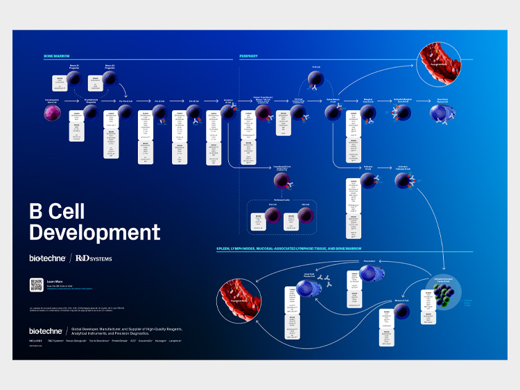
Simplify the characterization of different stages of human and mouse B cell development with our B cell markers poster.
Human Immune Cell Markers Poster
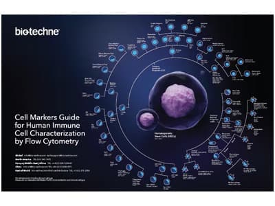
Explore this poster to find markers used to distinguish different immune cell types by flow cytometry.
Simple Reader™ Compact ELISA Plate Reader

Save space and costs with Simple Reader™, a compact, cost-effective plate reader offering precise results.
Periodic Table of Human Cytokine & Chemokine Families Poster
Learn about different cytokine families. This poster will serve as a great reference tool and a colorful addition to your lab.
Control Variables & Save with Bulk Reagents
Receive competitive discounts on bulk quantities of a single item or multiple items in one order.