Interleukin-4 (IL-4), also known as B cell-stimulatory factor-1, is a monomeric, approximately 13 kDa‑18 kDa Th2 cytokine that shows pleiotropic effects during immune responses (1‑3). It is a glycosylated polypeptide that contains three intrachain disulfide bridges and adopts a bundled four alpha -helix structure (4). Bovine IL-4 is synthesized with a 24 amino acid (aa) signal sequence. Alternate splicing generates two additional isoforms with internal deletions (5). Mature bovine IL-4 shares 60%, 91%, 93%, 78%, 55%, 39%, and 41% aa sequence identity with equine, goat, ovine, porcine, human, mouse, and rat IL-4, respectively. IL-4 exerts its effects through two receptor complexes (6, 7). The type I receptor, which is expressed on hematopoietic cells, is a heterodimer of the ligand binding IL-4 R alpha and the common gamma chain (a shared subunit of the receptors for IL-2, -7, -9, -15, and -21). The type II receptor on nonhematopoietic cells consists of IL-4 R alpha and IL-13 R alpha 1. The type II receptor also transduces IL-13 mediated signals. IL-4 is primarily expressed by Th2-biased CD4+ T cells, mast cells, basophils, and eosinophils (1, 2). It promotes cell proliferation, survival, and immunoglobulin class switch to IgE in B cells, acquisition of the Th2 phenotype by naïve CD4+ T cells, priming and chemotaxis of mast cells, eosinophils, and basophils, and the proliferation and activation of epithelial cells (8‑11). IL-4 plays a dominant role in the development of allergic inflammation and asthma (10, 12).

Key Product Details
Species Reactivity
Bovine
Applications
Western Blot, Immunocytochemistry
Label
Unconjugated
Antibody Source
Monoclonal Mouse IgG2B Clone # 701214
Loading...
Product Specifications
Immunogen
E. coli-derived recombinant bovine IL-4
His25-Cys135
Accession # P30367.2
His25-Cys135
Accession # P30367.2
Specificity
Detects bovine IL-4 in direct ELISAs and Western blots. In direct ELISAs, less than 25% cross-reactivity with
recombinant porcine, canine, rhesus, cotton rat, equine, or rabbit IL-4 is
observed and no cross-reactivity with recombinant human, mouse, rat, or feline
IL-4 is observed.
Clonality
Monoclonal
Host
Mouse
Isotype
IgG2B
Scientific Data Images for Bovine IL-4 Antibody (701214)
Detection of Bovine IL‑4 by Western Blot.
Western blot shows Recombinant Bovine IL-4 (Catalog # 2469-BL) (10 ng/lane). PVDF membrane was probed with 2 µg/mL of Mouse Anti-Bovine IL-4 Monoclonal Antibody (Catalog # MAB2469) followed by HRP-conjugated Anti-Mouse IgG Secondary Antibody (Catalog # HAF007). A specific band was detected for IL-4 at approximately 12 kDa (as indicated). This experiment was conducted under reducing conditions and using Immunoblot Buffer Group 1.
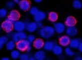
IL‑4 in Bovine PBMCs.
IL-4 was detected in immersion fixed bovine peripheral blood mononuclear cells (PBMCs) stimulated with concanavalin A using Mouse Anti-Bovine IL-4 Monoclonal Antibody (Catalog # MAB2469) at 25 µg/mL for 3 hours at room temperature. Cells were stained using the NorthernLights™ 557-conjugated Anti-Mouse IgG Secondary Antibody (red; Catalog # NL007) and counterstained with DAPI (blue). Specific staining was localized to cell cytoplasm. View our protocol for Fluorescent ICC Staining of Non-adherent Cells.Applications for Bovine IL-4 Antibody (701214)
Application
Recommended Usage
Immunocytochemistry
8-25 µg/mL
Sample: Immersion fixed bovine peripheral blood mononuclear bells (PBMCs) stimulated with concanavalin A
Sample: Immersion fixed bovine peripheral blood mononuclear bells (PBMCs) stimulated with concanavalin A
Western Blot
2 µg/mL
Sample: Recombinant Bovine IL‑4 (Catalog # 2469-BL)
Sample: Recombinant Bovine IL‑4 (Catalog # 2469-BL)
Reviewed Applications
Read 1 review rated 5 using MAB2469 in the following applications:
Formulation, Preparation, and Storage
Purification
Protein A or G purified from hybridoma culture supernatant
Reconstitution
Sterile PBS to a final concentration of 0.5 mg/mL. For liquid material, refer to CoA for concentration.
Loading...
Formulation
Lyophilized from a 0.2 μm filtered solution in PBS with Trehalose. *Small pack size (SP) is supplied either lyophilized or as a 0.2 µm filtered solution in PBS.
Shipping
Lyophilized product is shipped at ambient temperature. Liquid small pack size (-SP) is shipped with polar packs. Upon receipt, store immediately at the temperature recommended below.
Stability & Storage
Use a manual defrost freezer and avoid repeated freeze-thaw cycles.
- 12 months from date of receipt, -20 to -70 °C as supplied.
- 1 month, 2 to 8 °C under sterile conditions after reconstitution.
- 6 months, -20 to -70 °C under sterile conditions after reconstitution.
Calculators
Background: IL-4
References
- Benczik, M. and S.L. Gaffen (2004) Immunol. Invest. 33:109.
- Chomarat, P. and J. Banchereau (1998) Int. Rev. Immunol. 17:1.
- Heussler, V.T. et al. (1992) Gene 114:273.
- Redfield, C. et al. (1991) Biochemistry 30:11029.
- Waldvogel, A.S. et al. (2004) Vet. Immunol. Immunopathol. 97:53.
- Mueller, T.D. et al. (2002) Biochim. Biophys. Acta 1592:237.
- Nelms, K. et al. (1999) Annu. Rev. Immunol. 17:701.
- Paludan, S.R. (1998) Scand. J. Immunol. 48:459.
- Corthay, A. (2006) Scand. J. Immunol. 64:93.
- Ryan, J.J. et al. (2007) Crit. Rev. Immunol. 27:15.
- Grone, A. (2002) Vet. Immunol. Immunopathol. 88:1.
- Rosenberg, H.F. et al. (2007) J. Allergy Clin. Immunol. 119:1303.
Long Name
Interleukin 4
Alternate Names
BCDF, BCGF-1, BCGF1, BSF-1, BSF1, IL4, IL4E12
Entrez Gene IDs
Gene Symbol
IL4
UniProt
Additional IL-4 Products
Product Documents for Bovine IL-4 Antibody (701214)
Certificate of Analysis
To download a Certificate of Analysis, please enter a lot or batch number in the search box below.
Note: Certificate of Analysis not available for kit components.
Product Specific Notices for Bovine IL-4 Antibody (701214)
For research use only
Related Research Areas
Citations for Bovine IL-4 Antibody (701214)
 Powered by Bioz
Customer Reviews for Bovine IL-4 Antibody (701214) (1)
5 out of 5
1 Customer Rating
Have you used Bovine IL-4 Antibody (701214)?
Submit a review and receive an Amazon gift card!
$25/€18/£15/$25CAN/¥2500 Yen for a review with an image
$10/€7/£6/$10CAN/¥1110 Yen for a review without an image
Submit a review
Customer Images
Showing
1
-
1 of
1 review
Showing All
Filter By:
-
Application: Immunocytochemistry/ImmunofluorescenceSample Tested: Peripheral blood mononuclear cells (PBMCs)Species: BovineVerified Customer | Posted 06/27/2022

There are no reviews that match your criteria.
Protocols
Find general support by application which include: protocols, troubleshooting, illustrated assays, videos and webinars.
- Appropriate Fixation of IHC/ICC Samples
- Cellular Response to Hypoxia Protocols
- ClariTSA™ Fluorophore Kits
- Detection & Visualization of Antibody Binding
- ICC Cell Smear Protocol for Suspension Cells
- ICC Immunocytochemistry Protocol Videos
- ICC for Adherent Cells
- Immunocytochemistry (ICC) Protocol
- Immunocytochemistry Troubleshooting
- Immunofluorescence of Organoids Embedded in Cultrex Basement Membrane Extract
- Immunohistochemistry (IHC) and Immunocytochemistry (ICC) Protocols
- Preparing Samples for IHC/ICC Experiments
- Preventing Non-Specific Staining (Non-Specific Binding)
- Primary Antibody Selection & Optimization
- Protocol for VisUCyte™ HRP Polymer Detection Reagent
- Protocol for the Fluorescent ICC Staining of Cell Smears - Graphic
- Protocol for the Fluorescent ICC Staining of Cultured Cells on Coverslips - Graphic
- Protocol for the Preparation and Fluorescent ICC Staining of Cells on Coverslips
- Protocol for the Preparation and Fluorescent ICC Staining of Non-adherent Cells
- Protocol for the Preparation and Fluorescent ICC Staining of Stem Cells on Coverslips
- Protocol for the Preparation of a Cell Smear for Non-adherent Cell ICC - Graphic
- R&D Systems Quality Control Western Blot Protocol
- TUNEL and Active Caspase-3 Detection by IHC/ICC Protocol
- The Importance of IHC/ICC Controls
- Troubleshooting Guide: Western Blot Figures
- Western Blot Conditions
- Western Blot Protocol
- Western Blot Protocol for Cell Lysates
- Western Blot Troubleshooting
- Western Blot Troubleshooting Guide
- View all Protocols, Troubleshooting, Illustrated assays and Webinars
Loading...
Associated Pathways


