B7-1 and B7-2, together with their receptors CD28 and CTLA-4, constitute one of the dominant co-stimulatory pathways that regulate T- and B-cell responses. Although both CTLA-4 and CD28 can bind to the same ligands, CTLA-4 binds to B7-1 and B7-2 with a 20‑100 fold higher affinity than CD28 and is involved in the down‑regulation of the immune response. B7-1 is expressed on activated B cells, activated T cells, and macrophages. B7-2 is constitutively expressed on interdigitating dendritic cells, Langerhans cells, peripheral blood dendritic cells, memory B cells, and germinal center B cells. Additionally, B7-2 is expressed at low levels on monocytes and can be up‑regulated through Interferon gamma. B7-1 and B7-2 are both members of the Immunoglobulin superfamily. Human B7-1 is a 288 amino acid (aa) protein containing a 34 aa signal peptide, a 208 aa extracellular domain, a 21 aa transmembrane domain, and a 25 aa cytoplasmic domain. Human B7-1 and B7-2 share 26% aa sequenceidentity. Human and mouse B7-1 share 44% aa sequenceidentity. However, it has been observed that both human and mouse B7-1 and B7-2 can bind to either human or mouse CD28 and CTLA-4, suggesting that there are conserved amino acids which form the B7-1/B7-2/CD28/CTLA-4 critical binding sites.
Human B7-1/CD80 Antibody (37711)
R&D Systems | Catalog # MAB140

Key Product Details
Validated by
Orthogonal Validation, Biological Validation
Species Reactivity
Validated:
Human
Cited:
Human, Mouse
Applications
Validated:
Immunohistochemistry, ELISA Capture (Matched Antibody Pair), Neutralization, Flow Cytometry, Dual RNAscope ISH-IHC Compatible, CyTOF-ready
Cited:
Immunohistochemistry, Immunohistochemistry-Paraffin, Western Blot, Neutralization, Flow Cytometry, Immunocytochemistry, ELISA Capture, ELISA Development (Detection), Functional Assay, T Cell Activation
Label
Unconjugated
Antibody Source
Monoclonal Mouse IgG1 Clone # 37711
Loading...
Product Specifications
Immunogen
S. frugiperda insect ovarian cell line Sf 21-derived recombinant human B7‑1/CD80
Val35-Asn242 (predicted)
Accession # P33681
Val35-Asn242 (predicted)
Accession # P33681
Specificity
Detects human B7‑1/CD80 in ELISAs. In sandwich immunoassays, no cross-reactivity or interference with recombinant human (rh) B7-2, recombinant mouse (rm) B7-1, rmB7-2, rhB7-H1, rhB7-H2, rhB7-H3 or rmPD-L2 is observed.
Clonality
Monoclonal
Host
Mouse
Isotype
IgG1
Endotoxin Level
<0.10 EU per 1 μg of the antibody by the LAL method.
Scientific Data Images for Human B7-1/CD80 Antibody (37711)
Detection of B7‑1/CD80 in Raji Human Cell Line by Flow Cytometry.
Raji human Burkitt's lymphoma cell line was stained with Mouse Anti-Human B7-1/CD80 Monoclonal Antibody (Catalog # MAB140, filled histogram) or isotype control antibody (Catalog # MAB002, open histogram), followed by Fluorescein-conjugated Anti-Mouse IgG Secondary Antibody (Catalog # F0103B). View our protocol for Staining Membrane-associated Proteins.
IL‑2 secretion Induced by B7‑1/CD80 and Neutralization by Human B7‑1/CD80 Antibody.
Recombinant Human B7-1/CD80 Fc Chimera (Catalog # 140-B1) co-stimulates IL-2 secretion in the Jurkat human acute T cell leukemia cell line in the presence of PHA in a dose-dependent manner (orange line), as measured by the Human IL-2 Quantikine ELISA Kit (Catalog # D2050). IL-2 secretion elicited by Recombinant Human B7-1/CD80 Fc Chimera (1 µg/mL) and PHA (10 µg/mL) is neutralized (green line) by increasing concentrations of Mouse Anti-Human B7-1/CD80 Monoclonal Antibody (Catalog # MAB140). The ND50 is typically 0.2-1 µg/mL.
B7‑1/CD80 in Human Tonsil.
B7-1/CD80 was detected in immersion fixed paraffin-embedded sections of human tonsil using Mouse Anti-Human B7-1/CD80 Monoclonal Antibody (Catalog # MAB140) at 5 µg/mL overnight at 4 °C. Tissue was stained using the Anti-Mouse HRP-DAB Cell & Tissue Staining Kit (brown; Catalog # CTS002) and counterstained with hematoxylin (blue). Specific staining was localized to cytoplasm in lymphocytes. View our protocol for Chromogenic IHC Staining of Paraffin-embedded Tissue Sections.
B7-1/CD80 in Human Tonsil Using Dual RNAscope®ISH and IHC.
B7-1/CD80 mRNA was detected in formalin-fixed paraffin-embedded tissue sections of human tonsil probed with ACD RNAScope®Probe (Catalog # 42471) and stained using ACD RNAscope®2.5 HD Duplex Detection Reagents (red, Catalog # 322500). Adjacent tissue section was processed for immunohistochemistry using R&D Systems Mouse Anti-Human B7-1/CD80 Monoclonal Antibody (Catalog# MAB140) at 5ug/mL with overnight incubation at 4 °C followed by incubation with the Anti-Mouse IgG VisUCyte HRP Polymer Antibody (R&D Systems, Catalog # VC001) and DAB chromogen (lower image, brown). Tissues were counterstained with hematoxylin (blue).
Detection of Mouse B7-1/CD80 by Immunohistochemistry
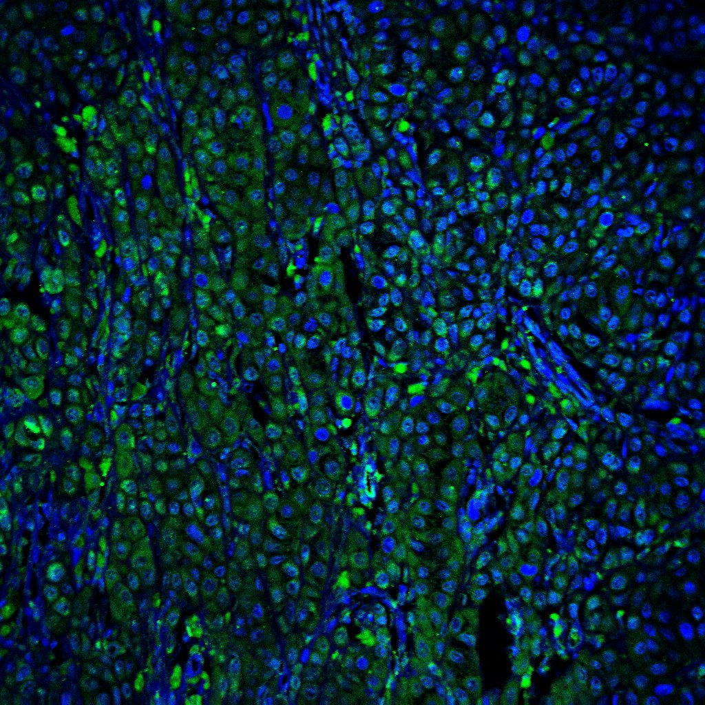
MNK inhibitors induce polarization of TAMs toward an M2 phenotype in ex vivo human PDAC slice cultures.(A) Serial sections of human PDAC tumors (n = 15) were stained for Arginase-1 and phosphorylated eIF4ES209 (p-eIF4E) by IHC. Scale bar: 200 μm. The staining positivity was analyzed by the TissueGnostics Imaging system using the HistoQuest, and the correlation analysis for Arginase-1 and p-eIF4E expression was performed using GraphPad. (B) Slice cultures (n = 1–4 replicates for each treatment group) from 6 different PDAC tumors were treated with DMSO (vehicle control) or CGP57380 for 5 days and stained for phosphorylated eIF4E (p-eIF4E), CD68, Arginase-1, CD163, MRC1, CD86, CD80, and MHCII. The percentage of positive cells, relative to the total number of nucleated cells, was analyzed by ImageJ. Scale bar: 50 μm. Data points represent individual patients. Paired t test was performed by GraphPad. *P < 0.05; **P < 0.01. (C) The relative expression of MKNK2, CD163, and MRC1 in TCGA studies and their transcript abundance from RNA-Seq data, quantified as RSEM, were downloaded from cBioPortal. Correlation analysis was performed in GraphPad to evaluate the relationship between MKNK2, CD163, and MRC1. Image collected and cropped by CiteAb from the following publication (https://pubmed.ncbi.nlm.nih.gov/35380995), licensed under a CC-BY license. Not internally tested by R&D Systems.
Human B7-1 / CD80 ELISA Standard Curve
Recombinant Human B7‑1/CD80 Fc Chimera (Catalog # 140-B1) was serially diluted and captured by Mouse Anti-Human B7‑1/CD80 Monoclonal Antibody (Catalog # MAB140) coated on a Clear Polystyrene Microplate (Catalog # DY990). Mouse Anti-Human B7‑1/CD80 Biotinylated Monoclonal Antibody (Catalog # ) was incubated with the protein captured on the plate. Detection of the standard curve was achieved by incubating Streptavidin-HRP (Catalog # DY998)Applications for Human B7-1/CD80 Antibody (37711)
Application
Recommended Usage
CyTOF-ready
Ready to be labeled using established conjugation methods. No BSA or other carrier proteins that could interfere with conjugation.
Dual RNAscope ISH-IHC Compatible
5-25 µg/mL
Sample: Immersion fixed paraffin-embedded sections of human tonsil tissue
Sample: Immersion fixed paraffin-embedded sections of human tonsil tissue
Flow Cytometry
0.25 µg/106 cells
Sample: Raji human Burkitt's lymphoma cell line
Sample: Raji human Burkitt's lymphoma cell line
Immunohistochemistry
5-25 µg/mL
Sample: Immersion fixed paraffin-embedded sections of human tonsil tissue
Sample: Immersion fixed paraffin-embedded sections of human tonsil tissue
Neutralization
Measured by its ability to neutralize B7‑1/CD80 and PHA-induced IL‑2 secretion in the Jurkat human acute T cell leukemia cell line. The Neutralization Dose (ND50) is typically 0.2-1 µg/mL in the presence of 1 µg/mL Recombinant Human B7‑1/CD80 Fc Chimera and 10 µg/mL PHA.
Human B7-1/CD80 Sandwich Immunoassay
Please Note: Optimal dilutions of this antibody should be experimentally determined.
Reviewed Applications
Read 2 reviews rated 3.5 using MAB140 in the following applications:
Flow Cytometry Panel Builder
Bio-Techne Knows Flow Cytometry
Save time and reduce costly mistakes by quickly finding compatible reagents using the Panel Builder Tool.
Advanced Features
- Spectra Viewer - Custom analysis of spectra from multiple fluorochromes
- Spillover Popups - Visualize the spectra of individual fluorochromes
- Antigen Density Selector - Match fluorochrome brightness with antigen density

Formulation, Preparation, and Storage
Purification
Protein A or G purified from hybridoma culture supernatant
Reconstitution
Reconstitute at 0.5 mg/mL in sterile PBS. For liquid material, refer to CoA for concentration.
Loading...
Formulation
Lyophilized from a 0.2 μm filtered solution in PBS with Trehalose. *Small pack size (SP) is supplied either lyophilized or as a 0.2 µm filtered solution in PBS.
Shipping
Lyophilized product is shipped at ambient temperature. Liquid small pack size (-SP) is shipped with polar packs. Upon receipt, store immediately at the temperature recommended below.
Stability & Storage
Use a manual defrost freezer and avoid repeated freeze-thaw cycles.
- 12 months from date of receipt, -20 to -70 °C as supplied.
- 1 month, 2 to 8 °C under sterile conditions after reconstitution.
- 6 months, -20 to -70 °C under sterile conditions after reconstitution.
Calculators
Background: B7-1/CD80
References
- Azuma, M. et al. (1993) Nature 366:76.
- Freeman, G.J. et al. (1993) Science 262:909.
- Freeman, G. et al. (1991) J. Exp. Med. 174:625.
- Selvakumar, A. et al. (1993) Immunogenetics 38:292.
- Chen, C. et al. (1994) J. Immunol. 152:4929.
- Freeman, G.J. et al. (1993) J. Exp. Med. 178:2185.
Alternate Names
B71, CD80
Gene Symbol
CD80
UniProt
Additional B7-1/CD80 Products
Product Documents for Human B7-1/CD80 Antibody (37711)
Certificate of Analysis
To download a Certificate of Analysis, please enter a lot or batch number in the search box below.
Note: Certificate of Analysis not available for kit components.
Product Specific Notices for Human B7-1/CD80 Antibody (37711)
For research use only
Related Research Areas
Citations for Human B7-1/CD80 Antibody (37711)
 Powered by Bioz
Customer Reviews for Human B7-1/CD80 Antibody (37711) (2)
3.5 out of 5
2 Customer Ratings
Have you used Human B7-1/CD80 Antibody (37711)?
Submit a review and receive an Amazon gift card!
$25/€18/£15/$25CAN/¥2500 Yen for a review with an image
$10/€7/£6/$10CAN/¥1110 Yen for a review without an image
Submit a review
Customer Images
Showing
1
-
2 of
2 reviews
Showing All
Filter By:
-
Application: Flow CytometrySample Tested: Dendritic cellsSpecies: HumanVerified Customer | Posted 10/11/2022
-
Application: Immunocytochemistry/ImmunofluorescenceSample Tested: Melanoma tissueSpecies: HumanVerified Customer | Posted 04/11/2022

There are no reviews that match your criteria.
Protocols
Find general support by application which include: protocols, troubleshooting, illustrated assays, videos and webinars.
- 7-Amino Actinomycin D (7-AAD) Cell Viability Flow Cytometry Protocol
- Antigen Retrieval Protocol (PIER)
- Antigen Retrieval for Frozen Sections Protocol
- Appropriate Fixation of IHC/ICC Samples
- Cellular Response to Hypoxia Protocols
- Chromogenic IHC Staining of Formalin-Fixed Paraffin-Embedded (FFPE) Tissue Protocol
- Chromogenic Immunohistochemistry Staining of Frozen Tissue
- ClariTSA™ Fluorophore Kits
- Detection & Visualization of Antibody Binding
- Extracellular Membrane Flow Cytometry Protocol
- Flow Cytometry Protocol for Cell Surface Markers
- Flow Cytometry Protocol for Staining Membrane Associated Proteins
- Flow Cytometry Staining Protocols
- Flow Cytometry Troubleshooting Guide
- Fluorescent IHC Staining of Frozen Tissue Protocol
- Graphic Protocol for Heat-induced Epitope Retrieval
- Graphic Protocol for the Preparation and Fluorescent IHC Staining of Frozen Tissue Sections
- Graphic Protocol for the Preparation and Fluorescent IHC Staining of Paraffin-embedded Tissue Sections
- Graphic Protocol for the Preparation of Gelatin-coated Slides for Histological Tissue Sections
- IHC Sample Preparation (Frozen sections vs Paraffin)
- ISH-IHC Protocol for Chromogenic Detection on Formalin Fixed Paraffin Embedded (FFPE) Tissue
- Immunofluorescent IHC Staining of Formalin-Fixed Paraffin-Embedded (FFPE) Tissue Protocol
- Immunohistochemistry (IHC) and Immunocytochemistry (ICC) Protocols
- Immunohistochemistry Frozen Troubleshooting
- Immunohistochemistry Paraffin Troubleshooting
- Intracellular Flow Cytometry Protocol Using Alcohol (Methanol)
- Intracellular Flow Cytometry Protocol Using Detergents
- Intracellular Nuclear Staining Flow Cytometry Protocol Using Detergents
- Intracellular Staining Flow Cytometry Protocol Using Alcohol Permeabilization
- Intracellular Staining Flow Cytometry Protocol Using Detergents to Permeabilize Cells
- Preparing Samples for IHC/ICC Experiments
- Preventing Non-Specific Staining (Non-Specific Binding)
- Primary Antibody Selection & Optimization
- Propidium Iodide Cell Viability Flow Cytometry Protocol
- Protocol for Heat-Induced Epitope Retrieval (HIER)
- Protocol for Liperfluo
- Protocol for Making a 4% Formaldehyde Solution in PBS
- Protocol for VisUCyte™ HRP Polymer Detection Reagent
- Protocol for the Characterization of Human Th22 Cells
- Protocol for the Characterization of Human Th9 Cells
- Protocol for the Preparation & Fixation of Cells on Coverslips
- Protocol for the Preparation and Chromogenic IHC Staining of Frozen Tissue Sections
- Protocol for the Preparation and Chromogenic IHC Staining of Frozen Tissue Sections - Graphic
- Protocol for the Preparation and Chromogenic IHC Staining of Paraffin-embedded Tissue Sections
- Protocol for the Preparation and Chromogenic IHC Staining of Paraffin-embedded Tissue Sections - Graphic
- Protocol for the Preparation and Fluorescent IHC Staining of Frozen Tissue Sections
- Protocol for the Preparation and Fluorescent IHC Staining of Paraffin-embedded Tissue Sections
- Protocol for the Preparation of Gelatin-coated Slides for Histological Tissue Sections
- Protocol: Annexin V and PI Staining by Flow Cytometry
- Protocol: Annexin V and PI Staining for Apoptosis by Flow Cytometry
- TUNEL and Active Caspase-3 Detection by IHC/ICC Protocol
- The Importance of IHC/ICC Controls
- Troubleshooting Guide: Fluorokine Flow Cytometry Kits
- Troubleshooting Guide: Immunohistochemistry
- View all Protocols, Troubleshooting, Illustrated assays and Webinars


